
A ratiometric fluorescence probe for detection of hydrogen sulfide in cells†
Bin Wang,
Na Jiang*,
Wei Sun,
Qiufen Wang and
Gengxiu Zheng*
School of Chemistry and Chemical Engineering, University of Jinan, 336 Nanxinzhuang West Road, 250022, Jinan, China. E-mail: chm_jiangn@ujn.edu.cn; chm_zhenggx@ujn.edu.cn
First published on 30th March 2016
Abstract
A near-infrared ratiometric fluorescence probe for detection of H2S has been designed, synthesized and evaluated. The probe features high selectivity and sensitivity. Confocal microscopy imaging experiments demonstrate that the probe has potential as a powerful tool for the imaging of H2S in living cells.
Recently, hydrogen sulfide (H2S), a venomous gas with a stinky smell, has been recognized as an important signaling molecule following carbon monoxide (CO) and nitric oxide (NO).1–3 H2S normally generates in mammalian cells by enzymes, including cystathionine β-synthase (CBS), cystathionine γ-lyase (CSE), 3-mercapto-pyruvate sulfurtransferase (3-MST), and cysteine, and derivatives serve as the substrates.4–8 The abnormity of the concentration level of H2S is generally one of the key factors, which both causes and develops in many diseases such as chronic kidney disease, liver cirrhosis, Down's syndrome and Alzheimer's disease.9 Therefore, detection of H2S is of great importance for better understanding its biological functions.
Near-infrared (NIR) fluorescence probes can markedly reduce background absorption and light scattering to allow in vivo imaging. Thus, it is still necessary to develop NIR fluorescence probes.10–16 Considering their non-destructiveness, simplicity and sensitive nature, fluorescence probes, which are used for analyzing H2S, have received widespread attention. To date, many fluorescence probes for H2S have been reported.17–19 Most of them have the capability to detect H2S of high sensitivity and selectivity; however, they mainly depend on intensity changes in a single-emission window, so low accuracy limits the development of this types of probe in a biological environment.20,21 On the other hand, the ratiometric fluorescence probe, which recognizes the analyte by self-calibration at two emission bands, can alleviate this shortcoming and provide more reliable analysis results.22 Although some available ratiometric fluorescence probes for H2S have been reported to date,23,24 they suffer from either a long response time or a relatively small fluorescence emission ratio enhancement. Thus, ratiometric fluorescence probes for H2S still need to be developed.
In this study, a NIR ratiometric fluorescence probe BOC (Fig. 1), which is based on intramolecular charge transfer (ICT) strategy, was reported. Coumarin was selected as the donor because of its high quantum yields, high extinction coefficients and ease of synthesis. Concerning the acceptor, we chose the benzothiazole group due to its electron-withdrawing aromatic system, which is also capable of extending electron conjugation. Moreover, the quaternized aromatic amino groups impart good water solubility to the probe and the benzothiazole C-2 atom is an effective nucleophilic addition target for selective detecting HS− in a medium of near-neutral pH value.24 Therefore, the probe was conveniently synthesized through a simple condensation of 7-diethylaminocoumarin-3-aldehyde with 3-((2′-cyanobiphenyl-4-yl)methyl)-2-methyl benzothiaz-ol-3-ium and favorable for the dual emission ratiometric imaging of H2S in living cells. BOC showed two well-resolved emission bands (492 nm and 652 nm, excited at 430 nm) in PBS buffer (10 mM, pH 7.4) containing 70% CH3CN. In addition, BOC features high selectivity and sensitivity with a response time of 45 seconds. All these indicate that it is a promising probe for detection of H2S.
 | ||
| Fig. 1 Synthesis of probe BOC. | ||
The synthetic route of BOC is outlined in Scheme S1.† The product was characterized by electrospray ionization mass spectrometry and nuclear magnetic resonance (NMR) spectroscopy. The spectral properties of BOC in various solvents are shown in Table S1.† BOC (10 μM) shows two absorption maxima (354 nm and 565 nm) and two emission bands centered at 482 nm and 653 nm in CH3CN (Fig. S1†).
The absorption spectra of BOC (10 μM) displays a strong ICT band at 562 nm (Fig. 2a). This band decreased gradually with one isosbestic point at 465 nm upon HS− titration, indicating the formation of a new compound with a growth of 400 nm and confirming the disruption of the ICT effect in the whole molecule caused by the nucleophilic attack of HS− group, which not only interrupted the π-electron conjugation, but also blocked the ICT progress. The solution turned from purple to faint yellow, thereby suggesting HS− can be detected with the naked eye when BOC is used. On the other hand, the distinct gap between the two emission bands is 160 nm, which makes this probe favorable for dual emission ratiometric imaging owing to the minimum overlap between the two bands. As shown in Fig. 2b, fluorescence titration of a BOC solution with HS− (0–366.7 μM) demonstrated that its emission band at 652 nm obviously decreased, whereas the band at 492 nm underwent a noticeable increase simultaneously. The intensity ratio of the two emission bands (I492/I652) increased from 0.77 to 9.14. Moreover, BOC showed a good linear relationship (R2 = 0.98) between fluorescence intensity (log![[thin space (1/6-em)]](https://www.rsc.org/images/entities/char_2009.gif) I492/I652) and concentrations of HS− (from 0 to 366.7 μM, Fig. 2c). Consequently, a ratiometric near-infrared fluorescence probe for H2S was achieved. As reported, the benzothiazole C-2 atom was a HS− addition site.24 For BOC, the same detection principles were proved by the 1H NMR titration experiment (Fig. S2†). In general, these results indicate that BOC can be used as an attractive ratiometric near-infrared fluorescence probe for reliable detection of H2S.
I492/I652) and concentrations of HS− (from 0 to 366.7 μM, Fig. 2c). Consequently, a ratiometric near-infrared fluorescence probe for H2S was achieved. As reported, the benzothiazole C-2 atom was a HS− addition site.24 For BOC, the same detection principles were proved by the 1H NMR titration experiment (Fig. S2†). In general, these results indicate that BOC can be used as an attractive ratiometric near-infrared fluorescence probe for reliable detection of H2S.
 | ||
| Fig. 2 (a) Absorption spectra of BOC (10 μM) in PBS (10 mM, pH 7.40, 70% CH3CN, v/v) obtained upon titration with HS− from 0 to 366.7 μM. (b) Fluorescence spectra. (c) The linear relationship between fluorescence intensity ratio (I492/I652) and concentration of HS−. Spectra were obtained 45 s after HS− addition, λex = 430 nm. | ||
Then, the selectivity of BOC to H2S was investigated (Fig. 3). Only HS− induces a dramatic increment of emission ratio I492/I652, whereas other ions and biothiols only trigger minor changes compared to that induced by HS−. Though the classical nucleophile CN− induced a minor change in the ratio, its low concentration under physical condition made the interference of CN− negligible. This high selectivity and sensitivity can be explained by the strong nucleophilic property of HS− and its relatively small size and a low pKa of around 6.8, whereas other thiols (Cys, Hcy and GSH) have higher pKa values, and the steric effect makes the nucleophilic reaction more difficult. In addition, an obvious enhancement of emission ratio I492/I652 was observed after subsequent addition of HS− to the BOC solution in the presence of various ions and biothiols (Fig. S3†). All these investigations indicate that the interference of these ions and thiol species is very small, and detection of HS− using BOC is still effective. Therefore, BOC was available for the detection of HS− under environmental conditions.
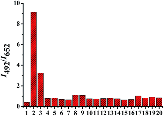 | ||
| Fig. 3 Emission ratio I492/I652 of BOC (10 μM) in PBS (10 mM, pH 7.40, 70% CH3CN, v/v) in the presence of HS−, various ions and biologically relevant species. (1) Probe alone, (2) HS− (333 μM), (3) CN−, (4) Na+, (5) K+, (6) Ca2+, (7) NH4+, (8) Ba2+, (9) Cu+, (10) F−, (11) Br−, (12) NO2−, (13) ClO4−, (14) SO42−, (15) S2O82−, (16) S2O32−, (17) SCN−, (18) Cys, (19) GSH, (20) Hcy (4–17: 5 mM; and 3, 18–20: 1 mM). λex = 430 nm. | ||
In order to determine the sensing performance of BOC under physiological conditions, we further investigated the influence of pH on the fluorescence behavior of BOC for the detection of HS−. It can be seen in Fig. S4† that the ratio I492/I652 for BOC was barely affected within a wide pH range from 4.0 to 9.0. Within the same pH range, enormous enhancement of I492/I652 was observed when BOC was treated with HS−. These results suggested that BOC was suitable as a ratiometric fluorescent probe for the detection of HS− under physiological conditions. The time-dependent fluorescence emission intensity changes (at 495 nm) of BOC in the presence of HS− (Fig. S5†) showed that the reaction can be completed within 45 seconds, which is faster than that of the reported probes.25
For the sake of assessing the photophysical behavior of the prepared coumarin–hemicyanine hybrids in biological environments, BOC was examined in HeLa and MCF-7 cells. HeLa or MCF-7 cells were incubated with BOC (5 μM) for 30 min at 37 °C, then washed with PBS three times. As shown in Fig. 4, BOC was almost gathered in the cytoplasm and the cells were stained with a weak fluorescence from the green channel and a clear fluorescence from the red channel (Fig. 4a and b). When treated with 200 μM HS− for another 30 min, a clear fluorescence was observed from the green channel and the fluorescence in the red channel was weakened under the same conditions (Fig. 4d and e). Furthermore, a histogram was made to better present the photophysical behavior of BOC treated with HS−, from which we can see that the addition of HS− turned the emission of BOC from red channel to green channel (Fig. 4g). The same results were obtained from MCF-7 cells (Fig. S6†). It is apparent that the staining results were consistent with the results of HS− titration in vitro. Taken together, these experiments demonstrate that BOC was cell permeable and could monitor the intracellular H2S in living samples.
 | ||
| Fig. 4 Confocal fluorescence images (a) and (b) of HeLa cells incubated with 5 μM BOC. Images (d) and (e) of the above HeLa cells after adding 200 μM HS−. Ratio images (c) and (f) of green channel compared to the red channel. (g) Ratio of 10 points drawn from (c) and (f), respectively; black bars represent BOC and red bars represent BOC + HS−. | ||
Conclusions
In conclusion, a ratiometric fluorescence probe for imaging H2S in living cells based on the coumarin–hemicyanine dye has been developed. This probe displayed high sensitivity and selectivity toward H2S. Significantly, BOC has a linear ratiometric fluorescence response to H2S in PBS (10 mM, pH 7.40, 70% CH3CN, v/v). Furthermore, preliminary biological experiments have demonstrated the value of this probe by monitoring the intracellular H2S in living biological samples.Notes and references
- L. Li, P. Rose and P. K. Moore, Annu. Rev. Pharmacol. Toxicol., 2011, 51, 169–187 CrossRef CAS PubMed.
- J. M. Fukuto, S. J. Carrington, D. J. Tantillo, J. G. Harrison, L. J. Ignarro, B. A. Freeman, A. Chen and D. A. Wink, Chem. Res. Toxicol., 2012, 25, 769–793 CrossRef CAS PubMed.
- T. L. Guidotti, Int. J. Toxicol., 2010, 29, 569–581 CrossRef CAS PubMed.
- G. K. Kolluru, X. Shen, S. C. Bir and C. G. Kevil, Nitric Oxide, 2013, 35, 5–20 CrossRef CAS PubMed.
- R. Wang, Physiol. Rev., 2012, 92, 791–896 CrossRef CAS PubMed.
- O. Kabil and R. Banerjee, J. Biol. Chem., 2010, 285, 21903–21907 CrossRef CAS PubMed.
- O. Kabil and R. Banerjee, Antioxid. Redox Signaling, 2014, 20, 770–782 CrossRef CAS PubMed.
- H. Kimura, Amino Acids, 2011, 41, 113–121 CrossRef CAS PubMed.
- (a) Y. Han, J. Qin, X. Z. Chang, Z. X. Yang and J. B. Du, Cell. Mol. Neurobiol., 2006, 26, 101–107 CrossRef PubMed; (b) K. Eto, T. Asada, K. Arima, T. Makifuchi and H. Kimura, Biochem. Biophys. Res. Commun., 2002, 293, 1485–1488 CrossRef CAS PubMed; (c) N. Gupta, S. I. Reja, V. Bhalla, M. Gupta, G. Kaur and M. Kumar, Chem. Commun., 2015, 51, 10875–10878 RSC; (d) P. Kamoun, M. C. Belardinelli, A. Chabli, K. Lallouchi and B. C. Vekemans, Am. J. Med. Genet., Part A, 2003, 116, 310–311 CrossRef PubMed; (e) Z. Lou, P. Li and K. Han, Methods Mol. Biol., 2015, 1208, 97–110 CrossRef CAS PubMed; (f) D. Maity, A. Raj, P. K. Samanta, D. Karthigeyan, T. K. Kundu, S. K. Pati and T. Govindaraju, RSC Adv., 2014, 4, 11147–11151 RSC.
- Z. Xu, L. Xu, J. Zhou, Y. F. Xu, W. P. Zhu and X. H. Qian, Chem. Commun., 2012, 48, 10871–10873 RSC.
- K. Kiyose, H. Kojima and T. Nagano, Chem.–Asian. J., 2008, 3, 506–515 CrossRef CAS.
- R. Weissleder, Nat. Biotechnol., 2001, 19, 316 CrossRef CAS PubMed.
- J. O. Escobedo, O. Rusin, S. Lim and R. M. Strongin, Curr. Opin. Chem. Biol., 2010, 14, 64–70 CrossRef CAS PubMed.
- S. A. Hilderbrand and R. Weissleder, Curr. Opin. Chem. Biol., 2010, 14, 71–79 CrossRef CAS PubMed.
- R. Wang, F. B. Yu, L. X. Chen, H. Chen, L. J. Wang and W. W. Zhang, Chem. Commun., 2012, 48, 11757–11759 RSC.
- D. Maity and T. Govindaraju, Org. Biomol. Chem., 2013, 11, 2098–2104 CAS.
- (a) M. K. Thorson, T. Majtan, J. P. Kraus and A. M. Barrios, Angew. Chem., Int. Ed., 2013, 52, 4641–4644 CrossRef CAS PubMed; (b) S. Chen, Z. Chen, W. Ren and H. W. Ai, J. Am. Chem. Soc., 2012, 134, 9589–9592 CrossRef CAS PubMed; (c) W. Sun, J. Fan, C. Hu, J. F. Cao, H. Zhang, X. Q. Xiong, J. Y. Wang, S. Cui, S. G. Sun and X. J. Peng, Chem. Commun., 2013, 49, 3890–3892 RSC.
- (a) C. Liu, J. Pan, S. Li, Y. Zhao, L. Y. Wu, C. E. Berkman, A. R. Whorton and M. Xian, Angew. Chem., Int. Ed., 2011, 123, 10511–10513 CrossRef; (b) Y. Qian, J. Karpus, O. Kabil, S. Y. Zhang, H. L. Zhu, R. Banerjee, J. Zhao and C. He, Nat. Commun., 2011, 2, 495 CrossRef PubMed.
- (a) X. Cao, W. Lin and L. He, Org. Lett., 2011, 13, 4716–4719 CrossRef CAS PubMed; (b) F. P. Hou, L. Huang, P. X. Xi, J. Cheng, X. F. Zhao, G. Q. Xie, Y. J. Shi, F. J. Cheng, X. J. Yao, D. C. Bai and Z. Z. Zeng, Inorg. Chem., 2012, 51, 2454–2460 CrossRef CAS PubMed; (c) K. Sasakura, K. Hanaoka, N. Shibuya, Y. Mikami, Y. Kimura, T. Komatsu, T. Ueno, T. Terai, H. Kimura and T. Nagano, J. Am. Chem. Soc., 2011, 133, 18003–18005 CrossRef CAS PubMed.
- A. P. Demchenko, J. Fluoresc., 2010, 20, 1099–1128 CrossRef PubMed.
- X. Zhang, Y. Xiao and X. Qian, Angew. Chem., Int. Ed., 2008, 47, 8025–8029 CrossRef CAS PubMed.
- M. H. Lee, J. S. Kim and J. L. Sessler, Chem. Soc. Rev., 2015, 44, 4185–4191 RSC.
- (a) Q. Q. Wan, Y. C. Song, Z. Li, X. H. Gao and H. M. Ma, Chem. Commun., 2013, 49, 502–504 RSC; (b) M. Y. Wu, K. Li, J. T. Hou, Z. Huang and X. Q. Yu, Org. Biomol. Chem., 2012, 10, 8342–8347 RSC.
- Y. C. Chen, C. C. Zhu, Z. H. Yang, J. J. Chen, Y. F. He, Y. Jiao, W. J. He, L. Qiu, J. J. Cen and Z. J. Guo, Angew. Chem., Int. Ed., 2013, 125, 1732–1735 CrossRef.
- (a) F. B. Yu, P. Li, P. Song, B. S. Wang, J. Z. Zhao and K. L. Han, Chem. Commun., 2012, 48, 2852–2854 RSC; (b) T. B. Liu, J. Lin, Z. Li, L. Lin, Y. N. Shen, H. L. Zhu and Y. Qian, Analyst, 2015, 140, 7165–7169 RSC.
Footnote |
| † Electronic supplementary information (ESI) available: Experimental details and spectroscopic analysis. See DOI: 10.1039/c6ra02579j |
| This journal is © The Royal Society of Chemistry 2016 |