DOI:
10.1039/C6RA02558G
(Paper)
RSC Adv., 2016,
6, 31196-31201
Formation of a robust Ru4O4 skeleton with two Ru2(CO)4 units in criss–cross configuration†
Received
28th January 2016
, Accepted 18th March 2016
First published on 21st March 2016
Abstract
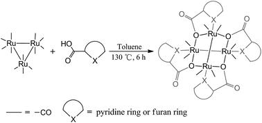
Thermolysis of triruthenium dodecacarbonyl Ru3(CO)12 with picolinic acid, 2-furoic acid and 2-thiophenecarboxylic acid was thoroughly studied. Three ruthenium carbonyl products, Ru4(CO)8(μ2-O, η1-N-pic)4 (1) (pic = picolinate), [Ru2(CO)4(fur)2]m (2) (fur = 2-furoate) and [Ru2(CO)4(thi)2]n (3) (thi = 2-thiophenecarboxylate) were isolated. Single crystal X-ray crystallography revealed that compound 1 is a tetraruthenium cluster with two Ru2(CO)4 units in an unexpected criss–cross geometry, while compounds 2 and 3 exhibit the classical sawhorse structure. Compound 1 was stable to common donor ligands and formed solvated compounds Ru4(CO)8(μ2-O, η1-N-pic)4·H2O (4) and Ru4(CO)8(μ2-O, η1-N-pic)4·CH3CN (5) in hydrous toluene and acetonitrile, respectively; compounds 2 and 3 converted into monomers Ru2(CO)4(fur)2(H2O)2·H2O (2b) and Ru2(CO)4(thi)2(CH3OH)2·CH3OH (3b) in hydrous dichloromethane and acetonitrile, respectively. [Ru2(CO)4(pic)2]2 (1a), Ru2(fur)2(CO)6 (2a) and Ru2(thi)2(CO)6 (3a) were proposed as the corresponding intermediates of 1–3 based on in situ FT-IR spectroscopy, LC-MS and the molecular structures of the known ruthenium carbonyl carboxylates.
Introduction
Reactions of the group 8 trimetallic carbonyl clusters Ru3(CO)12 with carboxylic acids have been known for a long time.1 The most diverse and best understood class of ruthenium carbonyl carboxylates consists of dinuclear ruthenium sawhorse type compounds, which have been devised as molecular catalysts,2 liquid crystals,3 and bio-active substances.4 The polydentate carboxylate ligands also assembled the dinuclear ruthenium units as one dimensional chains, tetranuclear loops, hexanuclear triangles, or octanuclear squares.5
With propionic acid or 1-adamantanecarboxylic acid as a bridging ligand, the weak Ru–O6 (Scheme 1a) or Ru–Ru7 interactions were strengthened. The dimeric “Ru2(CO)4(μ-OOCR)2L” fragments assembled as tetranuclear Ru complexes (Scheme 1b). The dicarboxylate ligands linked two Ru2(CO)4 units and constructed tetranuclear loops (Scheme 1c).5a–5c To explore the diversity of Ru2(CO)4 assembly, we set about to introduce heterocyclic carboxylic acids as linkers. To our surprise, unlike other carboxylate or pyridyl linkers,8 picolinic acid induced an unprecedented tetraruthenium compound Ru4(CO)8(μ2-O, η1-N-pic) (Scheme 1d).
 |
| | Scheme 1 Diverse diruthenium carbonyl carboxylates. | |
X-ray crystallography unveiled the relevant configuration, in which the four oxygen donors bridge two discrete Ru2(CO)4 moieties and four N donors chelate the ruthenium atoms. The coordination mode of μ2-O, η1-N heterocyclic carboxylate ligand stabilized such unusual tetrametallic structure. Herein, we reported the novel compound Ru4(CO)8(μ2-O, η1-N-pic)4 (1) containing a Ru4O4 skeleton with two criss–cross Ru2(CO)4 units in the thermolysis of picolinic acid with Ru3(CO)12 and the formation, geometric structure and the relevant coordination behavior were also investigated. Moreover, thermolysis of 2-furoic acid and 2-thiophenecarboxylic acid with Ru3(CO)12 were also investigated. Their formation mechanisms and reactivity were further examined.
Results and discussion
The thermolysis reaction of picolinic acid with Ru3(CO)12
In the preliminary experiment, Ru4(CO)8(μ2-O, η1-N-pic)4 (1) was obtained as yellow powder with 64% yield from the thermolysis reaction of picolinic acid with Ru3(CO)12 in toluene (130 °C) (Scheme 2). The experiments in lower temperatures (90 and 110 °C) were further performed and it was found that a longer reaction time was needed to form compound 1, which also obtained in o-xylene. However, compound 1 was not formed in n-heptane, due probably to the very low polarity of the solvent. IR analysis of 1 in dichloromethane exhibited its characteristic terminal carbonyl stretchings at 2029, 1984 and 1952 cm−1. In the solid state, compound 1 is very stable and no decomposition products were detected after storage in open air for months. Compound 1 did not react with 2e donor ligands, such as Ph3P, CH3CN and H2O. The molecular structure of 1 was identified as a discrete Ru4O4 cluster via single crystal X-ray diffraction (Fig. 1).
 |
| | Scheme 2 The formation of Ru4(CO)8(μ2-O, η1-N-pic)4. | |
 |
| | Fig. 1 Ball-and-stick model representation of single-crystal X-ray diffraction structure of Ru4(CO)8(μ2-O, η1-N-pic)4 (1). Selected bond lengths [Å] and angles [°]: N1–Ru1 = 2.192(4); Ru1–Ru1B = 2.758(7); Ru1–O1C = 2.132(3); Ru1–O1 = 2.179(3); Ru1A–O1A = 2.132(3); Ru1A–O1 = 2.179(3); Ru1A–Ru1C = 2.758(7); O1–Ru1–O1C = 82.79(9); O1–Ru1–N1 = 74.38(15); O1C–Ru1–N1 = 87.65(15); C8–Ru1–Ru1B = 88.19(16); C7–Ru1–Ru1 = 91.78(15); O1C–Ru1–Ru1A = 80.57(9); O1–Ru1–Ru1B = 97.82(8). Color code: green = Ru, red = O, blue = N, grey = C. Hydrogen atoms are omitted for clarity. | |
The crystal structure of 1 crystallizes in a tetragonal, P4(2)/n space group with four ruthenium atoms, four picolinate linkers and eight terminal carbonyl ligands. It contains a discrete Ru4O4 skeleton in which μ2-O donors linked two Ru2(CO)4 units in an unexpected criss–cross way. The chelation of N donors reinforced the whole cluster structure. Two species [Ga–Ga]4+ (ref. 9a) and [As–As]4+ (ref. 9b) exhibited the similar criss–cross assembly, but none of transition metal analogous were reported.
The coordination polyhedron of each Ru atom is a slightly distorted octahedron, with the equatorial position being occupied by two oxygen atoms of two picolinate ligands and two CO ligands and the axial position by nitrogen atom of the chelate picolinate ligand and a ruthenium atom of the same Ru2(CO)4 unit (angles O1C–Ru1–N1, O1–Ru1–O1C, O1–Ru1–N1, O1–Ru1–Ru1B, O1C–Ru1–Ru1B and N1–Ru1–Ru1B are 87.65, 82.80, 74.38, 97.81, 80.60 and 166.70°, respectively). The plane of Ru1N1C1 with the edge-shared pyridine ring is nearly in a plane (the dihedral angle is 0.61°). Each pentagon face of the Ru4O4 moiety was defined by three ruthenium atoms and two oxygen atoms and all faces were identical. In each pentagon, Ru–O bond lengths are in the range of 2.132 to 2.179 Å and the Ru–Ru bond length is 2.758 Å, which are in the typical ranges of Ru–O and Ru–Ru bond lengths in the dinuclear ruthenium sawhorse type complexes.6,7 Four pyridyl groups lie at the trans sites to each other. Each carboxylato oxygen atom bridges two adjacent ruthenium atoms belonging to different Ru2(CO)4 units. Terminal carbonyl ligands coordinated to Ru at cis-equatorial positions. The mean chelation angle of N–Ru(I)–O is 74.4°, similar to the corresponding values in [(η2-N, O-picolinate)2(η2-CO)2Ru(II).10
Interestingly, the small polar molecules, such as water and acetonitrile co-crystallize with compound 1. The compounds Ru4(CO)8(μ2-O, η1-N-pic)4·H2O (4) and Ru4(CO)8(μ2-O, η1-N-pic)4·CH3CN (5) were obtained when 1 was diffused into aqueous toluene and acetonitrile, respectively. Both 4 and 5 crystallize in a tetragonal, P4(2)/n space group (Fig. S4 and S5†). The packing diagrams of compounds 1 and 5 were shown in Fig. 2. In the packing structures of 1, 4 and 5, there is weak off-set stacking interactions between pyridine rings of adjacent molecules, with the plane distances being 3.51 Å in 1, 3.48 Å in 4 and 3.61 Å in 5.11 The similar distances in both 1 and 4 indicate that the presence of water molecules in the crystalline region has no effect on the cell parameters of 4. However, the existence of acetonitrile molecules enlarges the pore size in the solid structure of 5.
 |
| | Fig. 2 The packing diagram of compounds Ru4(CO)8(μ2-O, η1-N-pic)4 (1) (left) and Ru4(CO)8(μ2-O, η1-N-pic)4·CH3CN (5) (right). | |
When the thermolysis reaction of Ru3(CO)12 and picolinic acid in toluene at 130 °C was carefully examined, it was found that orange precipitate 1a was formed after 2 h and converted to 1 quantitatively after 5 h. IR analysis of 1a in dichloromethane found its characteristic terminal carbonyl stretchings at 2089 (m), 2017 (vs) and 1948 (m) cm−1, which is different from those found in compound 1. The 1H NMR spectrum of 1a showed two doublets centered at 8.80 and 7.67 ppm which were assigned as 6- and 3-H protons of a pyridine ring, and two triplets at 7.83 and 7.64–7.59 ppm ascribed to the resonance of pyridine's 4- and 5-H protons, respectively. ESI-HRMS analysis identified the anionic fragment of 1a as [M−] with m/z 1280.97, consistent with a molecule Ru4(CO)8(pic)4 and four acetonitrile molecules from the flowing phase of LC-MS, which has a calculated molecular weight of 1280.86. The molecular structure of 1a was proposed as a dimer [Ru2(CO)4(pic)2]2, shown in Scheme 3, based on the experimental data and analogous molecular structures reported in the literatures.8 Unfortunately, its single crystals could not be grown up.
 |
| | Scheme 3 Reaction of Ru3(CO)12 with heterocyclic carboxylic acid. | |
The thermolysis reaction of 2-furoic acid and 2-thiophenecarboxylic acid with Ru3(CO)12
The thermolysis reactions of Ru3(CO)12 with 2-furoic acid and 2-thiophenecarboxylic acid were then examined since they both have similar molecular structures to that of picolinic acid. The results showed that both carboxylic acids exhibited a different reaction process in the similar situation, however. The typical sawhorse compounds Ru2(CO)6(fur)2 (fur = 2-furoate) (2a) and Ru2(CO)6(thi)2 (thi = 2-thiophenecarboxylate) (3a) were proposed by means of in situ IR spectroscopy and reported molecular structures of diruthenium carbonyl carboxylates.8 The bridging carboxylate ligands showed C
O absorption at 1591–1564 cm−1, whilst terminal carbonyls appeared at the range of 2098–1941 cm−1. Under the thermal condition, 2a and 3a were stable in toluene, but they converted to polymers [Ru2(CO)4(fur)2]m (2) and [Ru2(CO)4(thi)2]n (3), respectively, by adding dry hexane (Scheme 3). Compound 2 was depolymerize into monomer Ru2(CO)4(fur)2(H2O)2·H2O (2b) in hydrous toluene. Whereas, the monomer Ru2(CO)4(thi)2(CH3OH)2·CH3OH (3b) was formed when 3 was dissolved in methanol.
Compound 2b was isolated as orange yellow crystals from hydrous toluene of 2, which was fairly soluble in dichloromethane. The crystal structure of compound 2b is shown in Fig. 3 and crystallizes in a tetragonal, P4(1)2(1)2 space group. The sawhorse geometry of 2b contains a Ru2(CO)4 backbone, two bridging carboxylate ligands and two water molecules occupying the axial positions. The O atoms in two furan rings arrange in a trans-position. The crystal structure of 3b crystallizes in a monoclinic C2/c space group with similar molecular geometry (Fig. 3) to that of 2b, although the two S atoms in the bridging carboxylate ligands being at cis-position according to the X-ray crystal analysis. Both 2b and 3b each contains a Ru2(CO)4 backbone arranged in a typical sawhorse geometry. All of the responding bond distances are similar in 1, 2b and 3b, of which the Ru–Ru distance are 2.758, 2.628 and 2.633 Å, respectively. The distances of Ru(1)–C(7) in compounds 1 and 2b are 1.883 and 1.827 Å, respectively, and Ru(1)–C(11) in 3b is 1.835 Å. All of the data indicated that the Ru2(CO)4 moieties in 1 is similar in bond lengths to the responding units in a typical sawhorse geometry.
 |
| | Fig. 3 Ball-and-stick model representation of single-crystal X-ray diffraction structure of Ru2(CO)4(fur)2(H2O)2·H2O (2b) and Ru2(CO)4(thi)2(CH3OH)2·CH3OH (3b); selected bond lengths [Å] and angles [°]: (A) (2b) Ru1–O1 = 2.126(5); Ru1–O6 = 2.141(5); Ru1–O1W = 2.277(5); Ru1–Ru2 = 2.628(8); Ru2–O7 = 2.117(5); Ru2–O2 = 2.123(5); Ru2–O2W = 2.234(5); H1W1–O6 = 2.810(7); H2W2–O3W = 2.734(12); O6–Ru1–O1W = 81.97(19); O7–Ru2–O2 = 83.24(19); (B) (3b) Ru1–O3 = 2.123(5); Ru1–O1 = 2.129(5); Ru1–O7 = 2.253(5); Ru1–Ru2 = 2.633(8); Ru2–O4 = 2.118(5); Ru2–O2 = 2.139(5); Ru2–O10 = 2.250(5); O11–H7A = 1.986(6); O3–Ru1–O1 = 83.3(2); O4– Ru2–O2 = 85.4(2). Color code: green = Ru, red = O, grey = C, yellow = S. | |
A DFT study at the B3LYP/LANL2DZ level was used to predict the existence possibility of the molecules Ru4(CO)8(μ2-O, η1-O-fur)4 (2c) and Ru4(CO)8(μ2-O, η1-S-thi)4 (3c) with structures similar to compound 1. The calculation results showed that the optimized structure of 2c (Fig. S2†) is feasible theoretically, but the optimized structure of 3c does not exist (Fig. S3†). Although 2c exists in theory, it was not obtained or observed in the experiment. The highest occupied molecular orbits (HOMOs) and the lowest unoccupied molecular orbits (LUMOs) of both 1 and 2c were illustrated in Fig. 4. The HOMOs of 1 and 2c are mainly localized on the dinuclear fragments of the Ru4O4 center. The LUMO of 1 is principally located on the two picolinate moieties, while for compound 2c it is located at the intersection between the Ru and O atoms of carboxylates. There is significant diversity in electron distribution in two optimized structures and this difference is possibly responsible for their stability distinction.
 |
| | Fig. 4 Calculated frontier molecular orbitals of compounds Ru4(CO)8(μ2-O, η1-N-pic)4 (1) and Ru4(CO)8(μ2-O, η1-O-fur)4 (2c). HOMO (left) and LUMO (right) calculated for 1 (top) and 2c (bottom). | |
Experimental
All reactions and manipulations were performed using standard Schlenk line techniques under an inert atmosphere of dry nitrogen. All reactants were purchased from Tokyo Chemical Industry. Solvents were purified, dried, and distilled under nitrogen atmosphere prior to use. Liquid FT-IR spectra were recorded on an EQUINX55 Fourier-transform spectrometer in toluene solution and 1H NMR spectra were per-formed on a Bruker Avance 400 MHz spectrometer unless indicated. ESI-MS was recorded on a Thermo Deca Max (LCMS) mass spectrometer with an ion-trap mass detector, while high-resolution mass spectra were recorded in ESI mode on a Waters UPLC-Q-TOF mass spectrometer.
Synthesis of Ru4(CO)8(μ2-O, η1-N-pic)4 (1)
A mixture of triruthenium dodecacarbonyl Ru3(CO)12 (0.29 g, 0.3 mmol) and picolinic acid (pic) (0.110 g, 0.9 mmol) in 10 mL of toluene was heated at 130 °C under nitrogen atmosphere in the sealed Schlenk tube for 6 h under stirring in which time the color of the reaction solution changed from orange to red-brown. The solution was cooled and toluene was then removed under vacuum. The residue was chromatographed on silica gel with dichloromethane as eluent and collected only one fraction. The solvent was evaporated under vacuum to yield a yellow powder as compound 1 (yield 64%). IR (νCO, cm−1, KBr): 2026 (vs), 1989 (m), 1945 (vs); 1H NMR (400 MHz, CDCl3) δ 9.11 (d, J = 4.9 Hz, 1H), 8.24 (d, J = 7.6 Hz, 1H), 7.97–7.94 (m, 1H), 7.64–7.62 (m, 1H); MS (m/z, ESI−) 1120.666 (M−). Anal. calcd for Ru4C34H16N4O16: 1120.676; elemental analysis calcd (%): C 34.42, H 1.44, N 5.02; found: C 34.36, H 1.56, N 4.85.
Synthesis of [Ru2(CO)4(pic)2]2 (1a)
A mixture of triruthenium dodecacarbonyl Ru3(CO)12 (0.192 g, 0.3 mmol) and picolinic acid (pic) (0.036 g, 0.3 mmol) in 10 mL of toluene was heated at 130 °C under nitrogen atmosphere in the sealed Schlenk tube for 2 h under stirring in which time the precipitate was formed. The solution was cooled and the precipitate was filtered, washed with toluene (3 × 10 mL) and afforded red powder under vacuum (yield: 88%). IR (νCO, cm−1, CH2Cl2): 2089 (m), 2017 (vs), 1948 (m); 1H NMR (400 MHz, CDCl3) δ 8.80 (d, J = 5.0 Hz, 1H), 7.83 (td, J = 7.8, 1.3 Hz, 1H), 7.67 (d, J = 7.6 Hz, 1H), 7.64–7.59 (m, 1H); MS (m/z, ESI−) 1280.97 (M−). Anal. calcd for Ru4(CO)8(pic)4(CH3CN)4: 1280.86.
Synthesis of [Ru2(CO)4(fur)2]2 (2)
A similar synthetic procedure to that of 1 was employed and 2-furoic acid was used instead of picolinic acid in the formation of compound 2. The obtained red solution was added hexane and the red orange powder was deposited. The precipitate was filtered and washed with cold hexane (3 × 20 mL), and then dried under vacuum (yield: 89%). IR (νCO, cm−1, toluene): 2040 (vs), 1992 (m), 1959 (vs); 1H NMR (400 MHz, CDCl3) δ 7.51 (s, 1H), 7.11 (d, J = 2.7 Hz, 1H), 6.46 (s, 1H); MS (m/z, ESI−) 1076.600 (M−). Anal. calcd for Ru4C28H12O20: 1076.613; elemental analysis calcd (%): C 31.35, H 1.13; found: C 31.40, H 1.16.
Synthesis of Ru2(CO)4(fur)2(H2O)2·H2O (2b)
This compound was obtained when compound 2 was crystallized in hydrous toluene (yield: 90%). IR (νCO, cm−1, CH2Cl2): 2046 (vs), 1995 (m), 1964 (vs); 1H NMR (400 MHz, CD3OD) δ 7.56 (s, 1H), 7.07 (d, J = 3.3 Hz, 1H), 6.47 (dd, J = 3.3, 1.7 Hz, 1H); MS (m/z, ESI−) 1117.575 (M−). Anal. calcd for Ru4(CO)8(fur)4(CH3CN): 1117.638; elemental analysis calcd (%) C14H12O13Ru2: C 28.46, H 2.51; found: C 28.46, H 2.36.
Synthesis of Ru2(CO)4(thi)2(CH3OH)2·CH3OH (3b)
A similar synthetic procedure to that of 1 was employed and 2-thiophecarboxylic acid was used instead of picolinic acid. To the obtained red solution was added hexane and the red orange powder was deposited. The precipitate was filtered and washed with cold hexane. The orange crude powder was then recrystallized in methanol/hexane to afford the orange crystalline product 3b (yield, 91%). IR (νCO, cm−1, CH2Cl2): 2046 (vs), 1994 (m), 1964 (vs); 1H NMR (400 MHz, CDCl3) δ 7.75 (d, J = 1.9 Hz, 1H), 7.54 (d, J = 4.7 Hz, 1H), 7.11–7.07 (m, 1H), 3.51 (s, 6H); MS (m/z, ESI−) 1181.482 (M−). Anal. calcd for Ru4(CO)8(thi)4(CH3CN): 1181.547; elemental analysis calcd (%) Ru2C17H18O11S2: C 30.72, H 2.73; found: C 30.80, H 2.74.
Crystallography
X-ray crystallographic data were collected from their single crystals mounted on Single-crystal X-ray structural measurements were carried out with a Bruker SMART APEX-II CCD detector using graphite monochromated MoKα radiation (λ = 0.71073). The data were collected by the ω–2θ scan mode, and absorption correction was applied by using Multi-Scan. The structure was solved by direct methods (SHELXS-97) and refined by full-matrix least squares against F2 using SHELXL-97 software.12 Non-hydrogen atoms were refined with anisotropic thermal parameters. All hydrogen atoms were geometrically fixed and refined using a riding model. Single crystals of 1 were obtained by slow evaporation of its toluene solution, and of compound Ru4(CO)8(μ2-O, η1-N-pic)4·H2O (4) was obtained if a small amount of water was added in toluene solution of 1 in the crystallization process. Single crystals of Ru4(CO)8(μ2-O, η1-N-pic)4·CH3CN (5) were obtained by slow evaporation of an acetonitrile solution of 1 at room temperature. Single crystals of 2b and 3b were obtained from their aqueous toluene and methanol solution, respectively. In the determination of the crystal structures of 2b and 3b, their single crystals were immediately coated with oil to prevent loss of solvent of crystallization. Relevant crystallographic data and details of measurements are given in Table S1 in the ESI.†
Crystallographic data for the structural analysis have been deposited with the Cambridge Crystallographic Data Centre: 1055805 Ru4(CO)8(μ2-O, η1-N-pic)4 (1); 1403694 Ru2(CO)4(fur)2-(H2O)2·H2O (2b); 1400328 Ru2(CO)4(thi)2(CH3OH)2·CH3OH (3b); 1449761 Ru4(CO)8(μ2-O, η1-N-pic)4·H2O (4); 1409343 Ru4(CO)8(μ2-O, η1-N-pic)4·CH3CN (5).
Conclusions
In this work, thermolysis reaction of Ru3(CO)12 with three heterocyclic carboxylic acids were investigated in detail. An unexpected tetranuclear ruthenium carbonyl carboxylate Ru4(CO)8(μ2-O, η1-N-pic)4 (1) containing unusual criss–cross assembly of two Ru2(CO)4 units was isolated. However, traditional sawhorse–structural polymeric complexes 2 and 3 were collected when 2-furoic acid and 2-thiophecarboxylic acid were used. The respective intermediates 1a, 2a, and 3a were rationally conjectured. Compound 1 was unreactive to common coordination solvents and ligands, but 2 and 3 were labile in coordination solvents, forming sawhorse–structural Ru2(CO)4(fur)2(H2O)2·H2O and Ru2(CO)4(thi)2(CH3OH)2·CH3OH. The formation and reactivity of 1–3 indicated that the atoms N, O, S of the heterocyclic rings in the carboxylic acids play very important roles during assembly of the diruthenium Ru2(CO)4 units. The DFT calculation results provided relevant supports for unusual geometry of compound 1. This work provided a feasible route to synthesize discrete molecules with Ru2(CO)4 units in criss–cross structure. The assembly of more sophisticated Ru2(CO)4 moieties was under investigation.
Computational details
For geometry optimization and the ground state electronic structure calculations, the DFT method with the Becke's three parameter hybrid functional and Lee Yang Parr's gradient corrected correlation functional (B3LYP) was used.13 The calculations were performed with the GAUSSIAN-03 program.14 The LanL2DZ basis set and effective core potential were used for the Ru atom and the 6-31G basis sets were used for all other atoms, respectively.15 The nature of all stationary points was confirmed by performing a normal-mode analysis. The input model molecule of 2c and 3c was based on the X-ray structure of 1, conveniently modified by manually adding or removing appropriate atoms.
Acknowledgements
This work is financially supported by the National Natural Science Foundation of China (21171112, 21371112, 21271124), the 111 Project (B14041), the Fundamental Funds Research for the Central Universities (GK201501005) and the program for Changjiang Scholars and Innovative Research Team in University (IRT_14R33).
Notes and references
- G. R. Crooks, B. F. G. Johnson, J. Lewis, I. G. Williams and G. Gamlen, J. Chem. Soc. A, 1969, 2761 RSC.
-
(a) A. Salvini, P. Frediani and F. J. Piacenti, J. Mol. Catal. A: Chem., 2000, 159, 185 CrossRef CAS;
(b) A. Salvini, P. Frediani and E. Rivalta, Inorg. Chim. Acta, 2003, 351, 225 CrossRef CAS;
(c) A. Salvini, P. Frediani, C. Giannelli and L. Rosi, J. Organomet. Chem., 2005, 690, 371 CrossRef CAS;
(d) Y. Sevryugina, B. Weaver, J. Hansen, J. Thompson, H. M. L. Davies and M. A. Petrukhina, Organometallics, 2008, 27, 1750 CrossRef CAS;
(e) J. Ziriakus, T. K. Zimmermann, A. Pothig, M. Drees, S. Haslinger, D. Jantke and F. E. Kuhn, Adv. Synth. Catal., 2013, 355, 2845 CrossRef CAS.
-
(a) S. Frein, M. Auzias, A. Sondenecker, L. Vieille-Petit, B. Guintchin, N. Maringa, G. Süss-Fink, J. Barberá and R. Deschenaux, Chem. Mater., 2008, 20, 1340 CrossRef CAS;
(b) R. Deschenaux, B. Donnio, G. Rheinwald, F. Stauffer, G. Süss-Fink and J. Velker, J. Chem. Soc., Dalton Trans., 1997, 4351 RSC.
-
(a) J. P. Johnpeter, F. Schmitt, E. Denoyelle-Di-Muro, G. Wagnières, L. Juillerat-Jeanneret and B. Therrien, Inorg. Chim. Acta, 2012, 393, 246 CrossRef CAS;
(b) F. Schmitt, P. Govindaswamy, G. Süss-Fink, W. H. Ang, P. J. Dyson, L. Juillerat-Jeanneret and B. Therrien, J. Med. Chem., 2008, 51, 1811 CrossRef CAS PubMed;
(c) F. Schmitt, P. Govindaswamy, O. Zava, G. Süss-Fink, L. Juillerat-Jeanneret and B. Therrien, J. Biol. Inorg. Chem., 2009, 14, 101 CrossRef CAS PubMed;
(d) K. Davia, D. King, Y. Hong and S. Swavey, Inorg. Chem. Commun., 2008, 11, 584 CrossRef CAS;
(e) P. Sweigert, Z. Xu, Y. Hong and S. Swavey, Dalton Trans., 2012, 41, 5201 RSC;
(f) T. Gianferrara, A. Bergamo, I. Bratsos, B. Milani, C. Spagnul, G. Sava and E. Alessio, J. Med. Chem., 2010, 53, 4678 CrossRef CAS PubMed.
-
(a) M. Bianchi, U. Matteoli, P. Frediani, F. Piacenti, U. Nardelli and G. Pelizzi, Chim. Ind., 1981, 63, 475 CAS;
(b) K. B. Shiu, H. C. Lee, G. H. Lee and Y. Wang, Organometallics, 2002, 21, 4013 CrossRef CAS;
(c) G. Süss-Fink, J. L. Wolfender, F. Neumann and H. Stoeckli-Evans, Angew. Chem., Int. Ed. Engl., 1990, 29, 429 CrossRef;
(d) M. Auzias, B. Therrien and G. Süss-Fink, Inorg. Chem. Commun., 2007, 10, 1420 CrossRef CAS;
(e) K. B. Shiu, H. C. Lee, G. H. Lee, B. T. Ko, Y. Wang and C. C. Lin, Angew. Chem., Int. Ed. Engl., 2003, 42, 2999 CrossRef CAS PubMed.
-
(a) Y. Sevryugina, B. Weaver, J. Hansen, J. Thompson, H. M. L. Davies and M. A. Petrukhina, Organometallics, 2008, 27, 1750 CrossRef CAS;
(b) Y. Sevryugina, A. V. Olenev and M. A. Petrukhina, J. Cluster Sci., 2005, 16, 217 CrossRef CAS;
(c) D. J. Morris, G. J. Clarkson and M. Wills, Organometallics, 2009, 28, 4133 CrossRef CAS;
(d) J. H. Reibenspies, B. Fontal and D. Darensbourg, Acta Crystallogr., Sect. C: Cryst. Struct. Commun., 1993, 49, 1619 CrossRef;
(e) T. K. Zimmermann, J. Ziriakus, E. Herdtweck, A. Pothig and F. E. Kühn, Organometallics, 2014, 33, 2667 CrossRef CAS.
-
(a) S. P. Oh, B. Y. Chor, W. Y. Fan, Y. Li and W. K. Leong, Organometallics, 2011, 30, 6774 CrossRef CAS;
(b) M. A. Petrukhina, Y. Sevryugina and K. W. Andreini, J. Cluster Sci., 2004, 15, 451 CrossRef CAS.
- B. Therrien and G. Süss-Fink, Coord. Chem. Rev., 2009, 253, 2639 CrossRef CAS.
-
(a) N. Wiberg, T. Blank, M. Westerhausen, S. Schneiderbauer, H. Schnöckel, I. Krossing and A. Schnepf, Eur. J. Inorg. Chem., 2002, 3, 351 CrossRef;
(b) See textbooks of Inorganic Chemistry.
-
(a) L. Xu and Y. Sasaki, J. Organomet. Chem., 1999, 585, 246 CrossRef CAS;
(b) D. Ooyama, T. Kobayashi, K. Shiren and K. Tanaka, J. Organomet. Chem., 2003, 665, 107 CrossRef CAS.
-
(a) C. A. Hunter and J. K. Sanders, J. Am. Chem. Soc., 1990, 112, 5525 CrossRef CAS;
(b) M. O. Sinnokrot and C. D. Sherrill, J. Am. Chem. Soc., 2004, 126, 7690 CrossRef CAS PubMed;
(c) A. L. Ringer and C. D. Sherrill, J. Am. Chem. Soc., 2009, 131, 4574 CrossRef CAS PubMed.
- G. M. Sheldrick, Acta Crystallogr., Sect. A: Found. Crystallogr., 2008, 64, 112 CrossRef CAS PubMed.
- P. J. Stephens, F. J. Devlin, C. F. Chabalowski and M. J. Frisch, J. Phys. Chem., 1994, 98, 11623 CrossRef CAS.
- M. J. Frisch, G. W. Trucks, H. B. Schlegel and G. E. Scuseria, Gaussian09, (Revision B.04,Gaussian) Inc., Wallingford, CT, 2003 Search PubMed.
- P. J. Hay and W. R. Wadt, J. Chem. Phys., 1985, 82, 270 CrossRef CAS.
|
| This journal is © The Royal Society of Chemistry 2016 |
Click here to see how this site uses Cookies. View our privacy policy here.