Synthesis of peptide nanofibers decorated with palladium nanoparticles and its application as an efficient catalyst for the synthesis of sulfides via reaction of aryl halides with thiourea or 2-mercaptobenzothiazole

Arash Ghorbani-Choghamarani* and Zahra Taherinia
Department of Chemistry, Ilam University, P.O. Box 69315516, Ilam, Iran. E-mail: arashghch58@yahoo.com; a.ghorbani@mail.ilam.ac.ir; Fax: +98 841 2227022; Tel: +98 841 2227022

Received 25th January 2016 , Accepted 15th June 2016

First published on 15th June 2016


Abstract

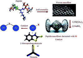
In this work supported Pd nanoparticles on a peptide nanofiber (PdNP–PNF) have been prepared via fabrication of self-assembled woven nanofiber from peptide, subsequently immobilization of palladium nanoparticles on this nanostructural compound. To obtain self-assembled woven nanofiber, we designed and synthesized a peptide using arginine as building block. The C-terminus of amino acid was protected as ethylester. Coupling was mediated by dicyclohexylecarbodiimide-1-hydroxybenzotriazole (DCC-HOBT). TEM, SEM, XRD, ICP and FT-IR techniques were employed to characterize prepared nanofiber materials. In this work, the effect of phosphate buffer solutions pH 8 and pH 11 (isoelectric point of arginine amino acid) on the structure of peptide nanofiber was investigated. Supported Pd nanoparticles on the peptide nanofiber (PdNP–PNF) were applied for the C–S coupling reaction using two different sulfur transfer reagents (thiourea and 2-mercaptobenzothiazole).


1 Introduction

The production of nanofibers ranging from micron to nanometer scales has been studied due to their special properties such as up grading human artificial tissue including bone,1 cartilage,2 ligament,3 skeleton muscle,4 skin,5 vascular tissue engineering,6 neural,7 and as carriers for the controlled delivery,8 protines9 and controlled DNA delivery.10 Nanofiber can be produced in many different ways, for example self-assembly,11 drawing,12 electrospinning,13 template synthesis.14 Among these methods, electrospinning is a well-known technique to make a layer of nanofibers. In this layer, though, each nanofiber cannot be separated without their destruction and electrospinning needs larger electric fields, sensitivity to variability in solution conductivity and low production speed. Template synthesis is an efficacious route to produce nanofibers or nanotubes using a nanoporous membrane as a template. Yet, a disadvantage of this method is that it cannot make continuous nanofibers one by one. However, the template method is possible by controlling parameters such as melt time and temperature. Self-assembling is a procedure by which molecules organize and arrange themselves into patterns or structures through non-covalent forces such as hydrogen bonding, hydrophobic forces, and this technique shows good potential for designing novel scaffolds for tissue engineering applications. Several factors including, the concentration of peptide molecules, pH, solvent polarity, sonication, ionic strength and interaction with anions such as phosphate have been used to tune the peptide self-assembly process. Da-Wei15 indicated that the self-assembling process is enhanced at a low concentration of phosphate and the length of the fibrils is longer than that in pure water. Goldberger16 designed a series of PAs that can self-assemble into nanofibers when the solution pH is decreased from the normal physiological condition 7.4 to 6.6, Smith17 saw that the overall length of the nanofibers increased as the initial concentration of insulin peptide increased. Prabhu18 has shown that ultrasonication-induced fibril-formation by a bolaform peptide. Shen19 demonstrated solvent effects for dissolved β-amyloid in different solvents of various concentrations, and found that the growth rate of the nanofibers was reduced. In the case of 100% dimethyl sulfoxide, no β-sheet content is visible, but in 10% dimethyl sulfoxide, nanofibers are seen to contain the rigid, hydrogen-bonded β-sheet structure. Therefore, nanofibers, are irrespective of their method of synthesis. Among different nanoparticles, self-assembling peptides are the best choice due to unique properties such as high surface area-to-volume and they are desirable in order to allow delivery of a high density of cells and tissue engineering. Among the applications of peptide nanofiber, their catalytic applications in organic reactions have attracted extensive attention; for example Indrajit Maity20 reported the fabrication of peptide capped Pd nano particles, which enhanced the catalytic activity of C–C coupling reactions in aerobic conditions. Khalily21 reported a supramolecular peptide nanofiber templated Pd nano catalyst for efficient Suzuki coupling reactions under aqueous conditions. Shao22 reported coupling reactions of aromatic halides in the presence of palladium catalyst immobilized on poly(vinylalcohol) nanofiber. We demonstrate for the first time, the use of peptide nanofibers decorated with Pd nanoparticles with sizes ranging from 7.1 to 10.28 nm for the C–S coupling reactions using two different sulfur transfer reagents. Sulfides have shown widely applications as potent drugs for HIV,23 cancer,24 Alzheimer25 and Parkinsons diseases.26 They are useful compounds in chemistry due to their role as important intermediates in organic synthesis.27 Therefore, there is still a great interest to find a new reagent or system to synthesis sulfides. Herein, we present for the first time the cross-coupling reactions of aryl/alkyl halides using 2-mercaptobenzothiazole or thiourea as efficient sulfur transfer reagent to afford symmetrical sulfides, which has been catalyzed by immobilized palladium nanoparticles on peptide nanofiber.

2 Results and discussion

In this work, to achieve an efficient and simple nanofiber support we performed the self-assembly of a peptide with arginine as building block. After converting of this peptide into nanofiber, palladium nanoparticles were immobilized on the surface of this nanostructural compound (Scheme 1).
image file: c6ra02264b-s1.tif
Scheme 1 Schematic synthesis of Pd nanoparticles supported on the peptide nanofiber.

We studied a simple, economic and environmentally friendly approach to the self-assembly of a peptide using phosphate buffer solution in an aqueous media. Since one way to control the density and growth rate of nanofibers is through the use of the solvent that destabilize the peptide structure, so we decided to use water as self-assembling media because when we used peptide powder, no obvious self-assembly of nanofiber was observed in the SEM image (Fig. 1). Since the peptides formed various hydrogels depending on amino acids charges, therefore we investigate the effect of phosphate buffer solution at pH 8 and pH 11 (isoelectric point of arginine amino acid) on the structure of peptide nanofiber. The pH value was changed by adjusting the ratio of [HPO42−]/H2PO4] while keeping the total concentration of phosphate constant. The effect of pH 11 solution was evaluated in two ways: (i) when peptide solution was investigated at the pH 11 without adding succinic anhydride, colloidal solution was formed and no obvious self-assembly was observed in the scanning electron microscopy image (Fig. 2), (ii) when succinic anhydride added to the peptide solution, the final pH of the solution was found to be 10 due to hydrolysis of succinic anhydride to succinic acid, microscopic images showed the formation of micro-crystals (Fig. 3). Surprisingly, at pH 8 by adding succinic anhydride to the peptide solution, which the final pH of the solution was found to be 7, the woven morphology of the nanofiber was observed in the SEM image (Fig. 4). The pH effect can be explained since a high pH environment dissociates H+ of HPO42−/H2PO4, it is known that one divalent anion have a charge of −2 and can effectively interact with two positively charged groups of the peptide molecule like a bridge. Since distance between two nearest arginine resides of the same peptide molecule is much larger than the size of HPO42−, one divalent anion can hardly interact with two arginine residues simultaneously, which leads to form a short nanofibers aggregated before the peptide molecules self-assembled to grow longer nanofibers.


image file: c6ra02264b-f1.tif
Fig. 1 SEM images of powder peptide.

image file: c6ra02264b-f2.tif
Fig. 2 SEM images of solution peptide at pH 11 without adding succinic anhydride.

image file: c6ra02264b-f3.tif
Fig. 3 SEM images of peptide solution at pH 11 in the presence of succinic anhydride.

image file: c6ra02264b-f4.tif
Fig. 4 SEM image of peptide solution at pH 8. In the presence of succinic anhydride.

When the pH is raised to 11, it results in a negative deprotonated carboxylate of arginine and one positive protonated nitrogen atom (the arginine residue to be natural), thus, there is no electrostatic interaction between HPO42− ion and arginine residues of the peptide molecule. But, when pH changes to 7, it results in a negative deprotonated carboxylate and two positive protonated nitrogen atoms (overall charge of +1) (Scheme 2). Since one monovalent anion takes one negative charge and can interact with one positively charged groups of the peptide molecule therefore the monovalent ions can distribute the charges between the polar head groups to reduce electrostatic repulsion, which makes the more ordered nano-fibrils (Scheme 3). However increase of pH value results in the increase of [HPO42−], which makes conditions unfavorable for the formation of nanofiber network. But at pH 7 the concentration of the HPO42− is lower than the concentration of the monovalent anion H2PO4; therefore the monovalent anions play a major role in the shielding of charges on the peptide molecules.


image file: c6ra02264b-s2.tif
Scheme 2 Arginine amino acid charge at the pH 7 and 11.

image file: c6ra02264b-s3.tif
Scheme 3 Schematic illustration of the proposed mechanism for the template-free formation of woven nanofiber in an aqueous solution.

2.1 Characterization of peptide nanofiber

The self-assembly study of the peptide was investigated using various spectroscopic and microscopic techniques. FT-IR spectroscopy was used to study the structure of the nanofiber solution; a peak that appear at 1644 cm−1 is correspondence to bending vibration of NH groups, while this peak for the peptide powder appear at 1629 cm−1, the observed shift is probably due to signifies self-assembling and hydrogen bonding between phosphate and peptide. As a general rule, hydrogen bonding decreases the frequency of stretching vibrations, since it decreases the restoring force, but increases the frequency of bending vibrations since it produces an additional restoring force28 (Fig. 5). The SEM images show that the peptide molecules self-assembled into woven nanofiber29 structures (Fig. 4).
image file: c6ra02264b-f5.tif
Fig. 5 FT-IR spectra of peptide powder (blue line); the peptide nanofiber (red line).

2.2 Characterization of supported Pd nanoparticles on the peptide nanofiber

The effect in the secondary structure of the peptide via the interaction between the metal nanoparticles and peptide was considered by FT-IR, SEM and TEM analysis. In the IR spectrum of supported Pd nanoparticles on the peptide nanofiber, the bending vibration of NH groups appeared at 1648 cm−1 (Fig. 6). The SEM and TEM images show that the Pd nanoparticles were regularly immobilized on the surface of woven peptide nanofibers (Fig. 7). The amount of Pd incorporated into the nanofiber found to be 250 ppm that determined by inductively coupled plasma optical emission spectrometry (ICP-OES). X-ray diffraction patterns of peptide nanofibers decorated with palladium is shown in (Fig. 8). The strong diffraction peaks about at 2θ values of 41°, 47°, and 69° (ref. 30 and 31) is related to Pd(0) nanoparticles; also the other peaks related to peptide nanofiber structure.
image file: c6ra02264b-f6.tif
Fig. 6 FT-IR spectra of the peptide solution without Pd nanoparticles (black line), peptide nanofibers decorated with Pd nanoparticles (blue line).

image file: c6ra02264b-f7.tif
Fig. 7 (a) SEM image of immobilized Pd nanoparticles on the surface of the woven nanofibers; (b–f) TEM surface of the woven nanofibers at pH 8.

image file: c6ra02264b-f8.tif
Fig. 8 The X-ray diffraction pattern of Pd immobilized on peptide nanofibers.

2.3 Catalytic studies

After preparation and characterization of Pd nanoparticle supported on the peptide nanofiber catalytic activity of this compound was investigated for C–S coupling reaction. A variety of symmetrical aryl/alkyl sulfides can be obtained in moderate to excellent yields (up to 90%). In this work thiourea and 2-mercaptobenzothiazole (as a new heterocyclic sulfur donor) has been utilized for the direct synthesis of organic sulfides from aryl/alkyl halides in the presence of a peptide nanofibers decorated with Pd nanoparticles (PdNP–PNF) that effectively led to the production of sulfides in high yield. In order to optimize the reaction conditions, the reaction of iodobenzene with 2-mercaptobenzothiazole in the presence of PdNP–PNF has been selected as model reaction and different parameters including the type of base, solvent and temperature has been studied (Table 1). It was found that the base and solvent significantly influenced the outcome of C–S coupling reaction. Also, the reaction rate was increased by rising reaction temperature. With optimal conditions in hand, a variety of symmetrical diaryl/alkyl sulfides were synthesized using 1.3 equivalents of 2-mercaptobenzothiazole or thiourea at 130 °C with high purity (Tables 2 and 3). Generally, the C–S coupling reactions of aryl halides with electron drawing groups preceded more rapidly and in high yields of products have been obtained. However, the C–S coupling reaction including aryl chlorides showed less reactivity than that of aryl iodides and bromides. Suggested mechanism for these transformations has been illustrated in Scheme 4 and 5. Initially aryl halide reacts with Pd by oxidative addition to form intermediate (a), then the intermediate (a) reacts with 2-mercaptobenzothiazole to produce intermediate (b), which is transformed into thiol anion in the presence of KOH. Then thiol anion reacts with intermediate (a) via reductive elimination reaction to afford sulfide and releases palladium nanoparticle. It should be noted that Gracia-Espino et al. reported when thiolated substrates are used, such as S-rGOx, the Pd nanoparticles exhibit smaller sizes, improved spatial distribution, and poor or null agglomeration because of the stronger interaction with the thiolated graphene surface.32 The thiol bridges play an active role in the adsorption mechanism of the Pd cluster, which would increase the reactivity. During the theoretical studies Gracia-Espino et al. observed the following chemical reactions:
 
C7H7SH + rGOx–Vc → rGOx–H–S–C7H7 (1)
 
C7H7SH + rGOx–OH → rGOx–S–C7H7 + H2O (2)
 
C7H7SH + rGOx–NH2 → rGOx–S–C7H7 + NH3 (3)
Table 1 Optimization of the reaction conditions for the C–S coupling using 2-mercaptobenzothiazole as sulfur transfer agenta

image file: c6ra02264b-u2.tif

Entry Solvent Temp. (°C) Base Time (h) Yieldb (%)
a Reaction conditions: iodobenzene 1 mmol, 2-mercaptobenzothiazole 1.3 mmol, PdNP–PNF (200 μL), base 1 g.b Isolated yield.
1 DMSO 130 KOH 2 90
2 DMF 130 KOH 2 N.R
3 PEG 130 KOH 2 N.R
4 CH3CN 130 KOH 2 N.R
5 H2O 130 KOH 2 N.R
6 EtOH 130 KOH 2 N.R
7 DMSO 130 NaOH 2 43
8 DMSO 130 K2CO3 2 N.R
9 DMSO 130 Na2CO3 2 N.R
10 DMSO 130 Et3N 2 N.R
11 DMSO 130 NaOEt 2 N.R
12 DMSO 100 KOH 2 63
13 DMSO 80 KOH 2 Trace


Table 2 Synthesis of symmetrical sulfides via reaction of 2-mercaptobenzothiazole and aryl/alkyl halides catalyzed by peptide nanofibers decorated with Pd nanoparticles (PdNP–PNF) in DMSOa

image file: c6ra02264b-u3.tif

Entry Ar-X Product Time (h) Yieldb (%)
a Reaction conditions: aryl halides (1 mmol), 2-mercaptobenzothiazole (1.3 mmol), PdNP–PNF (200 μL), base (1 g) and DMSO (2 mL).b Isolated yield.
1 image file: c6ra02264b-u4.tif image file: c6ra02264b-u5.tif 5 92
2 image file: c6ra02264b-u6.tif image file: c6ra02264b-u7.tif 6 87
3 image file: c6ra02264b-u8.tif image file: c6ra02264b-u9.tif 12 43
4 image file: c6ra02264b-u10.tif image file: c6ra02264b-u11.tif 8.5 63
5 image file: c6ra02264b-u12.tif image file: c6ra02264b-u13.tif 10 57
6 image file: c6ra02264b-u14.tif image file: c6ra02264b-u15.tif 1 83
7 image file: c6ra02264b-u16.tif image file: c6ra02264b-u17.tif 25 min 55
8 image file: c6ra02264b-u18.tif image file: c6ra02264b-u19.tif 50 min 41
9 image file: c6ra02264b-u20.tif image file: c6ra02264b-u21.tif 7 61
10 image file: c6ra02264b-u22.tif image file: c6ra02264b-u23.tif 9 53
11 image file: c6ra02264b-u24.tif image file: c6ra02264b-u25.tif 2 92
12 image file: c6ra02264b-u26.tif image file: c6ra02264b-u27.tif 10 70
13 image file: c6ra02264b-u28.tif image file: c6ra02264b-u29.tif 1 83


Table 3 Synthesis of symmetrical sulfides via reaction of thiourea and aryl/alkyl halides catalyzed by peptide nanofibers decorated with Pd nanoparticles (PdNP–PNF) in DMSOa

image file: c6ra02264b-u30.tif

Entry Ar-X Product Time (h) Yieldb (%)
a Reaction conditions: aryl halides (1 mmol), 2-mercaptobenzothiazole (1.3 mmol), PdNP–PNF (200 μL), base (1 g) and DMSO (2 mL).b Isolated yield.
1 image file: c6ra02264b-u31.tif image file: c6ra02264b-u32.tif 2 95
2 image file: c6ra02264b-u33.tif image file: c6ra02264b-u34.tif 3 88
3 image file: c6ra02264b-u35.tif image file: c6ra02264b-u36.tif 10 47
4 image file: c6ra02264b-u37.tif image file: c6ra02264b-u38.tif 7 65
5 image file: c6ra02264b-u39.tif image file: c6ra02264b-u40.tif 9 55
6 image file: c6ra02264b-u41.tif image file: c6ra02264b-u42.tif 30 min 87
7 image file: c6ra02264b-u43.tif image file: c6ra02264b-u44.tif 10 min 55
8 image file: c6ra02264b-u45.tif image file: c6ra02264b-u46.tif 40 min 50
9 image file: c6ra02264b-u47.tif image file: c6ra02264b-u48.tif 6 67
10 image file: c6ra02264b-u49.tif image file: c6ra02264b-u50.tif 9 53
11 image file: c6ra02264b-u51.tif image file: c6ra02264b-u52.tif 2 92
12 image file: c6ra02264b-u53.tif image file: c6ra02264b-u54.tif 10 70
13 image file: c6ra02264b-u55.tif image file: c6ra02264b-u56.tif 1 83


image file: c6ra02264b-s4.tif
Scheme 4 Proposed mechanism for the synthesis of sulfides through cross-coupling reactions of aryl halides with 2-mercaptobenzothiazole catalyzed by peptide nanofibers decorated with Pd nanoparticles.

image file: c6ra02264b-s5.tif
Scheme 5 Proposed mechanism for the synthesis of aryl sulfides through cross-coupling reactions of aryl halides with thiourea catalyzed by peptide nanofibers decorated with Pd nanoparticle.

The chemical reaction shown in eqn (1) was observed during the first adsorption event, while the chemical reactions in eqn (2) and (3) were not observed until Pd13 adsorption.30 Thus, according to results we can conclude that thiol anion is more willing to react with palladium. Because otherwise if that reacts with functional groups on the surface of peptide, no disulfide could be synthesized in eqn (4).

 
image file: c6ra02264b-u1.tif(4)

Finally, the recoverability and reusability of the Pd nanoparticle supported on the peptide nanofiber in the synthesis of sulfides via reaction of iodobenzene with thiourea over four successive runs, was investigated. Reaction was performed in DMSO at 130 °C, using 1 mmol iodobenzene, 1.3 mmol thiourea and 1 g KOH in the presence of PdNP–PNF (200 μl), upon completion of the reaction, the mixture was cooled to room temperature. 20 mL of ethyl acetate was added to the reaction mixture, which led to the precipitation of PdNP–PNF. The resulting precipitate was washed twice with ethyl acetate (2 × 10 mL), dried and applied for the next run. It was found that PdNP–PNF could be reused at least four times without a significant loss of its catalytic activity (Fig. 9).


image file: c6ra02264b-f9.tif
Fig. 9 Reusability of the Pd immobilized on peptide nanofiber for the synthesis of symmetrical sulfides via reaction of thiourea with iodobenzene.

In order to evaluate the catalytic activity of peptide nanofibers decorated with palladium nanoparticles, we compared the results for the synthesis of diphenyl sulfide in the presence of described catalyst with previously reported methods in the literature (Table 4). This catalyst leads to good reaction time and higher yield than the other catalysts. More importantly, compared with other catalysts, PdNP–PNF is easily prepared and can be reused at least four times without any significant loss of its catalytic activity.

Table 4 Comparison of PdNP–PNF for the synthesis of diphenyl sulfide with previously reported procedures
Entry Catalyst Yielda (%) Time (h) Ref.
a Isolated yield.
1 MCM-41-2N-CuI Trace 48 37
2 CuO nanoparticles 70 15 38
3 Nano copper oxide 63 20 39
4 CuI N.R 21 40
5 Pd2(dba)3/xantphos 85 15 41
6 PdNP–PNF 88 3 This work


3 Conclusions

In summary, we demonstrate an effective route to fabricate peptide nanofiber in an aqueous solution using phosphate buffer solutions with pH 8. This peptide nanofiber was used as nano support to immobilized palladium nanoparticles. PdNP–PNF with special properties such as large specific surface area and with sizes ranging from 7.1 to 10.28 nm was use as catalyst for the synthesis of alkyl/aryl sulfides. We describe for the first time the reactivity of 2-mercaptobenzothiazole as a new heterocyclic sulfur transfer agent for direct synthesis of symmetric sulfides with aryl/alkyl halides in the presence of this peptide nanofiber decorated with Pd nanoparticles.

4 Experimental

4.1 Preparation of arginine ethyl ester hydrochloride

Thionyl chloride (6.0 mL, 82.1 mmol) was added via dropping funnel to a stirred suspension of arginine (9.03 g, 54.7 mmol) in ethanol (100 mL) at 0 °C. The mixture was stirred for 24 h at room temperature, and then solvent was removed. The crude product was recrystallized from EtOAc/EtOH (95[thin space (1/6-em)]:[thin space (1/6-em)]5) to give a white solid of the title compound melting point 46–48 °C,33 FT-IR (KBr) νmax/cm−1: 3428, 3190, 2876, 2940, 2874, 1735, 1658, 1735.

4.2 Synthesis of compound 1

0.5 g (5 mmol) succinic anhydride in 3 mL of DMF were cooled in an ice-water bath and in another round bottom flask, 1.235 g (5 mmol) of arginine ethyl ester hydrochloride was neutralized in ethyl acetate (10 mL), which was then added to the reaction mixture, and 0.5 g (5 mmol) N-methyl morpholine was added to this mixture. The reaction mixture was stirred for overnight. After completion, ethyl acetate (50 mL) was added to the reaction mixture. Solvent was removed under reduced pressure to yield compound 1 as a white solid. FT-IR (KBr) νmax/cm−1: 3608, 3566, 1718, 1648.

4.3 Synthesis of compound 2

0.917 g (3.5 mmol) of compound 1 in 3 mL of DMF was cooled in an ice-water bath then arginine ethyl ester in ethyl acetate (10 mL) was added to this mixture, which was isolated from 1.72 g (7 mmol) of arginine ethyl ester hydrochloride via neutralization. Then 0.68 g (3.85 mmol) DCC and 0.520 g (3.85 mmol) of HOBt was added to this mixture. The reaction mixture was stirred for overnight. Then ethyl acetate (50 mL) was added and the DCU was filtered off. The filtrate was washed with sodium carbonate solution to yield compound 2 as a white solid. FT-IR (KBr) νmax/cm−1: 3336, 3173, 2931, 2855, 1735, 1660, 1526, 1460, 1380, 1303, 1223, 1174, 1104, 1025.

4.4 Synthesis of compound 3

To the 1.31 g (2.7 mmol) of compound 2, in 6 mL EtOH in a round bottom flask, 2 M NaOH (2 mL) was added drop wise. The reaction mixture was stirred for overnight. Then 5 mL of distilled water was added to the reaction mixture and EtOH was removed under vacuum. The residue was washed with diethyl ether (2 × 30 mL). Then it was cooled down under ice-water bath for 10 minute and then pH was adjusted to 1 by drop wise addition of 1 M HCl. Then ethyl acetate (50 mL) was added and filtered off. The filtrate was washed with sodium carbonate solution to yield compound 3 as a white solid. FT-IR (KBr) νmax/cm−1: 3440, 3288, 3176, 2978, 1706, 1641, 1571, 1453, 1413, 1339, 1053, 1021.

4.5 Preparation of peptide nanofiber (PNF)

In our experiment 30.14 mg (0.04 mmol) of peptide was dissolved in 0.2 mL of doubly distilled water and 0.8 mL phosphate buffer solution (pH 8). Then 32 mg (0.31 mmol) of succinic anhydride were added to the peptide solution. The mixture sonicated for a few minutes, and then heated at 80 °C overnight to form nanofiber solution.

4.6 Synthesis of Pd nanoparticle supported on the peptide nanofiber (PdNP–PNF)

Peptide 30.14 mg (0.04 mmol) was dissolved in 0.2 mL in doubly distilled water and 0.8 mL phosphate buffer solution (pH 8) was added. Then solution was sonicated for a few minutes. This mixture was stirred overnight at 80 °C. In the next step Pd(OAc)2 (2.5 mg, 0.01 mmol) was added to reaction mixture and stirred for 12 hour at 80 °C, then NaBH4 (6 mg, 0.15 mmol) was added and the mixture kept under stirring for 2 h to obtain PdNP–PNF quantitatively.

4.7 General procedure for the sulfides synthesis

A round bottom flask was charged with of 1.3 mmol of thiourea or 2-mercaptobenzothiazole, 1 mmol aryl/alkyl halide, 1 g KOH, 200 μL of the solution containing Pd nanoparticle decorated on nanofibers and DMSO (2 mL). Then reaction mixture was stirred at 130 °C. The progress of reaction was monitored by TLC. After reaction completion, the mixture was extracted with dichloromethane (2 × 20 mL). The organic extract was washed twice with water and dried with anhydrous Na2SO4, then filtered and the solvent was evaporated to achieve corresponding sulfide. In order to obtain high pure sulfide, the crude product purified by preparative TLC.

4.8 Selected spectral data bis-(3-pyridyl) sulfide34

Colorless oil; 1H NMR (CDCl3, 400 MHz) δ (ppm) = 7.29–7.32 (m, 2H), 7.56–7.57 (m, 2H), 8.56–8.57 (m, 2H), 8.58 (s, 2H); 13C NMR (CDCl3, 100 MHz), δ (ppm) = 124.1, 132.1, 139.2, 148.1, 151.0.
2,2′-Dimethoxy diphenyl sulfide35. Yellow liquid; 1H NMR (CDCl3, 400 MHz) δ (ppm) = 6.88–7.30 (m, 8H), 3.87 (s, 6H); 13C NMR (CDCl3, 100 MHz), δ (ppm) = 157.7, 132.5, 128.4, 122.3, 121.3, 110.8, 55.8.
Bis-(1-naphthyl) sulfide36. White solid, mp 156–158 °C; 1H NMR (CDCl3, 400 MHz) δ (ppm) = 7.47–8.49 (m, 2H), 7.37–7.94 (m, 12H); 13C NMR (CDCl3, 100 MHz) δ (ppm) = 134.2, 132.7, 132.6, 130.0, 128.7, 128.0, 126.5, 126.3, 126.0, 125.1.

Acknowledgements

Authors thank the research facilities of Ilam University, Ilam, Iran, for financial support of this research project.

References

  1. (a) N. T. Linh and B. T. Lee, J. Biomater. Appl., 2012, 27, 255–266 CrossRef PubMed; (b) M. D. Schofer, L. Tunnermann, H. Kaiser, P. P. Roessler, C. Theisen, J. T. Heverhagen and S. Fuchs-Winkelmann, J. Mater. Sci.: Mater. Med., 2012, 23, 2227–2233 CrossRef CAS PubMed.
  2. R. Vasita and S. K. Dhirendra, Int. J. Nanomed., 2006, 1, 15–33 CrossRef CAS PubMed.
  3. S. Sahoo, H. Ouyang, J. C. H. Goh, T. E. Tay and S. L. Toh, Tissue Eng., 2006, 12, 91–99 CrossRef CAS PubMed.
  4. M. C. Chen, Y. C. Sun and Y. H. Chen, Acta Biomater., 2013, 9, 5562–5572 CrossRef CAS PubMed.
  5. F. Babaeijandaghi, I. Shabani, E. Seyedjafari, Z. S. Naraghi, M. Vasei, V. Haddadi-Asl, K. K. Hesari and M. Soleimani, Tissue Eng., 2010, 16, 3526–3527 Search PubMed.
  6. C. Y. Xu, R. Inai, M. Kotaki and S. Ramakrishna, Biomaterials, 2004, 25, 877–886 CrossRef CAS PubMed.
  7. (a) T. B. Bini, S. Gao, Wang and S. Ramakrishna, J. Mater. Sci., 2006, 41, 6453–6459 CrossRef CAS; (b) T. Freier, R. Montenegro, H. S. Koh and M. S. Shoichet, Biomaterials, 2005, 26, 4624–4632 CrossRef CAS PubMed.
  8. (a) M. Dong, K. Tu and L. M. Zhang, Biomacromolecules, 2010, 11, 2204–2212 CrossRef PubMed; (b) J. Naskar, G. Palui and A. Banerjee, J. Phys. Chem. B, 2009, 113, 11783–11789 CrossRef PubMed; (c) L. Ma, C. Abney and W. Lin, Chem. Soc. Rev., 2009, 38, 1248–1256 RSC; (d) P. Koley and A. Pramanik, Soft Matter, 2012, 8, 5364–5374 RSC.
  9. (a) D. B. Khadka and D. T. Haynie, Nanomedicine, 2012, 8, 1242–1262 CAS; (b) K. Ramji and R. N. Shan, J. Biomater. Appl., 2014, 29, 411–422 CrossRef PubMed.
  10. (a) S. T. Sullivan, C. Tang, A. Kennedy, S. Talwar and S. Khan, Food Hydrocolloids, 2014, 35, 36–50 CrossRef CAS; (b) Y. Liu, J. Chen, N. T. Anh, C. O. Too, V. Misoska and G. J. Wallace, J. Electrochem. Soc., 2008, 155, K100–K103 CrossRef CAS.
  11. (a) J. Liu, J. Ding, L. Qiao, A. Guo, B. Dymov, J. T. Gleeson, T. Hashimoto and K. Saijo, Chem.–Eur. J., 1999, 5, 2740–2749 CrossRef; (b) G. M. Whitesides and B. Grzybowski, Science, 2002, 295, 2418–2421 CrossRef CAS PubMed.
  12. S. Amrinder, S. Nain, C. W. Joanna, A. Cristina and S. Metin, Appl. Phys. Lett., 2006, 89, 183105–183107 CrossRef.
  13. J. M. Deitzel, J. Kleinmeyer, J. K. Hirvonen and N. C. T. Beck, Polymer, 2001, 42, 8163–8170 CrossRef CAS.
  14. (a) L. Feng, S. Li, H. Li, J. Zhai, J. Y. Song, L. Jiang and D. Zhu, Angew. Chem., Int. Ed., 2002, 41, 1221–1222 CrossRef CAS; (b) C. R. Martin, Chem. Mater., 1996, 8, 1739–1746 CrossRef CAS.
  15. Z. Wei, T. Z. Xiu, Q. Meng, L. Chun-Mei and W. Wei, Chin. Phys. Lett., 2009, 26, 088103 CrossRef.
  16. J. Goldberger, J. Am. Chem. Soc., 2012, 134, 3647–3650 CrossRef PubMed.
  17. F. J. Smith, T. P. J. Knowles, C. M. Dobson, C. E. MacPhee and M. E. Welland, Proc. Natl. Acad. Sci. U. S. A., 2006, 103, 15806–15811 CrossRef PubMed.
  18. P. S. Yavvari, M. A. Reddy and A. Srivastava, RSC Adv., 2013, 3, 17244–17253 RSC.
  19. C. L. Shen and R. M. Murphy, Biophys. J., 1995, 69, 640–651 CrossRef CAS PubMed.
  20. I. Maity, D. B. Rasale and A. K. Das, RSC Adv., 2014, 4, 2984–2988 RSC.
  21. M. F. Khalily, O. Ustahuseyin, R. Garifullin, R. Genc and M. O. Guler, Chem. Commun., 2012, 48, 11358–11360 RSC.
  22. L. Shao, W. Ji, P. Dong, M. Zeng, C. Qi and X. M. Zhang, Appl. Catal., 2012, 413, 267–272 CrossRef.
  23. S. Pasquini, C. Mugnaini, C. Tintori, M. Botta, A. Trejos, R. K. Arvela, M. Larhed, M. Witvrouw, M. Michiels, F. Christ, Z. Debyser and F. Corelli, J. Med. Chem., 2008, 51, 5125–5129 CrossRef CAS PubMed.
  24. A. Gangjee, Y. Zeng, T. Talreja, J. J. McGuire, R. L. Kisliuk and S. F. Queener, J. Med. Chem., 2007, 50, 3046–3053 CrossRef CAS PubMed.
  25. S. F. Nielsen, E. O. Nielsen, G. M. Olsen, T. Liljefors and D. Peters, J. Med. Chem., 2000, 43, 2217–2226 CrossRef CAS PubMed.
  26. S. Sciabola, E. Carosati, M. Baroni and R. Mannhold, J. Med. Chem., 2005, 48, 3756–3767 CrossRef CAS PubMed.
  27. G. Liang, J. Chen, J. Chen, W. Li, J. Chen and H. Wu, Tetrahedron Lett., 2012, 53, 6768–6770 CrossRef CAS.
  28. N. B. Colthup, L. H. Daly and S. E. Wiberley, Introduction to Infrared and Raman Spectroscopy, New York, 1975 Search PubMed.
  29. (a) A. Kemp, J. Text. Inst., 1958, 49, T44–T48 CrossRef; (b) G. Odegard, K. Searlesb and M. Kumosa, Compos. Sci. Technol., 2001, 61, 2501–2510 CrossRef CAS.
  30. A. Ghorbani-Choghamarani and M. Norouzi, New J. Chem., 2016 10.1039/c6nj00088f.
  31. M. A. Khalily, O. Ustahuseyin, R. Garifullin, R. Genc and M. O. Guler, Chem. Commun., 2012, 48, 11358–11360 RSC.
  32. E. Gracia-Espino, G. Hu, A. Shchukarev and T. Wågberg, J. Am. Chem. Soc., 2014, 136, 6626–6633 CrossRef CAS PubMed.
  33. V. Natarajan, T. Sivanesan and S. Pandi, Indian J. Sci. Technol., 2010, 3, 897–899 CAS.
  34. P. Zhao, H. Yin, H. Gao and C. Xi, J. Org. Chem., 2013, 78, 5001–5006 CrossRef CAS PubMed.
  35. C. Combellas, S. Dellerue, G. Mathey and A. Thiébault, Tetrahedron Lett., 1997, 38, 539–542 CrossRef CAS.
  36. F. Ke, Y. Qu, Z. Jiang, Z. Li, D. Wu and X. Zhou, Org. Lett., 2011, 13, 454–457 CrossRef CAS PubMed.
  37. M. Cai, R. Yao, C. Lin and H. Zhao, J. Mol. Catal. A: Chem., 2014, 395, 349–354 CrossRef CAS.
  38. K. H. V. Reddy, V. Prakash Reddy, J. Shankar, B. Madhav, B. S. P. Anil Kumar and Y. V. D. Nageswar, Tetrahedron Lett., 2011, 52, 2679–2682 CrossRef CAS.
  39. K. H. V. Reddy, V. P. Reddy, A. A. Kumar, G. Kranthi and Y. V. D. Nageswar, Beilstein J. Org. Chem., 2011, 7, 886–891 CrossRef CAS PubMed.
  40. M. Soleiman-Beigi, M. Alikaramib, F. Mohammadi and A. Izadi, Lett. Org. Chem., 2013, 10, 622–625 CrossRef CAS.
  41. T. Itoh and T. Mase, Org. Lett., 2004, 6, 4587–4590 CrossRef CAS PubMed.

This journal is © The Royal Society of Chemistry 2016
Click here to see how this site uses Cookies. View our privacy policy here.