DOI:
10.1039/C6RA01777K
(Paper)
RSC Adv., 2016,
6, 44878-44887
Designing hierarchical NiO/PAni-MWCNT core–shell nanocomposites for high performance super capacitor electrodes
Received
8th February 2016
, Accepted 16th April 2016
First published on 20th April 2016
Abstract
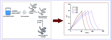
A novel type of nanocomposite material based on multi walled carbon nanotubes (MWCNT) and NiO nanoparticles coated with polyaniline (PAni) has been prepared by an in situ polymerization technique. Transmission electron microscopy confirms the core–shell structure of the composite. TG-analysis reveals that NiO/PAni-MWCNT nanocomposites exhibit better thermal stability than NiO-PAni. The synthesized nanocomposites have good conductivity and excellent electrochemical properties. The specific capacitance values of these composites were measured from their galvanostatic charge–discharge curves. The particles show a high value of specific capacitance and can find potential application as super capacitor electrodes.
Introduction
At present, designing multifunctional core–shell nanocomposites comprising organic and inorganic constituents represent a novel class of materials.1–3 The newly formed nanocomposite material possesses more superior properties than the individual constituent materials.4,5 Depending upon the composition of the core and shell materials, these particles create a class of materials with unique chemical as well as physical properties and they find applications in a wide range of fields.6 In recent years, electrochemical supercapacitors have gained considerable interest because of their high capacitance value, longer cycle life and low maintenance cost.7 An ideal electrode should have high specific capacitance value with enhance stability. It is observed that conducting polymer possesses good specific capacitance value but exhibits poor stability. The carbon based materials such as activated carbon, graphite (graphene) and carbon nanotubes (CNTs) etc. provide the realistic stability but specific capacitance is low enough.8–12 These problems can be minimized by preparing nanocomposite materials based on carbonaceous materials and conducting polymers. The supercapacitors obtained by this process have high capacitance as well as stability value. PAni is one of most prominent material for the redox capacitor application due to its ease of preparation using both chemical and electrochemical process; cost effectiveness, suitable conductivity in its doped form.13–15 PAni electrodes generally suffer from limited cycling stability because of swelling and shrinkage which occurs during doping and de-doping process respectively. This property leads PAni towards mechanical degradation of the electrode and decreases the electrochemical activity. In order to minimize such kinds of problem, researchers have tried to fabricate electrode tools with extremely electro active regions by monitoring the micro-structure.16,17 Recently, many works have reported the improvement of the electrochemical behaviour of PAni by introducing different carbon-based electro-active constituents.8,18 Xiao et al. reported the synthesis of supercapacitors composed of MWCNT with different content of conducting polymers and achieved specific capacitance of the composites in the range from 45 F g−1 to 87 F g−1.19 Mi et al. reported the preparation of PPy-PAni core–shell nanocomposites for the supercapacitors application and the value of the specific capacitance of the composite was 416 F g−1.20 Biswas et al. synthesized the multi layered nano architecture of graphene nano sheets and PPy nanowires for high performance supercapacitors electrodes. This multilayer composite electrode exhibits a high specific capacitance of 165 F g−1.21 Gao reported the synthesis of GO/PAni nanocomposites for the supercapacitor electrode material. The composite showed a maximum specific capacitance value of 329.5 F g−1 and outstanding cycle life.22 Bora et al. reported the synthesis of polypyrrole/sulfonated graphene composite and applied this composite as electrode material for supercapacitor. The composite revealed a maximum value of capacitance of 360 F g−1 at a current density of 1 A g−1. PPy losses 28% of its capacitance value (from 155 to 112 F g−1) whereas composite losses 10% of its capacitance value (from 360 to 323 F g−1) during 500 charge/discharge cycles at 1 A g−1 current density.23 Dhibar et al. reported the preparation of transition metal doped poly(aniline-co-pyrrole)/multi-walled carbon nanotubes nanocomposite for high performance supercapacitor electrode materials. Poly(An-co-Py) Cu CNT nanocomposite reached the highest specific capacitance value of 383 F g−1 at 0.5 A g−1 scan rate.24
In this work, we have reported the synthesis of NiO/PAni and NiO/PAni-MWCNT core–shell nanocomposites with different content of NiO nanoparticles and MWCNT. The structural, thermal, electrical and electrochemical properties of these core–shell composites were investigated and compared with their pristine counterpart. There are reports on synthesis of NiO/PAni however we have not found any report of NiO/PAni-MWCNT as super capacitor electrodes. Our study shows NiO/PAni-MWCNT as improved supercapacitor electrode material. The dependence of electrical conductivity on NiO nanoparticles in the core as well as the MWCNT content in the shell phase is examined. We also investigated the electro-chemical behaviour of the particles and calculated the super capacitance value of these materials from their galvanostatic charge–discharge curves. The study reveals that with the incorporation of NiO and MWCNT in the polymer matrix there is a significant increase in the super capacitance value as well as electrochemical stability of the material.
Experimental section
Materials
NiCl2·6H2O and NaHCO3, KCl, HCl, N-methyl-2-pyrrolidone (NMP), ammonium peroxodisulfate (APS) was purchased from Merck, India. Aniline monomer and acetonitrile were purchased from Sigma Aldrich. All the chemicals were of analytical grade. Multiwall carbon nanotube (MWCNT) was purchased from Redex nano lab, India. Doubled distilled water and purified ethanol were used as dispersion medium for all the chemical preparation as well as for washing.
Method
Preparation of NiO nanoparticles. NiO nanoparticles were prepared by two steps soft chemical synthesis method.25 At first, NiCl2·6H2O (2.4 g) and NaHCO3 (1.2 g) were added in 10 mL distilled water independently at room temperature. NiCl2·6H2O solution was stirred for 15 min and NaHCO3 solution was added drop wise to it. The stirring was continued upto the termination of gas formation from the reaction mixture. The resultant precipitated was separated by centrifugation and washed it with distilled water. Separated product was dried at 100 °C and then heated it for 2 h at 600 °C. The possible reactions for the synthesis of NiO nanoparticles are given below:
2NaHCO3(s) → 2Na+(aq) + 2HCO3−(aq) |
NiCl2·6H2O(s) → Ni2+(aq) + 2Cl−(aq) + 6H2O(aq) |
Ni2+(aq) + 2HCO3−(aq) + XH2O(aq) → Ni(HCO3)2·H2O(s) |
Preparation of NiO/PAni core–shell nanocomposites. NiO/Pani core–shell nanocomposites were prepared by in situ polymerization process. 45 mL of 0.25 M HCl was taken in a three necked round bottom flask. Then 1 mL aniline was added to flask and 45 mL of water was added. The stirring was continued under N2 environment. Temperature was maintained at 0–5 °C and pH 3.15. 0.2 g NiO was added and dispersed for half an hour. Then ammonium persulfate was added and stirring was carried out for 1 h in order to assist oxidative polymerization. After polymerization completion blackish green product was separated through centrifugation and washing several times with double distilled water. The resulting products were dried under vacuum at 60 °C for 24 h.
Preparation of NiO/PAni-MWCNT nanocomposites. 1 mL aniline monomer and 0.02 g MWCNT were added in 45 mL of 0.25 M HCl solution and sonicated until a proper dispersion is obtained. NiO (0.2 g) nanoparticles were suspended in 45 mL water sonicated for 1 h. Then NiO nanoparticles dispersion was added dropwise into the above solution and stirring continued for half an hour. The resultant mixture was transferred into a 150 mL round bottom flask and stirred under N2 atmosphere at 0–5 °C. After that, APS of 1 g was added gently into reaction mixture and the stirring was continued for another 1 h. A blackish green product was separated from the solution after centrifugation and it was sequentially washed with distilled water for several times. The resulting products were dried under vacuum at 60 °C for 24 h. Different core–shell particles of NiO/PAni-MWCNT were prepared by changing the amount of NiO and MWCNT from 0.02 g to 0.1 g keeping other ingredients constant.
Characterization
UV-Visible absorption spectra of the nanoparticles were recorded using Shimadzu UV-2550 UV-Visible spectrophotometer in the range of 200–800 nm at room temperature. FTIR spectrum of the nanoparticles, polymer, and the core–shell nanoparticles was recorded in the range of 400–4000 cm−1 using Nicolet Impact-410 IR spectrometer at room temperature. Crystalline structure of NiO nanoparticles, NiO/PAni core–shell and NiO/PAni-MWCNT core–shell were studied by X-ray diffractometry (Miniflex, Rigaku Japan) with CuKα radiation (λ = 0.15418 nm) at 30 kV and 15 mA with scanning rate of 0.005 S−1 in a 2θ range of 10–70°.The surface structure and energy dispersion spectroscopic analysis of the nanoparticles, polymer and the core–shell nanocomposite particles were determined by SEM (Jeol-JSM-6390LV) coupled with energy dispersive X-ray detector. Composite samples were coated with platinum thickness of 200 Å. The voltage and working distance was varied during the measurement. To obtain semi quantitative compositional information, EDX spectra was recorded. The particle size and morphology of the synthesized nanoparticles as well as the core–shell nanocomposites were determined with TEM (JEOL JEM 2100) at an acceleration voltage of 200 kV. TGA of the polymer as well as the core–shell composites were recorded in a Shimadzu TA50 thermal analyser. Heating of the samples was carried out in presence of nitrogen atmosphere at rate of 5 °C min−1 in the range of 25–700 °C. For the measurement of electrical conductivity of core–shell nanoparticles, hard round pellets (d × b = 1.5 cm × 2 mm) were made by a compression moulding machine equipped with hydraulic pressure. The electrical conductivity of NiO/PAni and NiO/PAni-MWCNT core–shell nanocomposites was measured using four probe techniques. I–V characteristic was studied by Keithley 2400 source meter at the room temperature with the frequency range of 102–106 Hz. CVmeasurements of polymer and core–shell nanocomposites were carried out on an electrochemical work station Biologic SP-150 with a platinum wire as a counter electrode, an Ag/AgCl as a reference electrode and PAni, NiO/PAni, NiO/PAni-MWCNT (varying NiO, MWCNT) deposited on ITO coated glass as working electrode. The CV measurements of the core–shell nanocomposite samples was carried out with a scanning rate of 50 mV s−1 and 0.1 M, 10 mL KCl in acetonitrile was used as supporting electrolyte.
Results and discussion
Fig. 1 is the schematic representation of the synthetic procedure as well as the plausible mechanism for the formation of NiO nanoparticles, NiO/PAni and NiO/PAni-MWCNT core–shell nanocomposites. NiO nanoparticles were prepared by two steps soft chemical synthesis route. NaHCO3 is used as a replacing agent and it replaces Cl− from NiCl2·6H2O to produce NiO nanoparticles. In the first step, formation of effervescences indicates the liberation of CO2 as a by-product from the reaction mixture. The dark grey coloured product formed after burning in the muffled furnace, indicates the formation of NiO nanoparticles. The NiO/PAni and the NiO/PAni-MWCNT core–shell nanocomposites were prepared by in situ chemical polymerization techniques. The polymerization is carried out in the acidic medium. A certain portion of aniline–hydrochloride solution may have been adsorbed on the surface of dispersed NiO nanoparticles as well as MWCNT and the remaining free aniline got polymerized in presence of an oxidizing agent to produce NiO/PAni and NiO/PAni-MWCNT. Whenever APS is added, the polymerization of aniline monomer starts and results the formation of dark green coloured products. The formation of dark green colour during the reaction gives the evidence towards the formation of PAni. Thus a core–shell structure was successfully achieved through the combination of two step soft chemical synthesis route, chemical oxidative polymerization with NiO as the magnetite core and PAni, PAni-MWCNT as the shell material.
 |
| Fig. 1 Schematic representation of formation of NiO nanoparticles, NiO/PAni and NiO/PAni-MWCNT core–shell nanocomposites. | |
FT-IR analysis
Fig. 2(a)–(d) depicts the FT-IR image of PAni, MWCNT, NiO/PAni-MWCNT core–shell nanocomposites and NiO nanoparticles respectively. The absorbance band at 474 cm−1 is due to the presence of Ni–O bond [Fig. 2(d) d1].6 Presence of this bond indicates the formation of NiO nanoparticles and incorporation of NiO nanoparticles into NiO/PAni as well as NiO/PAni-MWCNT nanocomposites. In the Fig. 2(a) the absorption band at 1570, 1460, 1306, 1272 and 589 cm−1 indicates the formation of PAni.26,27 The spectral band at 1570 and 1460 cm−1 are due to the C
C stretching vibration of quinoid and benziod rings respectively. The peak at 1306, 1272 and 589 cm−1 corresponds to the C–N stretching vibration of secondary aromatic amine, C–H in plane and out of plane deformation respectively. The absorption band at 3441 cm−1 is due to N–H stretching vibration [a2]. The absorption band at 2924 and 2867 cm−1 are due to the asymmetric C–H stretching and symmetric C–H stretching vibration. The absorption peaks at 1651 cm−1 is due to the C
C stretching vibration in aromatic nuclei [a3]. The bands observed in the region of 1600–1500 cm−1 are due to the C–H stretching vibration in aromatic compounds [a4]. The absorption band at 1468 and 1441 cm−1 are due to the C
N stretching vibration in the aromatic compounds [a5]. The absorption bands at 1300–1200 cm−1, are for the C–N stretching of primary aromatic amine [a6]. The bands at 1110 and 1139 cm−1 reveals the C–H bending vibrations [a7]. The absorption bands which were observed below the 1000 cm−1 are due to the characteristic mono substituted benzene. Presence of these peaks indicates the formation of PAni. These peaks are also observed in the Fig. 2(c), which is an indication that the core–shell nanocomposites also contain PAni. In case of MWCNT, two dominant peaks are observed at 3432 and 1620 cm−1 [b1 and b2], which are associated with –OH functional group. The peak at 1716 cm−1 is due to the presence of carbonyl group [b3]. Several peaks are observed in the range of 3000 cm−1, which are responsible for CHX groups. Presence of these peaks indicates incorporation of MWCNT in the final composites.25
 |
| Fig. 2 FTIR images of PAni, MWCNT, NiO/PAni-MWCNT core–shell nanocomposites and NiO nanoparticles. | |
UV-analysis
UV-Visible analysis of NiO nanoparticles, PAni, NiO/PAni and NiO/PAni-MWCNT nanocomposites are given in the Fig. 3(a)–(d). NiO nanoparticles showed the UV-Visible absorption maximum peak at 342 nm.6 The UV-Visible analysis of PAni, shows a broad peak at around 628–631 nm and a nearly sharp absorption peak at around 322–328 nm.28 The absorption band at 322–328 nm is associated with the π–π* transition of the conjugated polymer and peak at 628–631 nm is due to the charge-transfer exciton from the highest occupied energy level of the benzenoid rings to the lowest occupied energy level of the quinonoid rings.23 Here PAni and its composites are synthesized in the acidic medium and hence the absorption band of the benzenoid to quinonoid rings has been observed at more than 700 nm. Formation of hydrogen bonding between the C
O group of NMP and the –NH group of PAni prevents the acid-doping of PAni.
 |
| Fig. 3 UV-Visible spectra of (a) NiO nanoparticles, (b) PAni, (c) NiO/PAni core–shell nanocomposites, (d) NiO/PAni-MWCNT core–shell nanocomposites. | |
In the UV-Visible analysis of PAninanocomposites with NiO and MWCNT both peaks suffered a blue shift. These blue shifts and change of intensity of the samples are attributed to the interaction between NiO and MWCNT with PAni through hydrogen bonding. These changes contributes to a decrease in the degree of orbital overlapping between the π electrons of the phenyl rings with the lone pair of the nitrogen atom in the PAni molecules. These result in decrease in the extent of conjugation of PAni and henceforth leading to the shifting of peaks towards shorter wavelength.
TG-analysis
Fig. 4(a)–(e) depict the TG-curves of PAni, NiO/PAni and NiO/PAni-MWCNT nanocomposites. From the analysis, it is observed that the all samples exhibit weight loss at three stages. For PAni, the first weight loss occurs at about 100 °C and this may be due to the loss of residual moisture from the polymer; second weight loss at about 200–360 °C is due to the loss of dopant from the polymer matrix; weight loss observed at about 360–700 °C is due to the degradation of polymeric back bone. Thermal stability of the polymer matrix increases with the incorporation of NiO and the MWCNT into the polymer matrixes. Thermal degradation patterns of all the composites are found to be the similar. From the analysis it is observed that the major degradation for the composite materials starts at higher temperature in comparison to PAni. In NiO/PAni and NiO/PAni-MWCNT nanocomposites, the first weight loss occurs at about 200 °C, which is due to the removal of residual moisture from the composite. The major weight loss at 400–700 °C is due to the polymer degradation. With the increasing amount of incorporated NiO nanoparticles as well as the MWCNT in the polymer matrixes, the residual weight of the composites was found to be better in compared to PAni. The weight retention for NiO/PAni nanocomposites was found to be 20–30% and 25–35% for NiO/PAni-MWCNT at 700 °C. This improvement of thermal stability may be due to the well-ordered and compact structure which delays the thermal degradation of composites by restricting the thermal motion of the polymer chains.
 |
| Fig. 4 TG-analysis of synthesized (a) PAni, (b) NiO/PAni-7 core–shell nanocomposites, (c) NiO/PAni-10 core–shell nanocomposites, (d) NiO/PAni-MWCNT-7 core–shell nanocomposites, (e) NiO/PAni-MWCNT-10 core–shell nanocomposites. | |
X-ray diffraction analysis
XRD patterns of synthesized NiO nanoparticles, PAni, MWCNT, NiO/PAni and NiO/PAni-MWCNT are shown in the Fig. 5(a)–(d). Three sharp peaks observed [Fig. 5(c)] at about (2θ) 38° (111), 44° (200) and 64° (220) are due the formation of NiO nanoparticles. Pure PAni is amorphous in nature and hence it gives a broad peak (2θ) 20°–30° [Fig. 5(a)]. By using Williamson Holl plot, the average crystallite size of the synthesized nanoparticles is found to be 5.96 Å. In the XRD spectra of MWCNT [Fig. 5(b)] the peaks are observed at about (2θ) 26° (002). In case of NiO/PAni-MWCNT nanocomposites, three sharp peaks are also observed at about (2θ) 38°, 44° and 64° for NiO particles along with the broad XRD peak for PAni [Fig. 5(d)]. Presence of these peaks gives the evidence of successful incorporation of PAni into NiO nanoparticles. Moreover, characteristic peaks of MWCNT are also observed along with the three sharp peaks of NiO nanoparticles and the broad peaks of PAni. This confirms the incorporation of MWCNT into the polymer matrixes.
 |
| Fig. 5 XRD analysis of (a) PAni, (b) MWCNT, (c) NiO nanoparticles, (d) NiO/PAni-MWCNT core–shell nanocomposites. | |
Surface morphology analysis
Scanning electron microscope (SEM) analysis. SEM images of NiO nanoparticles, MWCNT, NiO/PAni and NiO/PAni-MWCNT are given in the Fig. 6. From the Fig. 6(a), the shape of the NiO nanoparticles are found to be uniform. Tubular shapes of MWCNT are observed in the Fig. 6(b). In the Fig. 6(c) size of NiO/PAni nanocomposites is slightly greater than that of the NiO nanoparticles. The composite particles are found to be nearly uniform in size. This indicates the successful coating of PAni over NiO nanoparticles surface. In the same image of NiO/PAni-MWCNT [Fig. 6(d)], it is observed that the width of the MWCNT has been enhanced than that of the pure MWCNT. This indicates that NiO/PAni has grown on the MWCNT surface.
 |
| Fig. 6 SEM images of (a) NiO nanoparticles, (b) MWCNT, (c) NiO/PAni core–shell nanoparticles, (d) NiO/PAni-MWCNT. | |
Energy dispersive X-ray (EDX) analysis. Fig. 7 depicts the EDX patterns of the NiO/PAni and NiO/PAni-MWCNT respectively. Fig. 7(a); Ni, O peaks confirms the presence of NiO and C, N peaks confirms the formation of PAni which is successfully coated over NiO nanoparticles. Fig. 7(b); Ni, O, C, N peaks give the evidence of presence of NiO and PAni. The enhancement of C peaks in NiO/PAni-MWCNT is additional evidence towards the successful incorporation of MWCNT into the polymer matrixes.
 |
| Fig. 7 EDX pattern of synthesized (a) NiO/PAni core–shell nanocomposites, (b) NiO/PAni-MWCNT core–shell nanocomposites. | |
TEM analysis. Fig. 8 depicts the TEM images of the synthesized materials. Fig. 8(a), it is observed that the synthesized NiO nanoparticles are nearly uniform in shape and the remains in the range of 15–25 nm. In the TEM image [Fig. 8(c)] of the NiO/PAni, the dark dotted spot depicts the NiO nanoparticles and the slightly transparent layer over the NiO nanoparticles represents the PAni. It confirms the formation of NiO/PAni nanocomposites with well-defined core–shell morphology. The average size and shell thickness of the core–shell nanocomposites are found to be 65 nm and 15 nm respectively. In the TEM images [Fig. 8(d)] of NiO/PAni-MWCNT, it is observed that NiO/PAni core–shell particles are homogeneously grown on MWCNT taking it as the support. Thickness of the MWCNT is found to be 20 nm and the size of the core–shell nanocomposites are found to be similar which is obtained in the NiO/PAni core–shell nanocomposites.
 |
| Fig. 8 TEM images of (a) NiO nanoparticles, (b) MWCNT, (c) NiO/PAni core–shell nanocomposites, (d) NiO/PAni core–shell nanocomposites [inset NiO/PAni core–shell with two particles], (e) NiO/PAni-MWCNT core–shell nanocomposites. | |
Electrical behaviour
DC-electrical conductivity. The DC-electrical conductivity of PAni and the core–shell nanocomposites with different the amount of NiO nanoparticles as well as MWCNT are determined at room temperature and their conductivities are listed in the Table 1. From the conductivity measurements, it is observed that the conductivity of NiO/PAni or NiO/PAni-MWCNT increases dramatically in comparison with pure PAni. With the increasing amount of NiO nanoparticles content in the core, the electrical conductivity of NiO/PAni core–shell nanocomposites vary widely in the range from 0.67 × 10⁻³ to 1.71 × 10⁻³ S cm−1. In case of NiO/PAni-MWCNT it varies from 1.94 × 10⁻³ S cm−1 to 8.23 × 10⁻³ S cm−1. The increase in conductivity for NiO/PAni may be due to the increase in compactness of the core–shell nanocomposites as the growing polymer chains are supported on NiO nanoparticles. The improvement in conductivity of NiO/PAni-MWCNT core–shell nanocomposites may be due to the π–π stacking between the polymer backbone and MWCNT. MWCNT itself is conducting in nature and when the polymerization of PAni takes place on its surface the pore sites eventually controlling the twisting of the polymer backbone away from the planarity. The polymer chains become well-organized and linking between the polymer chains are significantly improved. Thereby increases conductivity both of NiO/PAni and NiO/PAni-MWCNT nanocomposites. Thus, NiO and MWCNT play a significant role to enhancement of the conductivity of PAni.
Table 1 DC-electrical conductivity of PAni, NiO/PAni and NiO/PAni-MWCNT core–shell nanocomposite
Core–shell nanocomposites |
NiO-content (%) |
MWCNT content (%) |
Resistivity, ρ (Ω cm) |
Conductivity σ (S cm−1) |
PAni |
0 |
0 |
1.49 × 10³ |
0.67 × 10⁻³ |
NiO/PAni-2 |
2 |
0 |
1.09 × 10³ |
0.92 × 10⁻³ |
NiO/PAni-5 |
5 |
0 |
0.87 × 10³ |
1.15 × 10⁻³ |
NiO/PAni-7 |
7 |
0 |
0.68 × 10³ |
1.46 × 10⁻³ |
NiO/PAni-10 |
10 |
0 |
0.58 × 10³ |
1.71 × 10⁻³ |
NiO/PAni-MWCNT-2 |
2 |
2 |
0.52 × 10³ |
1.94 × 10⁻³ |
NiO/PAni-MWCNT-5 |
5 |
5 |
0.25 × 10³ |
4.01 × 10⁻³ |
NiO/PAni-MWCNT-7 |
7 |
7 |
0.16 × 10³ |
6.11 × 10⁻³ |
NiO/PAni-MWCNT-10 |
10 |
10 |
0.12 × 10³ |
8.23 × 10⁻³ |
Electrochemical property. Fig. 9 represents the CV graphs of PAni, NiO/PAni and NiO/PAni/MWCNT core–shell nanocomposites with different NiO and MWCNT content. From the electrochemical analysis it is observed that the shapes of the CV graphs of the nanocomposites are different from the pure PAni. The composite particles possess comparatively lower electrochemical band gap than that of the PAni. The electrochemical band gaps of NiO/PAni decreases with the increase of NiO content in the core. When MWCNT is incorporated in the composite the band gap is further decreased. The peak currents for the composite were also higher compared to PAni. This enhancement in current for the nanocomposites may be attributed to the special nanostructure of this composite. From the analysis, it may be concluded that with incorporation of PAni into the NiO nanoparticles as well as the MWCNT results in change in electronic band structure.
 |
| Fig. 9 CV graph of (a) PAni, (b) NiO (2%)/PAni, (c) NiO (10%)/PAni, (d) NiO (2%)/PAni-MWCNT (5%), (e) NiO (2%)/PAni-MWCNT (7%). | |
Fig. 10 represents the charging–discharging curve [E (V) vs. time] of the composites upto 6 cycles. From the figure it is seen that there is no change in potential drop during the experiment which signify that columbic efficiency is nearly 100%. Fig. 11 gives the galvanostatic charge–discharge curves of NiO/PAni and NiO/PAni/MWCNT composites having different NiO and MWCNT content at a current density of 5 mA cm−2. The specific capacitance (Cm) of the materials is calculated from these curves using the following equation.29
where
I is the charge–discharge current, Δ
t is the discharge time, Δ
V is the electrochemical window and
m is the mass of active material within the electrode (5 mg). The specific capacitance calculated from the figure was 145.87 F g
−1 for composites with 2% NiO which increases to 172.47 F g
−1 when percentage of NiO is increased to 10%. We further investigated the effect of incorporation of MWCNT on the super capacitance value of the composite particles. It is found from the figure that for the composite (with 2% NiO in the shell phase) the super capacitance value with 5% MWCNT is 337.04 F g
−1 where as it is 356.54 F g
−1 with 7% MWCNT (
Tables 2 and
3).
 |
| Fig. 10 Charging–discharging curve of NiO/PAni-MWCNT (10%) upto 6 cycles. | |
 |
| Fig. 11 Galvanostatic charge–discharge curves of (a) NiO (2%)/PAni (b) NiO (10%)/PAni (c) NiO (2%)/PAni/MWCNT (5%) (d) NiO (2%)/PAni/MWCNT (7%). | |
Table 2 Variation of specific capacitance values of the nanocomposites with percentage of NiOa
% of NiO in the nanocomposite |
Specific capacitance (F g−1) |
Results are in form: mean ± standard deviation, where each of the experiment were performed in triplicate and analyzed in triplicate as well. |
2 |
145.87 ± 0.469681 |
5 |
148.72 ± 0.12083 |
7 |
152.70 ± 0.071414 |
10 |
172.47 ± 0.049699 |
Table 3 Variation of specific capacitance with scan rate NiO/PAni-MWCNT (7%)a
Scan rate (mV s−1) |
Specific capacitance (F g−1) |
Results are in form: mean ± standard deviation, where each of the experiment were performed in triplicate and analyzed in triplicate as well. |
5 |
356.54 ± 0.049193 |
10 |
199.9 ± 0.039623 |
20 |
100.13 ± 0.038471 |
30 |
66.68 ± 0.037683 |
This significant increase in super capacitance value with the incorporation of MWCNT may be attributed to the fact that the presence of MWCNT facilitates the oxidation or reduction process of α-cabon or β-carbon of PAni. Further attachment of PAni on the surface of MWCNT further reduces the diffusion and migration length and hence helps in the electrochemical utilization of PAni. The average energy density (E) and the power density (P) of the composites were evaluated from the following expressions:10
For the composite with 7% MWCNT these values are found to be 55.9 W h kg−1 and 1996.42 W kg−1 respectively. A reasonably high value of energy density and power density indicates that the composite material be potentially used as a super capacitor electrode.
The variation in the specific capacitance of 7% MWCNT incorporated composites as a function of scan rate is shown in Fig. 12. The specific capacitance is 356.543 F g−1 at scan rate of 5 mV s−1 which decreases to 66.68 F g−1 when scan rate is increases upto 30 mV s−1. The high value of specific capacitance at a lower scan rate may be attributed to the fact that at low scan rate the ions get enough time to migrate freely in the PAni matrix. The discharge curves observed are not rectangular irrespective of the applied current density, are characteristics of pseudocapacitance behavior of the NiO/PAni/MWCNT samples. It is observed that charging discharging curves are not triangular due to kinetic irreversibility in diffusion of OH− ions. The initial sudden change in voltage response with respect to time while charging and discharging has been found due to voltage drop (IR) across the internal resistance of the cells. Moreover at relatively low scan rate the oxidation or reduction of α-C or β-C may occur rather smoothly.
 |
| Fig. 12 Variation of specific capacitance with scan rate NiO/PAni-MWCNT (7%). | |
Charge capacity. To investigate the charge capacity of the nanocomposites, the samples were subjected to 1000 repeated charge–discharge cycle and the loss in capacitance was monitored and presented in the Fig. 13. It is observed that PAni losses 39.2% of its capacity (from 102 to 62.1 F g−1) during 1000 charge–discharge cycle at a current density of 5 A g−1. In case of NiO (2%)/PAni composite and NiO (2%)/PAni/MWCNT (7%) composite the loss in capacitance are 18.7% (145.2 to 118 F g−1) and 6.6% (356.54 to 333 F g−1) respectively. Thus it may be inferred that there is a significant improvement of the electrochemical stability of the composite in comparison to the pristine polymer. The increase in stability with the incorporation of the MWCNT may be attributed to the fact that sheets of MWCNT prevent the PAni chains to swell and shrink during the charge–discharge cycle.
 |
| Fig. 13 Cycling stabilities of (a) PAni and (b) NiO (2%)/PAni (c) NiO (2%)/PAni/MWCNT (7%) to 1000 charge/discharge cycles (current density, 5 A g−1). | |
Conclusion
A successful approach towards the synthesis of NiO/PAni-MWCNT core–shell nanocomposites by two steps in situ polymerization has been carried out. The effectiveness of the nanocomposite as a high performance super capacitor electrode has also been explored. The surface behaviour and the core–shell structure of the nanocomposites were confirmed by SEM and TEM analysis. TGA shows the improved thermal stability of the composite material in comparison to the pristine polymer. Electro-chemical study reveals that the material is sufficiently stable to be used in electronic devices. The specific-capacitance of the material is to be 356.54 F g−1 with 7% of MWCNT. These competent features make the newly synthesised nanocomposite material as a promising material for super capacitor electrode.
Acknowledgements
The authors would like to acknowledge the help and support of Tezpur University authority.
References
- Y. S. Lin, S. H. Wu, Y. Hung, Y. H. Chou, C. Chang, M. L. Lin, C. P. Tsai and C. Y. Mou, Chem. Mater., 2006, 18, 5170–5172 CrossRef CAS.
- V. S. Maceira and M. A. Correa-Duarte, Adv. Mater., 2007, 19, 4131–4144 CrossRef.
- S. Xuan, Y. Xiang, J. Wang, J. C. Yu and K. C. F. Leung, Langmuir, 2009, 25, 11835–11843 CrossRef CAS PubMed.
- L. Wang, H. Wei, Y. Fan, X. Liu and J. Zhan, Nanoscale Res. Lett., 2009, 4, 558–564 CrossRef CAS PubMed.
- Y. Zhao, X. Yang, J. Tian, F. Wang and L. Zhan, Int. J. Hydrogen Energy, 2010, 35, 3249–3257 CrossRef CAS.
- D. Das, B. C. Nath, P. Phukon, B. J. Saikia, I. R. Kamrupi and S. K. Dolui, Mater. Chem. Phys., 2013, 142, 61–69 CrossRef CAS.
- Q. Xiao and X. Zhou, Electrochim. Acta, 2003, 48, 575–580 CrossRef CAS.
- M. Z. Gao, W. Yang, J. Wang, B. Wang, Z. Li, Q. Liu, M. Zhang and L. Liu, Energy Fuels, 2013, 27, 568–575 CrossRef.
- L. Z. Fan, Y. S. Hu, J. Maier, P. B. Adelhelm and M. Smarsly, Adv. Funct. Mater., 2007, 17, 3083–3087 CrossRef CAS.
- J. Yan, T. Wei, B. Shao, Z. Fan, W. Qian, M. Zhang and F. Wei, Carbon, 2010, 48, 487–493 CrossRef CAS.
- L. Mao, K. Zhang, H. S. O. Chan and J. S. Wu, J. Mater. Chem., 2012, 22, 80–85 RSC.
- X. J. Lu, H. Dou, S. D. Yang, L. Hao, L. J. Zhang, L. F. Shen, F. Zhang and X. G. Zhang, Electrochim. Acta, 2011, 56, 9224–9232 CrossRef CAS.
- K. S. Ryu, K. M. Kim, N. G. Park, Y. J. Park and S. H. Chang, J. Power Sources, 2002, 103, 305–309 CrossRef CAS.
- F. Montilla, M. A. Cotarelo and E. Morallon, J. Mater. Chem., 2009, 19, 305–310 RSC.
- S. J. Choi and S. M. Park, J. Electrochem. Soc., 2002, 149, 26–34 CrossRef.
- N. A. Kumar, H. J. Choi, Y. R. Shin, D. W. Chang, L. Dai and J. B. Baek, ACS Nano, 2012, 6, 1715–1723 CrossRef CAS PubMed.
- L. L. Zhang, S. Li, J. Zhang, P. Guo, J. Zheng and X. S. Zhao, Chem. Mater., 2009, 22, 1195–1202 CrossRef.
- J. Li, H. Q. Xie, Y. Li, J. Liu and Z. X. Li, J. Power Sources, 2011, 196, 10775–10781 CrossRef CAS.
- Q. Xiao and X. Zhou, Electrochim. Acta, 2003, 48, 575–580 CrossRef CAS.
- H. Mi, X. Zhang, X. Ye and S. Yang, J. Power Sources, 2008, 176, 403–409 CrossRef CAS.
- S. Biswas and L. T. Drzal, Chem. Mater., 2010, 22, 5667–5671 CrossRef CAS.
- Z. Gao, W. Yang, J. Wang, B. Wang, Z. Li, Q. Liu, M. Zhang and L. Liu, Energy Fuels, 2013, 27, 568–575 CrossRef CAS.
- C. Bora, A. Kalita, D. Das, S. K. Dolui and P. K. Mukhopadhyay, Polym. Int., 2014, 63, 445–452 CrossRef CAS.
- S. Dhibar, P. Bhattacharya, G. Hatui and C. K. Das, J. Alloys Compd., 2015, 625, 64–75 CrossRef CAS.
- S. Chakrabarty and K. Chatterjee, J. Phys. Sci., 2009, 13, 245–250 Search PubMed.
- R. Singha, C. Dhand, G. Suman, R. Verma, S. Sood, R. K. Gupta and B. D. Malhotr, J. Mol. Recognit., 2010, 23, 472–479 CrossRef PubMed.
- J. Vivekanandan, V. Ponnusamy, A. Mahudeswaran and P. S. Vijayan, Arch. Appl. Sci. Res., 2011, 3, 147–153 CAS.
- L. J. Borthakur, S. Sharma and S. K. Dolui, J. Mater. Sci.: Mater. Electron., 2011, 22, 949–958 CrossRef CAS.
- S. Bose, N. H. Kim, T. Kuila and K. L. Lee, Nanotechnology, 2011, 22, 1–9 Search PubMed.
|
This journal is © The Royal Society of Chemistry 2016 |
Click here to see how this site uses Cookies. View our privacy policy here.