DOI:
10.1039/C6RA01527A
(Paper)
RSC Adv., 2016,
6, 33636-33642
Palladium nanoparticles modified electrospun CoFe2O4 nanotubes with enhanced peroxidase-like activity for colorimetric detection of hydrogen peroxide†
Received
18th January 2016
, Accepted 20th March 2016
First published on 22nd March 2016
Abstract
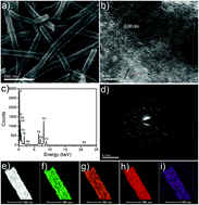
Herein, we report a simple procedure to decorate small palladium nanoparticles (Pd NPs) on the surface of CoFe2O4 nanotubes; the decorated nanotubes possess intrinsic peroxidase-like activity for the sensitive detection of H2O2. The CoFe2O4 nanotubes are prepared via an electrospinning technique, followed by a calcination process in air. The functionalization of Pd NPs on the surface of CoFe2O4 nanotubes was achieved through an in situ reduction process using ascorbic acid (AA) as a reducing agent. The synthesized Pd nanoparticles, which are small in size, are evenly dispersed on the surface of the CoFe2O4 nanotubes. The hollow structure of the CoFe2O4 nanotubes and the uniform distribution of the Pd nanoparticles enhance peroxidase-like activity toward the catalytic oxidation of the peroxidase substrate 3,3′,5,5′-tetramethylbenzidine (TMB) in the presence of H2O2. The peroxidase-like property of the Pd/CoFe2O4 composite nanotubes provides a facile approach for the colorimetric detection of H2O2 with a low detection limit. This work provides great potential for Pd/CoFe2O4 nanotubes as enzyme-like nanocatalysts to sensitively detect H2O2 in biological systems.
Introduction
Artificial enzyme mimics are materials that possess catalytic activity similar to natural enzymes but that display many advantages over said enzymes, such as superior stability, easy preparation, good recyclability and low cost.1–3 Over the past few decades, artificial enzyme mimics have been shown to have diverse applications in biosensing, medical diagnosis, food processing and environmental monitoring. Early reports on artificial enzyme mimics mainly include cyclodextrins, porphyrins, dendrimers, biomolecules, and supramolecular complexes.1–8 Recently, nanomaterial-based artificial enzyme mimics have attracted increasing attention owing to their large surface area, high and tunable catalytic activity, good selectivity, and facile preparation at low cost.9–11 For example, since the first report of Fe3O4 nanoparticles,12 various other nanomaterials, including metal oxide nanoparticles and nanowires,13–17 metal and metal alloy nanoparticles,18–20 carbon nanomaterials,21–23 chalcogenide nanomaterials,24–26 polymer nanoparticles,27–29 and multiferroic nanomaterials,30–32 have also been shown to possess intrinsic peroxidase-like activity. To enhance peroxidase-like catalytic performance, much study has been performed on the fabrication of nanocomposites with synergistic catalytic effects. For instance, conducting polyaniline (PANI)/Cu9S5 nanocomposites exhibited higher catalytic activity as artificial peroxidase mimics than individual PANI and Cu9S5 nanoparticles due to their strong interactions and synergistic effect.33 Other systems, such as Au nanoparticles/graphene composite,34 dumbbell-like Pt48Pd52–Fe3O4 nanoparticles,35 Co3O4/reduced graphene oxide composite,36 and CuS/graphene nanocomposite37 have also displayed superior peroxidase-like catalytic activity. Therefore, much effort should be devoted to design and prepare new nanocomposites with improved enzyme-like catalytic performance.
Noble metal nanoparticles, especially Au, Pd and Pt, have attracted widespread interest due to their unique optoelectronic properties and large surface area, which display potential applications in catalysis, electronics, energy storage and conversion, sensors, and biomedical research.38 Recently, noble metal nanoparticles have also been shown to possess intrinsic enzyme-like catalytic activity. However, individual noble metal nanoparticles usually aggregate during the catalytic process, limiting their catalytic activity. Therefore, it is necessary to anchor metal nanoparticles on specific supports. Recently, Zhao, Quan and co-workers reported that Au nanoparticles supported on the surface of graphene nanosheets exhibit superior intrinsic peroxidase-like activity over individual Au nanoparticles and graphene nanosheets alone, demonstrating a synergistic coupling effect between these two components.34 Similarly, Au nanoparticles supported on the surface of graphene oxide and carbon nanotubes,39,40 Au/Pd nanoparticles supported on the surface of graphene,41 and porous Pt nanoparticles supported on the surface of graphene oxide42 have also been found to possess excellent intrinsic peroxidase-like catalytic activities. In addition to carbon-based nanocomposites, metal oxide-based nanocomposites have also been found to exhibit high enzyme-like activity.43–46 By combining them with other active components, enhanced catalytic activity can also be achieved.
Recently, spinel ferrite compounds such as CoFe2O4 and ZnFe2O4 nanomaterials have been reported to display intrinsic peroxidase-like activity.31,32 However, the catalytic activities of pure CoFe2O4 and ZnFe2O4 nanoparticles as peroxidase mimics are relatively weak. Therefore, GE/CoFe2O4 and CNT/ZnFe2O4 nanocomposites were prepared as peroxidase mimics, which showed enhanced catalytic activities over independent CoFe2O4 and ZnFe2O4 nanomaterials.47,48 In this study, we demonstrate that the modification of noble metal nanoparticles on the surface of CoFe2O4 nanotubes provides a new platform as an artificial enzyme for self-activated peroxidase-like catalysis. In addition, the Pd/CoFe2O4 composite nanotubes have also been used to construct a colorimetric assay for the detection of H2O2 based on their peroxidase-like activity; the assay exhibits high sensitivity and a low detection limit.
Experimental
Chemicals and materials
Poly(vinyl pyrrolidone) (PVP, weight-average molecular weight, Mw = 1
300
000 g mol−1) was purchased from Sigma-Aldrich. Cobalt(II) acetate tetrahydrate (Co(Ac)2·4H2O) and 3,3′,5,5′-tetramethylbenzidine (TMB) were obtained from Sinopharm Chemical Reagent Beijing Co., Ltd. Iron nitrate (Fe(NO3)3·9H2O) was purchased from Tianjin East China Reagent Factory. Na2PdCl4 was obtained from Aladdin. All other chemicals were of analytical grade and were used without further purification.
Preparation of CoFe2O4 nanotubes by electrospinning technique
The CoFe2O4 nanotubes were prepared through an electrospinning technique. In a typical procedure, 1.64 g of PVP was dissolved in a mixture containing 11.4 g of ethanol and 7.8 g of N,N′-dimethylformamide (DMF), followed by the addition of 0.50 g of Fe(NO3)3·9H2O and 0.154 g of Co(Ac)2·4H2O. A homogeneous mixture solution was obtained after vigorous stirring at room temperature for about 12 h. The as-prepared mixture solution was electrospun using an applied voltage of 15 kV. A piece of aluminum foil was used to collect the PVP/Fe(NO3)3/Co(Ac)2 nanofibers, and the distance between the nozzle and the collector was fixed at about 15 cm. The as-fabricated PVP/Fe(NO3)3/Co(Ac)2 nanofibers were then calcined under air atmosphere at 550 °C for 3 h to remove the PVP matrix and produce the CoFe2O4 nanotubes.
Preparation of Pd nanoparticles modified CoFe2O4 nanotubes
5 mg of the obtained CoFe2O4 nanotubes were dispersed in 8 mL of water, then 45 mg of sodium citrate was added to the above solution. After shaking for several minutes, 2 mL of aqueous solution containing 3 mg of Na2PdCl4 was added to the mixture solution. Finally, 0.4 mL of ascorbic acid (AA) with a concentration of 0.1 M was added to the above solution and heated at 100 °C for 2.5 h under mechanical stirring. After cooling to room temperature, the obtained product was separated from the reactive solution by a magnet. After washing several times with distilled water and ethanol, the sample was dried at room temperature. The Pd NPs were also synthesized by a similar procedure in the absence of CoFe2O4 nanotubes.
Investigation of the peroxidase-like activity of Pd/CoFe2O4 composite nanotubes and H2O2 detection
The peroxidase-like activity of the as-prepared Pd/CoFe2O4 composite nanotubes was evaluated by testing the catalytic oxidation of TMB by H2O2. In a typical experiment, 20 μL of TMB solution (15 mM in dimethylsulfoxide, DMSO) and 20 μL of H2O2 (30%) were added to 3 mL of acetate buffer solution (pH = 4.0) in a spectrophotometer cell. Then 20 μL of catalyst suspension (2.81 mg mL−1) was added to the above mixture, and a blue color quickly appeared. The peroxidase-like activity during the catalytic reaction was monitored in time course mode by recording the absorption spectra at 650 nm. For the detection of H2O2, the same concentration of TMB (final concentration of 100 μM) in acetate buffer was used. Then H2O2 solution with a given concentration was added to the TMB solution. After the addition of the catalyst (20 μL 2.81 mg mL−1), UV-vis absorption spectral measurement was used to monitor the catalytic activity in time course mode. In regard to the monitoring of H2O2 concentration, a dose–response curve was obtained by recording the absorption of the spectra at 650 nm in the presence of different concentrations of H2O2. Hence, the detection limit can be calculated from the dose–response curve, which is similar to previous reports.15,37
Characterization
The morphology of the products was determined by scanning electron microscopy (SEM, Nova NanoSEM 450) and transmission electron microscopy (TEM, JEOL JEM-1200 EX) operated at 5.0, 15 and 100 kV, respectively. High-resolution TEM (HRTEM) experiments were performed on a FEI Tecnai G2 F20 electron microscope with an acceleration voltage of 200 kV. X-ray diffraction (XRD) data were collected using an X-ray diffractometer (Empyrean, PANalytical B.V.) based on Cu-Kα radiation. X-ray photoelectron spectra (XPS) were performed on a Thermo Scientific ESCALAB250 instrument. UV-vis spectra were recorded on Shimadzu UV-3101 and 2501 PC spectrometers. The weight percentage of Pd in the Pd/CoFe2O4 composite nanotubes was determined by inductively coupled plasma (ICP) atomic emission spectrometric analysis (PerkinElmer OPTIMA 3300DV).
Results and discussion
Morphology and composition
In the present work, CoFe2O4 nanotubes were prepared by electrospinning a solution containing Fe(NO3)3 and Co(Ac)2 (as precursors to CoFe2O4), PVP (as a polymer carrier), ethanol and DMF (as solvents), followed by a calcination process at 550 °C in air. The as-electrospun PVP/Fe(NO3)3/Co(Ac)2 composite nanofibers were collected in the form of a nonwoven mat on aluminum foil. As shown in Fig. 1a, the PVP/Fe(NO3)3/Co(Ac)2 composite nanofibers show a uniform fiber-like morphology throughout the entire length, and there are no beads or irregular particles in the product. After calcination, the composite nanofibers are converted into CoFe2O4 nanotubes with rough surfaces and outer diameters in the range of 50 to 200 nm (Fig. 1b). The hollow structure of the CoFe2O4 nanofibers was further investigated by TEM (Fig. 1c and d); the wall thickness of the hollow CoFe2O4 nanotubes is about 10 to 20 nm. The formation of the hollow structure of the CoFe2O4 nanotubes through the single spinneret and calcination process is likely due to the phase separation between the precursors of CoFe2O4 and polymer carrier along with solvent evaporation during the electrospinning process, which is in accordance with previous reports.49,50
 |
| Fig. 1 (a) SEM image of the electrospun PVP/Co(Ac)2/Fe(NO3)3 composite nanofibers; (b) SEM image of the synthesized CoFe2O4 nanotubes; (c and d) TEM images of CoFe2O4 nanotubes at low and high magnifications. | |
The functionalization of Pd NPs on the surface of CoFe2O4 nanotubes was achieved by placing the CoFe2O4 nanotubes in an aqueous reduction bath containing Na2PdCl4, sodium citrate and AA at a certain temperature. Fig. 2a and b present typical SEM and HRTEM images of the as-prepared Pd/CoFe2O4 composite nanotubes. The highly crystalline nature of the Pd nanoparticles on the surface of the CoFe2O4 nanotubes can be observed. The HRTEM image shows that the lattice fringe spacing, measured to be about 0.22 nm, is consistent with the (111) crystallographic plane of face-centered cubic (fcc) Pd,51 while the lattice fringe spacing of 0.25 nm is attributed to the (311) plane of inverse spinel CoFe2O4 with a cubic lattice.52 Furthermore, the energy dispersive X-ray (EDX) spectrum also demonstrates the existence of Pd, Fe, Co, O, and C elements in the as-prepared Pd/CoFe2O4 composite nanotubes (Fig. 2c). It should be noted that the signals of Cu and Si in the EDX spectrum are attributed to the carbon-coated copper grid and the TEM instrument. This result indicates the formation of Pd nanoparticles on the surface of the CoFe2O4 nanotubes. The selective area electron diffraction (SAED) pattern displays several discernible concentric rings, demonstrating the polycrystalline nature of the CoFe2O4 nanotubes (Fig. 2d). The element mapping analysis reveals that Pd atoms are distributed on the entire surface of the CoFe2O4 nanotubes (Fig. 2e–i). The weight percentage of Pd in the Pd/CoFe2O4 composite nanotubes is calculated to be about 7.73%, as determined by ICP measurement.
 |
| Fig. 2 (a) SEM image, (b) HRTEM image, (c) EDX spectrum of the synthesized Pd/CoFe2O4 nanotubes; (d) SAED pattern of the Pd/CoFe2O4 nanotubes; (e) HAADF-STEM pattern and EDX element mapping images of (f) Co–K, (g) Fe–K, (h) O–K and (i) Pd–L in Pd/CoFe2O4 nanotubes. | |
The crystalline structure of the synthesized Pd/CoFe2O4 composite nanotubes was also investigated by XRD measurements. As shown in Fig. 3, before the modification of Pd nanoparticles, the XRD pattern of individual CoFe2O4 nanotubes can be indexed to inverse spinel CoFe2O4 with a cubic lattice (JCPDS # 22-1086).52 The diffraction peaks at 2θ = 18.4, 30.3, 35.6, 43.0, 53.5, 57.1, 62.6, and 74.1° can be designed to the (111), (220), (311), (400), (422), (511), (440) and (533) planes. There are no other obvious diffraction peaks in the XRD pattern of CoFe2O4 nanotubes, indicating the complete removal of PVP and the good phase purity. After the modification with Pd nanoparticles, another obvious peak appears at about 2θ = 40.3°, which corresponds to the (111) Bragg reflection of the Pd nanoparticles. Using the Scherrer equation, the average crystalline size of the Pd nanoparticles is estimated to be about 8.3 nm.
 |
| Fig. 3 XRD patterns of the synthesized (a) CoFe2O4 nanotubes and (b) Pd/CoFe2O4 nanotubes. | |
X-ray photoelectron spectra (XPS) were further used to describe the chemical composition and surface electronic state of the as-prepared Pd/CoFe2O4 composite nanotubes. The spectrum clearly shows that the characteristic signals of Fe 2p bands are observed at about 712.1 and 725.9 eV, which are attributed to Fe 2p3/2 and Fe 2p1/2, respectively (Fig. 4a). This result indicates the presence of Fe3+ in the Pd/CoFe2O4 composite nanotubes.53 In Fig. 4b, two Co 2p3/2 and Co 2p1/2 peaks with binding energies of 781.2 and 796.6 eV are observed, respectively. In addition, two satellite peaks that are related to shake-up peaks of the Co 2p3/2 and Co 2p1/2 edges appear at around 786.6 and 804.7 eV, suggesting the presence of Co2+ species in the Pd/CoFe2O4 composite nanotubes.53 In the O 1s spectrum (Fig. 4c), the higher binding energy band at 532.4 eV is ascribed to hydroxyl groups, while the other peak at about 530.7 eV is related to the lattice oxygen. In addition to Fe, Co and O elements, Pd and C elements are also displayed in the XPS spectrum, among which the signal of C 1s may originate from the carbonaceous contaminant. As shown in Fig. 4d, the characteristic signals for Pd 3d5/2 and Pd 3d3/2 appear at 335.9 eV (Pd 3d5/2) and 341.2 eV (Pd 3d3/2) in the XPS spectrum; these are attributed to metallic Pd.53 From the XPS spectrum, it can be also concluded that almost no oxidation of Pd is observed by using this reduction strategy, which may be due to the protection of sodium citrate. The existence of Pd, Fe, Co and O elements suggests the successful modification of Pd nanoparticles on the surface of CoFe2O4 nanotubes.
 |
| Fig. 4 XPS spectra of the synthesized Pd/CoFe2O4 nanotubes. (a) Fe 2p, (b) Co 2p, (c) O 1s, and (d) Pd 3d. | |
Peroxidase-like activity of Pd/CoFe2O4 composite nanotubes
It has been reported that spinel ferrite compounds such as CoFe2O4 and ZnFe2O4 nanomaterials possess intrinsic peroxidase-like activity; however, their catalytic activity is relatively low. By modification with noble metal nanoparticles, it is anticipated that their catalytic performance will be greatly enhanced. In this study, we evaluated the peroxidase-like catalytic activity of the as-prepared Pd/CoFe2O4 composite nanotubes using a model reaction involving the oxidation of TMB in the presence of H2O2. As shown in Fig. 5a, the synthesized Pd/CoFe2O4 composite nanotubes can quickly catalyze the oxidation of TMB in the presence of H2O2, which produces a typical blue color in the mixture solution. The UV-vis spectra of TMB solution were used to evaluate the catalytic process, and a maximum absorption band at around 650 nm was observed when we mixed the Pd/CoFe2O4 composite nanotubes with TMB and H2O2 in acetate buffer for 10 min. This band is related to the charge transfer complex (CTC), which demonstrates the oxidation of TMB. Control experiments were performed to show that no blue color was generated in mixtures of TMB + H2O2, TMB + Pd/CoFe2O4 composite nanotubes, and H2O2 + Pd/CoFe2O4 composite nanotubes. Accordingly, no obvious absorption peaks for the oxidation of TMB appear in the UV-vis spectra for these three systems. This result suggests that Pd/CoFe2O4 composite nanotubes can catalyze the oxidation of TMB in the presence of H2O2.
 |
| Fig. 5 (a) UV-vis absorption curves of TMB solutions of different systems (TMB + H2O2 + Pd/CoFe2O4, TMB + Pd/CoFe2O4, H2O2 + Pd/CoFe2O4, TMB + H2O2); the inset shows the corresponding photographs of these systems. (b) Time-dependent absorption changes at 650 nm with the addition of different types of catalysts (CoFe2O4 nanotubes, Pd NPs, physical mixture of CoFe2O4 nanotubes and Pd NPs, Pd/CoFe2O4 composite nanotubes). (c) Dependence of the peroxidase-like activity of the synthesized Pd/CoFe2O4 composite nanotubes on the pH values and (d) the corresponding line chart. | |
In comparison, we have evaluated the catalytic activities of Pd/CoFe2O4 composite nanotubes, pure CoFe2O4 nanotubes, individual Pd nanoparticles fabricated by the same method, and even the physical mixture of CoFe2O4 nanotubes and Pd nanoparticles, respectively. As shown in Fig. 5b, pure CoFe2O4 nanotubes and Pd nanoparticles alone show very low peroxidase-like activity. Although the catalytic activity of the physical mixture of CoFe2O4 nanotubes and Pd nanoparticles is higher than that of the individual CoFe2O4 nanotubes and Pd nanoparticles, it is much lower than that of the Pd/CoFe2O4 composite nanotubes. The superior peroxidase-like activity of the Pd/CoFe2O4 composite nanotubes is likely due to the small size of the Pd nanoparticles and the synergistic effect between the Pd nanoparticles and the CoFe2O4 nanotubes. We have also studied the relationship between the catalytic activity of the Pd/CoFe2O4 composite nanotubes and the pH of the solution. As shown in Fig. 5c, the absorbance at 650 nm increases more rapidly at pH 4.0 than at other pH values; thus, the optimal pH for the peroxidase-like catalytic reaction is chosen as 4.0. Accordingly, the line chart of the relative activity at varied pH values is shown in Fig. 5d.
In addition, the recyclability of Pd/CoFe2O4 composite nanotube catalysts towards the oxidation of TMB in the presence of H2O2 has been investigated. Here, the Pd/CoFe2O4 composite nanotube catalyst can be easily separated from the solution after the reaction by using a magnet. As shown in Fig. S1,† it is found that the catalytic activity decreases dramatically after the first cycle. However, the activity does not change much after the following three cycles. The decrease of the catalytic activity may be due to the loss of the catalyst during the recycling process and the adsorption of the product on the surface of the Pd/CoFe2O4 composite nanotube catalyst after the reaction. The TEM images in Fig. S2† shows the morphology of the Pd/CoFe2O4 composite nanotube catalyst after five cyclic catalytic reactions. It is clearly seen that the tube-like morphology and the size of the nanoparticles does not change greatly after the fifth catalytic reaction. However, it appears that a thin organic layer is adsorbed on the surface of the Pd/CoFe2O4 composite nanotubes, which may have led to a decrease in their peroxidase-like catalytic activity.
Performance for H2O2 detection
On the basis of the peroxidase-like activity, the accurate determination of H2O2 could be achieved by a colorimetric strategy. As the concentration of H2O2 is proportion with the intensity of the absorption band at 650 nm corresponding to the oxidation of TMB, H2O2 could be quantitatively detected by monitoring the absorption spectrum of the TMB solution after its catalytic oxidation by Pd/CoFe2O4 composite nanotubes. Fig. 6a displays the time-dependent absorption changes at 650 nm in the presence of H2O2 with various concentrations under optimal conditions (pH = 4.0). It is obvious that the reaction rate increases with increasing H2O2 concentration, resulting in an increased absorption at 10 min. The typical H2O2 concentration–response curve in time course mode shows that H2O2 can be detected in a linear range of 10 to 100 μM. Also, the limit of detection is calculated to be about 1.68 μM at a signal to noise ratio (S/N) of 3. This result indicates that the as-prepared Pd/CoFe2O4 composite nanotubes provide potential applications in biosensors and environmental monitoring.
 |
| Fig. 6 (a) The time-dependent absorbance changes of TMB solution at 650 nm catalyzed by Pd/CoFe2O4 composite nanotubes in the absence or presence of H2O2 with varied concentrations in acetate buffer solution (pH = 4.0); (b) the dose–response curve for the detection of H2O2. The inset shows the linear calibration plot for H2O2, which exhibits the relationship between low concentrations of H2O2 and the corresponding absorbance as well as the capacity for the detection of H2O2. | |
Conclusions
In summary, we have successfully fabricated Pd nanoparticles modified CoFe2O4 composite nanotubes using a strategy involving electrospinning, calcination, and an in situ reduction process. The small Pd nanoparticles were distributed uniformly on the surface of CoFe2O4 nanotubes.
When employed as nanocatalysts for a peroxidase-like reaction, the obtained Pd/CoFe2O4 composite nanotubes with unique structures exhibited superior catalytic activity over the individual CoFe2O4 nanotubes and Pd NPs alone. Significantly, the obtained Pd/CoFe2O4 composite nanotubes with peroxidase-like catalytic activity have been exploited for the colorimetric detection of H2O2 with high sensitivity. It is anticipated that this work will be useful for the fabrication of new nanocomposite-based biomimetic catalysts and for extending their applications in biosensing and environmental monitoring.
Acknowledgements
This study was financially supported by research grants from the National Natural Science Foundation of China (51273075, 51473065, 21274052 and 21474043), the Fok Ying Tung Education Foundation of China.
Notes and references
- R. Breslow, Artificial enzymes, Wiley-VCH Verlag GmbH & Co. KGaA, Weinheim, New York, 2008 Search PubMed.
- R. Breslow, Acc. Chem. Res., 1995, 28, 146–153 CrossRef CAS.
- Y. Murakami, J.-i. Kikuchi, Y. Hisaeda and O. Hayashida, Chem. Rev., 1996, 96, 721–758 CrossRef CAS PubMed.
- R. Breslow and S. D. Dong, Chem. Rev., 1998, 98, 1997–2012 CrossRef CAS PubMed.
- M. C. Feiters, A. E. Rowan and R. J. M. Nolte, Chem. Soc. Rev., 2000, 29, 375–384 RSC.
- T. Darbre and J. L. Reymond, Acc. Chem. Res., 2006, 39, 925–934 CrossRef CAS PubMed.
- C. M. Thomas and T. R. Ward, Chem. Soc. Rev., 2005, 34, 337–346 RSC.
- Z. Y. Dong, Q. Luo and J. Q. Liu, Chem. Soc. Rev., 2012, 41, 7890–7908 RSC.
- H. Wei and E. K. Wang, Chem. Soc. Rev., 2013, 42, 6060–6093 RSC.
- Y. H. Lin, J. S. Ren and X. G. Qu, Acc. Chem. Res., 2014, 47, 1097–1105 CrossRef CAS PubMed.
- W. W. He, W. Wamer, Q. S. Xia, J. J. Yin and P. P. Fu, J. Environ. Sci. Health, Part C: Environ. Carcinog. Ecotoxicol. Rev., 2014, 32, 186–211 CrossRef CAS PubMed.
- L. Z. Gao, J. Zhuang, L. Nie, J. B. Zhang, Y. Zhang, N. Gu, T. H. Wang, J. Feng, D. L. Yang, S. Perrett and X. Yan, Nat. Nanotechnol., 2007, 2, 577–583 CrossRef CAS PubMed.
- A. Asati, S. Santra, C. Kaittanis, S. Nath and J. M. Perez, Angew. Chem., Int. Ed., 2009, 48, 2308–2312 CrossRef CAS PubMed.
- R. André, F. Natálio, M. Humanes, J. Leppin, K. Heinze, R. Wever, H.-C. Schröder, W. E. G. Müller and W. Tremel, Adv. Funct. Mater., 2011, 21, 501–509 CrossRef.
- G. D. Nie, L. Zhang, J. Y. Lei, L. Yang, Z. Zhang, X. F. Lu and C. Wang, J. Mater. Chem. A, 2014, 2, 2910–2914 CAS.
- J. F. Yin, H. Q. Cao and Y. X. Lu, J. Mater. Chem., 2012, 22, 527–534 RSC.
- W. Chen, J. Chen, Y. B. Feng, L. Hong, Q. Y. Chen, L. F. Wu, X. H. Lin and X. H. Xia, Analyst, 2012, 137, 1706–1712 RSC.
- Y. Jv, B. Li and R. Cao, Chem. Commun., 2010, 46, 8017–8019 RSC.
- W. W. He, X. C. Wu, J. B. Liu, X. N. Hu, K. Zhang, S. Hou, W. Y. Zhou and S. S. Xie, Chem. Mater., 2010, 22, 2988–2994 CrossRef CAS.
- W. W. He, Y. Liu, J. S. Yuan, J. J. Yin, X. C. Wu, X. N. Hu, K. Zhang, J. Liu, C. Y. Chen, Y. L. Ji and Y. T. Guo, Biomaterials, 2011, 32, 1139–1147 CrossRef CAS PubMed.
- Y. J. Song, X. H. Wang, C. Zhao, K. G. Qu, J. S. Ren and X. G. Qu, Chem.–Eur. J., 2010, 16, 3617–3621 CrossRef CAS PubMed.
- Y. Song, K. Qu, C. Zhao, J. Ren and X. Qu, Adv. Mater., 2010, 22, 2206–2210 CrossRef CAS PubMed.
- W. B. Shi, Q. L. Wang, Y. J. Long, Z. L. Cheng, S. H. Chen, H. Z. Zheng and Y. M. Huang, Chem. Commun., 2011, 47, 6695–6697 RSC.
- Z. H. Dai, S. H. Liu, J. C. Bao and H. X. Ju, Chem.–Eur. J., 2009, 15, 4321–4326 CrossRef CAS PubMed.
- W. W. He, H. M. Jia, X. X. Li, Y. Lei, J. Li, H. X. Zhao, L. W. Mi, L. Z. Zhang and Z. Zheng, Nanoscale, 2012, 4, 3501–3506 RSC.
- F. M. Qiao, L. J. Chen, X. N. Li, L. F. Li and S. Y. Ai, Sens. Actuators, B, 2014, 193, 255–262 CrossRef CAS.
- Y. Tao, E. G. Ju, J. S. Ren and X. G. Qu, Chem. Commun., 2014, 50, 3030–3032 RSC.
- J. Tian, S. Liu, Y. Luo and X. Sun, Catal. Sci. Technol., 2012, 2, 432–436 CAS.
- S. Liu, L. Wang, J. Zhai, Y. Luo and X. Sun, Anal. Methods, 2011, 3, 1475–1477 RSC.
- W. Luo, Y. S. Li, J. Yuan, L. H. Zhu, Z. D. Liu, H. Q. Tang and S. S. Liu, Talanta, 2010, 81, 901–907 CrossRef CAS PubMed.
- W. B. Shi, X. D. Zhang, S. H. He and Y. M. Huang, Chem. Commun., 2011, 47, 10785–10787 RSC.
- L. Su, J. Feng, X. M. Zhou, C. L. Ren, H. H. Li and X. G. Chen, Anal. Chem., 2012, 84, 5753–5758 CrossRef CAS PubMed.
- X. F. Lu, X. J. Bian, Z. C. Li, D. M. Chao and C. Wang, Sci. Rep., 2013, 3, 2955 Search PubMed.
- M. Liu, H. M. Zhao, S. Chen, H. T. Yu and X. Quan, Chem. Commun., 2012, 48, 7055–7057 RSC.
- X. L. Sun, S. J. Gou, C. S. Chung, W. L. Zhu and S. H. Sun, Adv. Mater., 2013, 25, 132–136 CrossRef CAS PubMed.
- J. X. Xie, H. Y. Cao, H. Jiang, Y. J. Chen, W. B. Shi, H. Z. Zheng and Y. M. Huang, Anal. Chim. Acta, 2013, 796, 92–100 CrossRef CAS PubMed.
- G. D. Nie, L. Zhang, X. F. Lu, X. J. Bian, W. N. Sun and C. Wang, Dalton Trans., 2013, 42, 14006–14013 RSC.
- Y. N. Xia, Y. J. Xiong, B. Lim and S. E. Skrabalak, Angew. Chem., Int. Ed., 2009, 48, 60–103 CrossRef CAS PubMed.
- Y. Tao, Y. H. Lin, Z. Z. Huang, J. S. Ren and X. G. Qu, Adv. Mater., 2013, 25, 2594–2599 CrossRef CAS PubMed.
- Y. F. Zhang, C. L. Xu, B. X. Li and Y. B. Li, Biosens. Bioelectron., 2013, 43, 205–210 CrossRef CAS PubMed.
- H. Y. Chen, Y. Li, F. B. Zhang, G. L. Zhang and X. B. Fan, J. Mater. Chem., 2011, 21, 17658–17661 RSC.
- L. N. Zhang, H. H. Deng, F. L. Lin, X. W. Xu, S. H. Weng, A. L. Liu, X. H. Lin, X. H. Xia and W. Chen, Anal. Chem., 2014, 86, 2711–2718 CrossRef CAS PubMed.
- H. Y. Sun, X. L. Jiao, Y. Y. Han, Z. Jiang and D. R. Chen, Eur. J. Inorg. Chem., 2013, 2013, 109–114 CrossRef CAS.
- J. W. Lee, H. J. Jeon, H. J. Shin and J. K. Kang, Chem. Commun., 2012, 48, 422–424 RSC.
- C. O. Song, J. W. Lee, H. S. Choi and J. K. Kang, RSC Adv., 2013, 3, 20179–20185 RSC.
- M. G. Zhao, J. Y. Huang, Y. Zhou, X. H. Pan, H. P. He, Z. Z. Ye and X. Q. Pan, Chem. Commun., 2014, 49, 7656–7658 RSC.
- J. H. Hao, Z. Zhang, W. S. Yang, B. P. Lu, X. Ke, B. L. Zhang and J. L. Tang, J. Mater. Chem. A, 2013, 1, 4352–4357 CAS.
- W. Y. Liu, H. M. Yang, Y. N. Ding, S. G. Ge, J. H. Yu, M. Yan and X. R. Song, Analyst, 2014, 139, 251–258 RSC.
- K. Zhang, X. Wang, Y. Yang, L. Wang, M. Zhu, B. S. Hsiao and B. Chu, J. Polym. Sci., Part B: Polym. Phys., 2010, 48, 1118–1125 CrossRef CAS.
- W. N. Sun, X. F. Lu, Y. P. Xue, Y. Tong and C. Wang, Macromol. Mater. Eng., 2014, 299, 361–367 CrossRef CAS.
- X. F. Lu, X. J. Bian, G. D. Nie, C. C. Zhang, C. Wang and Y. Wei, J. Mater. Chem., 2012, 22, 12723–12730 RSC.
- P. P. Jing, L. N. Pan, J. L. Du, J. B. Wang and Q. F. Liu, Phys. Chem. Chem. Phys., 2015, 17, 12841–12848 RSC.
- X. F. Lu, L. Yang, X. J. Bian, D. M. Chao and C. Wang, Part. Part. Syst. Charact., 2014, 31, 245–251 CrossRef CAS.
Footnote |
† Electronic supplementary information (ESI) available. See DOI: 10.1039/c6ra01527a |
|
This journal is © The Royal Society of Chemistry 2016 |
Click here to see how this site uses Cookies. View our privacy policy here.