β-Cyclodextrin included coumarin derivatives as selective fluorescent sensors for Cu2+ ions in HeLa cells

Raihana Imran Khana and Kasi Pitchumani*ab
aSchool of Chemistry, Madurai Kamaraj University, Madurai 625 021, India. E-mail: pit12399@yahoo.com
bCenter for Green Chemistry Processes, School of Chemistry, Madurai Kamaraj University, Madurai, India

Received 18th January 2016 , Accepted 11th February 2016

First published on 12th February 2016


Abstract

A highly sensitive and selective fluorescent sensor for Cu2+ ions in water medium is reported using 4-((benzo[d]thiazol-2-ylthio)methyl)-5,7-dihydroxy-2H-chromen-2-one (1) included β-cyclodextrin, as a probe. The fluorogenic supramolecule has displayed good selectivity and affinity towards Cu2+ ions over other cations after examining in biological systems with intracellular Cu2+ ions, especially in cultured HeLa cells using fluorescence microscopic imaging. The lowest detection limit of Cu2+ ions observed using this probe is as low as 2.52 × 10⁻¹⁰ M. The observed on-off fluorescence with the periodic addition of Cu2+ ion is explained via an Intramolecular Charge Transfer mechanism (ICT) and the inclusion of 1 in β-cyclodextrin is characterised by 1H-NMR molecular modeling studies. The results show that the present β-CD:1 system, studied in HeLa cells, can be potentially used in monitoring the biological functions of Cu2+ ions.

1. Introduction

Development of highly selective and efficient signaling systems to detect biologically relevant analytes has attracted significant attention in recent years.1–3 Highly selective chemosensors for the recognition of metal ions such as Cu2+, Cd2+, Fe3+, Hg2+, Pb2+ and Zn2+ has always been a very important goal in view of their environmental relevance.4 Determination of copper ions is of tremendous interest owing to the significant role of copper in biological, environmental, and chemical fields.5 Copper is a trace metal nutrient essential for most forms of life and is the third most abundant transition metal in humans. Copper serves as a structural and catalytic cofactor for many proteins and enzymes including important metabolic factors such as cytochrome c oxidase and copper–zinc superoxide dismutase.6–10 The redox activity of copper is critical for several key physiological processes. However, unregulated levels of copper can induce oxidative stress and toxicity in cells. Deregulation of copper homeostasis is associated with the following neurodegenerative disorders; Alzheimer's disease,11 amyotrophic lateral sclerosis,12 Menke's disease,13–15 Parkinson's disease,16–20 and Wilson's disease.21 Cells must maintain optimal concentrations and speciation of copper by tightly regulating the uptake, distribution, storage, mobility, and efflux of this ion.22–24 Much of the total cellular copper is associated with high affinity binding proteins, and what is considered labile copper is effectively buffered by a plethora of cellular ligands that minimize free copper ions.25,26 Live-cell fluorescence microscopy using copper selective sensors provides a valuable method to better understand the complex handling of copper in cells.27–29 However, there are added challenges posed by targeting copper ions over Zn2+ due to the need for selectivity between different oxidation states, the fluorescence quenching activity of Cu2+, and the fact that sensors must have high enough affinities to compete for copper within its biological window. As a result, copper sensors are limited known for biologically accessible copper.30–37

Coumarin and its derivatives are biological important and are useful intermediates in synthesis of many heterocyclic compounds. Coumarin derivatives have attracted attention as some of the most popular fluorophores amenable to novel sensor design. Recently, coumarin derivatives are also widely utilized as excellent chromogenic and fluorogenic dyes.38 In addition, the thiazole ring is biologically significant and prevalent as the chemically active centre of coenzyme A, as in luciferin of fireflies, etc., it also serves as a fungicide (thiabendazole), NSAID drug (meloxicam), etc., for the better design of a chemosensor with biological importance, it is relevant to consider the biocompatibility of the probe. Thus coumarin and thiazoline units, which are widely occurring in nature, will be free from cytotoxicity. This biocompatibility prompted us to choose the probe 1 as a rational design for the present study.

Cyclodextrins are oligosaccharides composed of six, seven or eight glucose units, are toroidal in shape with a hydrophobic inner cavity and a hydrophilic exterior. These characteristics facilitate them to bind selectively various organic and biological guest molecules into their hydrophobic cavities to form stable host–guest inclusion complexes. The selective association of target molecules to the hydrophobic cavity of cyclodextrin has been used to develop unique benign sensors as cyclodextrins are environmentally friendly, water soluble and can improve the solubility and stability of functional materials.39–43 Cyclodextrins (CDs) have also been extensively used in molecular recognition of neutral molecules, and are able to discriminate between guests of different shapes and dimensions. In recent times, cyclodextrins are also useful in drug delivery and the ability of cyclodextrins to include aromatic fluorophores, induces an increase in the fluorescence intensity,44 and these aspects have been exploited in several analytical applications.45 Ueno and co-workers have shown that this property could be utilized for achieving shape selective optical chemosensors based on cyclodextrins46 which are also modified with several other fluorescent groups.47–49 Among these, coumarin has been widely used,50 because of its spectroscopic and fluorescent properties.51 On account of all these excellent optical and biological properties, cyclodextrin was adapted as the template for our probe synthesis.

Our ongoing efforts to realize suitable supramolecular sensing probes52–58 with better optical and biological properties, prompted us to develop an efficient and highly selective sensing system for Cu2+ using β-cyclodextrin as a supramolecular host and 4-((benzo[d]thiazol-2-ylthio)methyl)-5,7-dihydroxy-2H-chromen-2-one (1) as a fluorescent guest molecule. We also present here the biological applications of β-CD:1 to monitor Cu2+ in cultured cells.

2. Materials and methods

The cations Ag+, Ca2+, Fe3+, Co2+, Cu2+, Ni2+, Zn2+, Hg2+, Cd2+, Al3+, Cr3+, In3+, and Mg2+ were obtained (either as the corresponding chlorides and acetates) from Sigma-Aldrich and Merck and used without further purification. Doubly distilled water and HPLC grade solvents were used for all spectral measurements. All other chemicals and solvents were purchased from either Aldrich (99% purity) or Merck (GR grade) and used without further purification.

UV-vis absorption spectra were recorded using JASCO-(V-550) double beam spectrophotometer using quartz cell with a volume of 2 mL and 1 cm path length at room temperature. All fluorescence measurements were recorded on a Fluoromax-4 Spectrofluorometer (HORIBA JOBIN YVON) with excitation and emission slit width set at 5.0 nm band pass in 1 cm × 1 cm quartz cell. Electrospray ionization mass spectrometry (ESI-MS) analysis was performed in the negative ion mode on a liquid chromatography-ion trap mass spectrometer (LCQ Fleet, Thermo Fisher Instruments Limited, USA). Nuclear magnetic resonance (NMR) spectra were acquired on a Bruker DRX-300 (300 MHz) instrument using TMS as an internal standard.

General procedure for syntheses of probes 1–3

In the experimental section, three sets of coumarin derivatives were prepared which differ in the position of OH groups. The intermediate products formed were termed as (A), (B) and (C) and the final products as (1), (2) and (3). The molecular structures for the intermediates and the final products are given in Scheme 1. The synthetic procedure for each of these three derivatives is given below:
image file: c6ra01522k-s1.tif
Scheme 1 Synthetic pathways to 1–3.
Step I.
Preparation of the intermediates A, B and C. Benzene-1,3,5-triol was used as the precursor for the preparation of (1). 0.5 g of benzene-1,3,5-triol (3 mmol) was added gradually to ethyl-4-chloroacetoacetate (1 mL, 4 mmol) in H2SO4[thin space (1/6-em)]:[thin space (1/6-em)]water (3[thin space (1/6-em)]:[thin space (1/6-em)]1) mixture. This mixture was then stirred at 20 °C for 45 minutes. The reaction mixture was then poured into ice cooled water immediately after the completion of the reaction. The white precipitate obtained was filtered using gravimetric filtration and then dried. The white color solid corresponds to 4-(chloromethyl)-5,7-dihydroxy-2H-chromen-2-one (A) was collected with an yield of 90%. The same chemicals and procedure was used to synthesize (B) (85%) and (C) (90%) except the starting material, where for (B) and (C), the precursors used are benzene-1,4-diol and benzene-1,3-diol respectively.
Step II.
Syntheses of probe 1, 2 and 3. 4-((Benzo[d]thiazol-2-ylthio)methyl)-7-hydroxy-2H-chromen-2-one (0.5 g, 3 mmol), was added gradually to benzothiazolidine-2-thiol (3 mmol) in DMF the presence of Et3N (5 mmol). The reaction mixture was stirred for 8 h at room temperature. The progress of the reaction was followed by thin layer chromatography. After completion of the reaction, the DMF was removed using rotovapor, and extracted twice with 5 mL of dichloromethane. The organic layer was washed twice with 5 mL of distilled water, and then dried over sodium sulfate. The filtrate was concentrated under vacuum and the residue was purified by column chromatography on silica (20% ethyl acetate in petroleum ether). Detail NMR spectrum for probe 1, 2 and 3 are in ESI Fig. S1–S11. A brown colored solid were obtained with 75%, 80% and 90% yields, melting point 230 °C, 210 °C and 210 °C.
The fluorescent quantum yields (Φ). Fluorescent quantum yields (Φ) of the β-CD:1 (Φ1) and β-CD:1:Cu2+ complex (Φ1:Cu2+) were compared, with the fluorescent quantum yield of quinine sulfate (0.1 M H2SO4) solution with as reference compound (ΦQs = 0.54%), via eqn (1) and (2).59
 
Φ1 = Φ(GradQs/Grad1) (η12/ηQs2) (1)
 
Φ1:Cu2+ = (GradQs/Gradβ-CD:1:Cu2+)(ηβ-CD:1:Cu2+2/ηQs2) (2)
where, Φ1 and Φ1:Cu2+ are the quantum yields of β-CD:1 and β-CD:1:Cu2+ complex; ΦQs is the quantum yield of reference dye quinine sulfate (0.1 M H2SO4) solution (ΦQs = 0.54), respectively. η1 and η1:Cu2+ were the refractive indexes of the solvents of β-CD:1and β-CD:1:Cu2+ complex; ηQs is the refractive index of the reference solvent respectively.

Cell incubation. HeLa cells were cultured in Dulbecco's modified Eagle's medium (DMEM) supplemented with 10% fetal calf serum, 50 μg mL−1 penicillin/streptomycin at 37 °C in a 5/95% CO2/air incubator. The cells were cultured on a well plate covered cover slip. The cultured cells were treated with CD:1 (10 micro molar in PBS buffer) for 30 minutes. After the incubation the cells were washed with PBS buffer for 5 times to ensure the non-availability of the CD:1 in extra cellular region. The CD:1 treated cells were again incubated with Cu2+ ions in two different concentrations (10 micromolar and 50 micromolar) for 10 minutes and the cells were subjected to the fluorescence imaging. Fluorescence imaging experiments were performed on an Olympus FV-1000 fluorescence microscopy system.

3. Results and discussion

Compounds 1–3 were synthesized in 75%, 76%, and 85% yields respectively via a two-step procedure (Scheme 1). The structures of 1–3 were confirmed by 1H-NMR, 13C-NMR, and ESI-MS data.

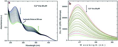
The absorption and fluorescence properties of compound 1, 2 and 3 were studied in water, which showed the dual absorption band at 278 nm and 336 nm (Fig. S12). Remarkably, the fluorescence bands of 1 were located at 465 nm when excited at 346 nm (Fig. S12). Compound 1 undergoes inclusion complex formation with β-cyclodextrin (β-CD:1), which can induced Intramolecular Charge Transfer (ICT). Fig. 1 shows changes in absorption and fluorescence spectra of an aqueous solution for β-CD:1 spectra upon addition of Cu2+. Addition of 50 equivalents of Cu2+ produced a ratiometric response in its absorption. The absorption maxima increased at 278 nm and decreased at 336 nm, with an isosbestic point at 295 nm. Also the absorbance at 278 nm remains constant, upon further addition of copper ions. On the other hand, the fluorescence maximum at 465 nm was quenched upon addition of Cu2+ ions in contrast to other metal cations, where no significant fluorescence changes were observed.


image file: c6ra01522k-f1.tif
Fig. 1 (a) Absorption and (b) fluorescence spectra of 1 (20 and 5 μM respectively) upon addition of various concentration of CuCl2 [0, 5, 10, 15, 20, 25, 30, 35, 40, 45, and 50 μM respectively] in aqueous solution with excitation at 350 nm.

The fluorescence changes of β-CD:1 were recorded in the range of 0–50 μM of [Cu2+] and it was observed that a detection limit of 0.5 μM is sufficient to sense Cu2+ ion in the blood samples. Using the fluorescence titration data, the binding constant of β-CD:1 with Cu2+ in aqueous solution was found to be (1 × 10⁻⁶ mol L−1). The detailed absorption and emission changes are shown in Fig. S12–S16 in ESI.

To have a clear insight into the role of 1, which behaves as an additional binding site for Cu2+ ion along with β-cyclodextrin, we have synthesized β-CD:2 and β-CD:3 complexes, bearing 6-hydroxy and 7-hydroxy units on the coumarin moiety, respectively. The above complex was tested for fluorescence changes upon addition of copper ion as well. As seen in Fig. 2, unlike β-CD:1, neither of them showed any distinct absorption or fluorescence changes upon addition of Cu2+ ion. This powerfully supports that the hydroxyl group in the 5th-position of the coumarin moiety plays an important role for the Cu2+ complexation.


image file: c6ra01522k-f2.tif
Fig. 2 (a) Fluorescence spectra of 1–3 (5 μM) their β-CD complexes with addition of CuCl2 (10 equiv. respectively) in aqueous solution with excitation at 350 nm. (b) Relative response at 475 nm of 1–3 (5 μM respectively) with addition of CuCl2 (10 equiv. respectively) in aqueous solution with excitation at 350 nm.

The binding constant of CD:1 with Cu2+ ions was estimated to be 5.5 × 10² mol L−1 from the fluorescence titration and the limit of detection (LoD) was found to be 2.52 × 10⁻¹⁰ mol L−1 (Fig. S18 in ESI). The other metal ions such as Ag+, Mg2+, Ca2+, Mn2+, Fe2+, Co2+, Ni2+, Cu2+, Zn2+, Hg2+, Pd2+, Cd2+, Sn2+, Pb2+, Al3+, Cr3+, Fe3+ and In3+ were added to CD:1 but insignificant changes were observed in the fluorescence spectra. To elucidate the binding stoichiometry between CD:1 and Cu2+, a continuous variation method (Job's plot) was employed using the fluorescence emission data. The maxima are observed when the mole fraction of Cu2+ reached 0.5, which indicates 1[thin space (1/6-em)]:[thin space (1/6-em)]1 binding (Fig. S17 and S18). The heavy metal ions such as Cu2+, Cd2+, Hg2+, and Pb2+ tend to quench the luminescence through electron- and/or energy-transfer processes.10 Additional evidence was also obtained from ESI-MS spectrum (Fig. S19) wherein initially a peak at 395.22 [M + K]+ value was observed which corresponds to 1. When 1.0 equiv. of β-cyclodextrin and Cu2+ ions (1 × 10⁻⁶ C) was added, the peak at 395.22 [M + K]+ disappeared and a new peak appeared at 1592.09 corresponding to [β-CD:1 + Cu2+ ion + K+ adduct].

Complexation of β-CD:1 was also confirmed by NMR titration studies (Fig. S20). In that aromatic protons in 1 and β-CD:1 are compared, where in β-CD:1 a continuous downfield shift from 6.526 to 8.323 ppm occurs when 1 undergoes inclusion into β-cyclodextrin cavity.

As selectivity is an important parameter for evaluating the performance of any fluorescence sensing system, the selectivity studies were also extended to various other metal ions, such as Ag+, Mg2+, Ca2+, Mn2+, Fe2+, Co2+, Ni2+, Cu2+, Zn2+, Hg2+, Pd2+, Cd2+, Sn2+, Pb2+, Al3+, Cr3+, Fe3+ and In3+. From the results, shown in Fig. 3a, it can be seen that Cu2+ ions induced a fluorescence quenching, whereas other metal ions led to a very slight or no fluorescence change. This result indicates clearly that the present β-CD:1 based sensor has good selectivity towards Cu2+ ion over other competitive cations.


image file: c6ra01522k-f3.tif
Fig. 3 (a) Bar chart illustrating fluorescence response of CD:1 and one equiv. of various metal ions in aqueous solution (λex = 350 nm, λem = 475 nm, slit: 5 nm/5 nm). (b) Bar chart illustrating fluorescence response in the selectivity of CD:1 (6.0 × 10⁻⁶ mol L−1) for Cu2+ ion in the presence of other metal ions (6.0 × 10⁻⁶ mol L−1). The blue bars represent the fluorescence intensity of CD:1 in the presence of one equivalent of the other metal ions. The brown bars represent the change in fluorescence intensity that occurs upon subsequent addition of one equivalent of Cu2+ to the solution containing CD:1 and the other metal ions in aqueous solution (λex = 350 nm, λem = 475 nm, slit: 5 nm/5 nm).

The effect of other coexisting cations on copper ion sensing was also determined. The fluorescence responses of the sensing system toward Cu2+ ions in the presence of alkali, alkaline earth and other transition metal ions are shown in Fig. 3b. The presence of the selected metal ions does not interference with Cu2+ binding to the probe, indicating that these co-existing ion have negligible interference effects on Cu2+ sensing by the β-CD:1 system.

The proposed structure for β-CD:1 also finds strong support from molecular modeling studies. In the proposed structure of β-CD:1, the mode of binding of 1 to β-CD plays a crucial role. Inclusion in which the thiazolidine group penetrates inside the primary side of β-CD cavity (mode A, Fig. 4) and coumarin group of 1 stays outside (mode B, Fig. 4) has a lower complexation energy (−E = −83.4251 kcal M−1) is preferred over the other mode such as coumarin group penetrates inside the primary side of β-CD cavity (mode B, Fig. 4), (−E = −72.8448 kcal M−1). The relevant details are given (Fig. S21) in ESI.


image file: c6ra01522k-f4.tif
Fig. 4 (a) CVFF optimized inclusion complex of β-CD:1. In mode A. Inclusion of thiazolidine part in β-cyclodextrin. In mode B: inclusion of coumarin part in β-cyclodextrin. (b) HOMO and LUMO of 4-((benzo[d]thiazol-2-ylthio)methyl)-5,7-dihydroxy-2H-chromen-2-one (1) calculated with DFT/TD-DFT at B3LYP/6-31G (d) level using Gaussian 03.

The geometry of probe 1 was optimized using DFT-B3LYP 6-31G level respectively using Gaussian 03 package. The sensor β-CD:1 effective binding sides to form a 1[thin space (1/6-em)]:[thin space (1/6-em)]1 complex with Cu2+ and this supports the experimental finding obtained from Job's plot and ESI-MS analysis of the complex. DFT-calculations,60 show that the HOMO of β-CD:1 rests with the thiazoline unit of the 1 (Fig. 4b) whereas LUMO is with the coumarin part of the 1 (Fig. 4b) indicating that the intramolecular charge transfer (ICT) takes place from thiazolidine unit to the coumarin unit. Thus when Cu2+ binds with sulphur atom in 2-position of thiazolidine ring, thiazolidine nitrogen, the 5-hydroxyl group of coumarin moiety and a primary hydroxyl group of β-cyclodextrin, the ICT is inhibited resulting in remarkable fluorescence quenching. In the titration of β-CD:1 with Cu2+, the quenching process is consistent with the coordination sulphur atom in 2-position of thiazolidine ring, thiazolidine nitrogen, the 5-hydroxyl group of coumarin moiety and a primary hydroxyl group of β-cyclodextrin. DFT calculations on 1 and its HOMO, LUMO form indicated that electron transfer takes place from thiazolidine group to coumarin group by Intramolecular Charge Transfer (ICT) process.

The proposed sensing of β-CD:1 in present system based on ICT mechanism. Before the addition of β-CD, compound 1 shows the emission at 465 nm. After addition of β-CD, the emission intensity has increased and also a red shift has occurred (from 465 nm to 475 nm), owing to the interaction between the probe 1 with β-CD. The (ICT) from the electron rich portion (thiazolidine group) to the electron withdrawing portion coumarin aided by the presence of β-CD takes place and the whole process is shown in Scheme 2.


image file: c6ra01522k-s2.tif
Scheme 2 Proposed mechanism of Cu2+ sensing by β-CD probe 1 system.

The red shift maybe interpreted as an indication of the existence of charge transfer in the excited state involving the promotion of the lone pair of electrons of thiazolidine group into a π–π* anti bonding orbital of the coumarin ring with help of β-CD. When Cu2+ is added, it forms a complex involving sulphur atom in 2-position of thiazolidine ring, thiazolidine nitrogen, the 5-hydroxyl group of coumarin moiety and a primary hydroxyl group of β-cyclodextrin. Due to this complexation, the intramolecular charge transfer (ICT) is suppressed. Control experiments show that when hydroxyl groups are present in the 6th and 7th position of coumarin moiety as in probe (2) and (3), respectively, no change in fluorescence intensity is observed when Cu2+ ion is added. This indicates that only with β-CD:1, strong binding of Cu2+ ion has occurred.

Upon addition of β-cyclodextrin to 1, the absorption band at 278 nm has slightly increased, and this indicates formation of inclusion complex β-CD:1. Similarly the intensity of the band at 336 nm has decreased which undergoes a significant increase, compared to other metal ions indicating a more efficient coordination of Cu2+ ion with sulphur atom in 2-position of thiazolidine ring, thiazolidine nitrogen, the 5-hydroxyl group of coumarin moiety and a primary hydroxyl group of β-cyclodextrin. Control experiments in the absence of β-cyclodextrin there is with 1 alone indicate that, there is no negligible change in fluorescence intensity, when Cu2+ is added.

When Cu2+ ion is added the charge transfer was arrested due to their strong complexation of Cu2+ with β-CD:1. In the case of other metal ions, the absence of significant binding with the sulphur atom is the main reason for the absence of any significant emission response. In addition, it is well known that Cu2+ ion (a borderline acid), preferentially interacts with the nitrogen, sulphur and hydroxyl atom according to Pearson's HSAB theory. Thus the changes of fluorescence spectrum, which is induced through the interaction of Cu2+ ion with the lone-pair of electrons on the nitrogen, sulphur and –OH atom are clearly justified.

In order to find out ICT suppression process, fluorescent quantum yields (Φ) of the β-CD:1 (Φ1) and β-CD:1:Cu2+ complex (Φ1:Cu2+) were compared, which were calculated using the fluorescent quantum yield of quinine sulfate as reference, in 0.1 M H2SO4 solution (ΦQs = 0.54). It was found that the Φ1 had decreased from 0.52 to 0.06 (Φ1:Cu2+) upon addition of equimolar Cu2+ ion. These observations clearly support the suppression of ICT process upon binding of Cu2+ ion to β-CD:1.

The potential of biosensing molecules to selectively examine a guest species in living cells is of great significant for biological applications.12 The cytotoxicity of CD:1 to HeLa cells was evaluated by standard MTT (3-(4,5-dimethylthiazol-2-yl)-2,5-diphenyltetrazolium bromide) assay. The results clearly indicate that the CD:1 was non-toxic to the HeLa cells under the experimental conditions (Fig. S24). The HeLa cell culture is utilized to find out the Cu2+ sensing by fluorescence microscope imaging technique (Fig. 5a–d). The CD:1 treated cells showed enhanced fluorescence in the intracellular region, which implicates the fluorescence behavior of the CD:1. After treatment using 10 micromolar solution of Cu2+, the fluorescence in the cellular region decreased, which shows that after forming complex of Cu2+, the probe becomes non-fluorescent complex. Similarly, addition of 50 micromolar solution of Cu2+, also leads to the quenching of fluorescence.


image file: c6ra01522k-f5.tif
Fig. 5 Bright field images and fluorescence images of HeLa cells. (a) Bright-field image of HeLa cells, (b) fluorescence image of HeLa cells were treated with CD:1 (10 micromole in PBS buffer) for 30 minutes, (c) the CD:1 treated cells were again incubated with Cu2+ ions in 10 micromole, (d) 50 micromole concentrations for 10 minutes.

The above results clearly represent that β-CD:1 is helpful for increasing the cell viability, which was decreased by the addition of Cu2+. Thus, the present β-CD:1 system can be employed as a suitable fluorescence chemosensing probe for Cu2+ ions in biological systems.

4. Conclusions

A highly sensitive and selective fluorescent sensing of Cu2+ ion is reported using the inclusion complex of 4-((benzo[d]thiazol-2-ylthio)methyl)-5,7-hydroxy-2H-chromen-2-one 1 with β-cyclodextrin (β-CD:1) in aqueous medium. To the best of our knowledge, this is the first report for selective fluorescent sensing of Cu2+ with 4-((benzo[d]thiazol-2-ylthio)methyl)-5,7-hydroxy-2H-chromen-2-one (1) included in β-cyclodextrin in water medium. Cyclodextrin functions not only as a supramolecular host to probe 1, but also provides a binding site to Cu2+ ion. Using confocal microscopic imaging, the present system is also found to be effective for sensing Cu2+ ions in HeLa cells. The red shifted on-off fluorescence quenching by the addition of Cu2+ ion is explained via an Intramolecular Charge Transfer mechanism (ICT) upon Cu2+ binding (which restricts the charge transfer process from thiazole group to coumarin moiety of 1) and a detection limit as low as 2.52 × 10⁻¹⁰ M of Cu2+ ions sensing is realized. In the context of water solubility, membrane permeability, and nontoxic nature, the present β-CD:1 system could be also potentially employed as a sensing probe for the detection of Cu2+ in living cells.

Acknowledgements

KP thanks DST, India for financial support, RI thanks to UGC-UPE (Madurai Kamaraj University) for Instrumentation facility.

Notes and references

  1. D. T. Quang and J. S. Kim, Chem. Rev., 2010, 110, 6280 CrossRef CAS PubMed.
  2. X. Chen, Y. Zhou, X. Peng and J. Yoon, Chem. Soc. Rev., 2010, 9, 2120 RSC.
  3. C. Yin, F. Huo, J. Zhang, R. Martinez-Manez, Y. Yang, H. Lv and S. Li, Chem. Soc. Rev., 2013, 42, 6032 RSC.
  4. J. T. Yeh, W. C. Chen, S. R. Liu and S. P. Wu, New J. Chem., 2014, 38, 4434 RSC.
  5. D. Maity, A. K. Manna, D. Karthigeyan, T. K. Kundu, K. Swapan and P. T. Govindaraju, Chem.–Eur. J., 2011, 17, 11152 CrossRef CAS PubMed.
  6. J. S. Valentine and P. J. Hart, Proc. Natl. Acad. Sci. U. S. A., 2003, 100, 3617 CrossRef CAS PubMed.
  7. L. I. Bruijn, T. M. Miller and D. W. Cleveland, Annu. Rev. Neurosci., 2004, 27, 723 CrossRef CAS PubMed.
  8. K. J. Barnham, C. L. Masters and A. I. Bush, Nat. Rev. Drug Discovery, 2004, 3, 205 CrossRef CAS PubMed.
  9. D. R. Brown and H. Kozlowski, Dalton Trans., 2004, 1907 RSC.
  10. E. Gaggelli, H. Kozlowski, D. Valensin and G. Valensin, Chem. Rev., 2006, 106, 1995 CrossRef CAS PubMed.
  11. T. Finkel, M. Serrano and M. A. Blasco, Nature, 2007, 448, 767 CrossRef CAS PubMed.
  12. Y. J. Kang, Pharmacol. Ther., 2011, 129, 321 CrossRef CAS PubMed.
  13. D. J. Waggoner, T. B. Bartnikas and J. D. Gitlin, Neurobiol. Dis., 1999, 6, 221 CrossRef CAS PubMed.
  14. C. Vulpe, B. Levinson, S. Whitney, S. Packman and J. Gitschier, Nat. Genet., 1993, 3, 7 CrossRef CAS PubMed.
  15. P. C. Bull, G. R. Thomas, J. M. Rommens, J. R. Forbes and D. W. Cox, Nat. Genet., 1993, 5, 327 CrossRef CAS PubMed.
  16. R. F. H. Viguier and A. N. A. Hulme, J. Am. Chem. Soc., 2006, 128, 11370 CrossRef CAS PubMed.
  17. Y. Q. Weng, Y. L. Teng, F. Yue, Y. R. Zhong and B. H. Ye, Inorg. Chem. Commun., 2007, 10, 443 CrossRef CAS.
  18. Y. Q. Weng, F. Yue, Y. R. Zhong and B. H. Ye, Inorg. Chem., 2007, 46, 7749 CrossRef CAS PubMed.
  19. P. Xi, J. Dou, L. Huang, M. Xu, F. Chen, Y. Wu, D. Bai, W. Li and Z. A. Zeng, Sens. Actuators, B, 2010, 148, 337 CrossRef CAS.
  20. C. Y. Tsai and Y. W. Lin, Analyst, 2013, 138, 1232 RSC.
  21. T. Hirayama, G. C. Van de Bittner, L. W. Gray, S. Lutsenko and C. J. Chang, Proc. Natl. Acad. Sci. U. S. A., 2012, 109, 2228 CrossRef CAS PubMed.
  22. B. Chen and P. Zhong, Anal. Bioanal. Chem., 2005, 381, 986 CrossRef PubMed.
  23. L. Shang and S. J. Dong, J. Mater. Chem., 2008, 18, 4636 RSC.
  24. G. Y. Lan, C. C. Huang and H. T. Chang, Chem. Commun., 2010, 46, 1257 RSC.
  25. Y. H. Chan, J. X. Chen, Q. S. Liu, S. E. Wark, D. H. Son and J. D. Batteas, Anal. Chem., 2010, 82, 3671 CrossRef CAS PubMed.
  26. M. Liu, H. M. Zhao, X. Quan, S. Chen and H. T. Yu, Chem. Commun., 2011, 47, 7749 RSC.
  27. Y. Zhou, S. Wang, K. Zhang and X. Jiang, Angew. Chem., Int. Ed., 2008, 47, 7454 CrossRef CAS PubMed.
  28. X. Xu, W. L. Daniel, W. Wei and C. A. Mirkin, Small, 2010, 6, 623 CrossRef CAS PubMed.
  29. Q. Shen, W. Li, S. Tang, Y. Hu, Z. Nie, Y. Huang and S. Yao, Biosens. Bioelectron., 2013, 41, 663 CrossRef CAS PubMed.
  30. A. J. Cotruvo, T. A. Aron, K. M. Ramos-Torresa and C. J. Chang, Chem. Soc. Rev., 2015, 44, 4400 RSC.
  31. W. Y. Liu, H. Y. Li, B. X. Zhao and J. Y. Miao, Analyst, 2012, 137, 3466 RSC.
  32. W. Y. Liu, H. Y. Li, B. X. Zhao and J. Y. Miao, Org. Biomol. Chem., 2011, 9, 4802 CAS.
  33. M. M Li, S. Y. Huang, H. Ye, F. Ge, J. Y. Miao and B. X. Zhao, J. Fluoresc., 2013, 23, 799 CrossRef PubMed.
  34. H. Ye, F. Ge, Y. M. Zhou, J. T. Liu and B. X. Zhao, Spectrochim. Acta, Part A, 2013, 112, 132 CrossRef CAS PubMed.
  35. W. Y. Liu, H. Y. Li, H. S. Lv, B. X. Zhao and J. Y. Miao, Spectrochim. Acta, Part A, 2012, 95, 658 CrossRef CAS PubMed.
  36. M. Li, H. S. Lv, J. Z. Luo, J. Y. Miao and B. X. Zhao, Sens. Actuators, B, 2013, 188, 1235 CrossRef CAS.
  37. F. Ge, H. Ye, J. Z. Luo, S. Wang, Y. J. Sun, B. X. Zhao and J. Y. Miao, Sens. Actuators, B, 2013, 181, 215 CrossRef CAS.
  38. G. Wenz, Angew. Chem., Int. Ed., 1994, 33, 803 CrossRef.
  39. I. Y. Tabushi, T. Kuroda and T. Mizutani, J. Am. Chem. Soc., 1986, 108, 4514 CrossRef CAS.
  40. T. Kinoshita, F. Iinuma and A. Tsuji, Chem. Pharm. Bull., 1974, 22, 2413 CrossRef CAS PubMed.
  41. Li. Song and W. C. Pordy, Chem. Rev., 1992, 92, 1457 CrossRef.
  42. A. Ueno, Adv. Mater., 1993, 5, 132 CrossRef CAS.
  43. K. Hamasaki, H. Ikeda, A. Nakamura, A. Ueno, F. Toda, I. Suzuki and T. Osa, J. Am. Chem. Soc., 1993, 115, 5035 CrossRef CAS.
  44. A. Ueno, ACS Symposium Series 538, ed. A. W. Czarnik, American Chemical Society, Washington, DC, 1992, ch. 6, p. 74 Search PubMed.
  45. A. Ueno, Supramol. Sci., 1996, 1, 31 CrossRef.
  46. Y. Wang, T. Ikeda, H. Ikeda, A. Ueno and F. Toda, Bull. Chem. Soc. Jpn., 1994, 67, 1598 CrossRef CAS.
  47. M. Nakamura, T. Ikeda, A. Ikeda, N. Ise, H. Ikeda, F. Toda and A. Ueno, J. Chem. Soc., Chem. Commun., 1995, 721 RSC.
  48. H. Ikeda, M. Nakamura, N. Ise, N. Oguma, A. Nakamura, T. Ikeda, F. Toda and A. Ueno, J. Am. Chem. Soc., 1996, 118, 10980 CrossRef CAS.
  49. Y. H. Li, L. M. Chan, L. Tyer, R. T. Moody, C. M. Himel and D. M. Hercules, J. Am. Chem. Soc., 1975, 97, 3118 CrossRef CAS.
  50. I. A. Azath and K. Pitchumani, Sens. Actuators, B, 2013, 188, 59 CrossRef CAS.
  51. P. Suresh, I. A. Azath and K. Pitchumani, Sens. Actuators, B, 2010, 146, 273 CrossRef CAS.
  52. K. Kanagaraj, K. Bavanidevi, T. J. Chow and K. Pitchumani, RSC Adv., 2014, 4, 11714 RSC.
  53. I. A. Azath, P. Suresh and K. Pitchumani, Sens. Actuators, B, 2011, 155, 909 CrossRef CAS.
  54. K. Kanagaraj, A. Affrose, S. Sivakolunthu and K. Pitchumani, Biosens. Bioelectron., 2012, 35, 452 CrossRef CAS PubMed.
  55. H. S. Jung, P. S. Kwon, J. Won Lee, J. I. Kim, C. S. Hong, J. W. Kim, S. Yan, J. Y. Lee, J. H. Lee, T. Joo and J. S. Kim, J. Am. Chem. Soc., 2009, 131, 2008 CrossRef CAS PubMed.
  56. A. Affrose, S. D. Shahida Parveen, B. Suresh Kumar and K. Pitchumani, Sens. Actuators, B, 2015, 206, 170 CrossRef CAS.
  57. S. D. Shahida Parveen, B. Suresh Kumar, S. Raj Kumar, R. Imran Khan and K. Pitchumani, Sens. Actuators, B, 2015, 221, 75 CrossRef CAS.
  58. S. D. Shahida Parveen, A. Affrose and K. Pitchumani, Sens. Actuators, B, 2015, 221, 521 CrossRef CAS.
  59. W. H. Melhuish, J. Phys. Chem., 1961, 65, 229 CrossRef CAS.
  60. V. Tharmaraj, S. Devi and K. Pitchumani, Analyst, 2012, 137, 5320 RSC.

Footnote

Electronic supplementary information (ESI) available: Detailed 1H-NMR, 13C-NMR, ESI-MS spectra, emission, absorption spectra, molecular modeling and confocal images. See DOI: 10.1039/c6ra01522k

This journal is © The Royal Society of Chemistry 2016
Click here to see how this site uses Cookies. View our privacy policy here.