A robust and easily integrated plasma separation chip using gravitational sedimentation of blood cells filling-in high-aspect-ratio weir structure

Yao Xiea, Di Chen*a, Shujing Lina, Zhigang Wangb and Daxiang Cuia
aKey Lab. for Thin Film and Microfabrication Technology of Ministry of Education, Shanghai Intelligent Diagnosis and Treatment Instrument Engineering Technology Research Center, Department of Instrument Science and Engineering, School of Electronic Information and Electrical Engineering, Shanghai Jiao Tong University, 800 Dongchuan Road, Shanghai 200240, P. R. China. E-mail: dchen@sjtu.edu.cn; Tel: +86-21-3420-6992
bDepartment of General Surgery, The Sixth People's Hospital, Shanghai Jiao Tong University, Shanghai 200240, P. R. China

Received 17th January 2016 , Accepted 12th March 2016

First published on 16th March 2016


Abstract

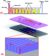
A robust and easily integrated plasma separation chip is very important for integration with microfluidic chips. Robust means that the system can tolerate small deviations in the boundary conditions (flow rate, temperature, position, hematocrit, dilutions and so on) without failure of the system. If a separation technique requires a precise flow rate and delivers other results, then the approach is not robust. Easily integrated means that the processing of fabrication and operation of the separation chip must be compatible with other parts of the microfluidic chip. In this work, a microfluidic plasma separation chip that uses the gravitational sedimentation effect to separate plasma from whole blood has been designed, fabricated and evaluated. The weir structure was constructed by using high-aspect-ratio SU-8 thick-photoresist molds, and then a double-polydimethylsiloxane (PDMS) layer was fabricated by casting, demolding and bonding with the PDMS layer. The high-aspect-ratio weir structure is beneficial for retaining blood cells in the gap of the weir structure. The conventional soft lithography process and use of PDMS materials makes it easy to integrate with other microfluidic technology. The simple weir structure is clogging-free and robust for the majority of plasma requirements. 20 μL of plasma was extracted from 65 μL of diluted blood (diluted factors ≥ 1[thin space (1/6-em)]:[thin space (1/6-em)]9) and the separation efficiency was above 90%.


1. Introduction

Many research groups have already successfully performed blood component analyses in microfluidic chips.1 Most blood tests are performed using blood plasma or serum. Plasma separation is an essential preliminary step in the preparation for biological detection and analysis on a microfluidic chip.2 The traditional procedures used to separate plasma from blood cells include mechanical separation by centrifugation and membrane-based filtration.3 Centrifugation is performed manually with a multi-step process at macroscale. As a result, the processed samples are often obtained hours after sampling in laboratories, require a larger volume of blood even for simple diagnoses, and there is a potential risk of contamination. The high cellular fractions with membrane-based filtration lead to membrane clogging and compromise separation efficiency. Moreover, the conventional method is more applicable for cases with large amounts of samples than those with samples on the micro- or nano-scale that cannot be miniaturized directly.

Automation and integration of a blood plasma separation step in microfluidic chips is ideal for point-of-care (POC) diagnostics. According to the presence or absence of an additional external field, plasma separation microfluidic chips are classified into two categories: the passive methods that separate without external fields and the active methods that require an additional external field. The passive separating techniques for a microfluidic chip include flow rate control,4–8 micro-filtration,9–12 geometrical obstacles,13,14 inertial force and gravitational sedimentation.13,15–18 The active separating techniques for a microfluidic chip include CD type centrifugation,19 acoustic separation and electrical separation.20–22 Especially, making use of gravity to realize plasma separation will greatly simplify the chip system and bring blood tests on microfluidic chips a step closer to real application. The gravitational sedimentation in a micro-scale channel/chamber can cause an obvious blood delamination within a short time. For example, plasma separation has been obtained by combining cross-flow filtration and a gravitational sedimentation effect on an interchannel microstructure at a slow injection flow rate of 1 μL min−1.23 In another report, a self-powered microfluidics blood analysis system based on sedimentation was developed and used to realize instantaneous detection of protein in plasma from a single drop of blood.16 Although the chip-based plasma pheresis systems that make use of the blood delamination effect are suitable for disposable application purposes, their long term performance may cause clogging of the chip with narrow channels. Therefore, because of the inherent limitations of these methods, they are prone to issues, including microchannel clogging, low efficiencies and long separation times.3 Furthermore, these methods have rarely been applied in integrated microfluidic chips as they are lacking in robustness and universality of integration.24 Some improvements include reducing the complexity of the microfluidic design and the amount of external support equipment required, while simultaneously reducing the number of components (such as pump, valves and membrane), or changing the fabrication processing and materials used.16,25,26

In this paper, we developed a robust and easily integrated plasma separation chip by gravitational sedimentation of blood cells. The weir structure was constructed by using high-aspect-ratio SU-8 thick-photoresist molds, and then the double-polydimethylsiloxane (PDMS) layer was fabricated by casting, demolding and bonding with the PDMS layer. The SU-8 molds can be reused for pouring and molding PDMS, this method was easy and could also decrease the fabrication cost. The high-aspect-ratio weir structure has the advantage of retaining blood cells in the gap of the weir structure. The absolutely soft lithography process is easy to integrate with other microfluidic technologies. The simple weir structure is clogging-free and robust for the majority of plasma requirements.

2. Experimental

2.1 Device design

The plasma separation chip was designed with three rows in the main chamber to combine with a series of high-aspect-ratio weir structures (Fig. 1). The device consists of two layers: one for the blood chamber (height: 700 μm) in patterned PDMS and the other for the plasma separation structure (height: 600 μm) in upside-down patterned PDMS. The blood sample enters the blood chamber by using a micro-control pump from the right outlet and flows through the weir structure. Simultaneously, the blood cells can be blocked in the gap of the weir structure, and the upper plasma can be extracted from the chamber. Typically, the volume of plasma sample required for an integrated microfluidic chip does not exceed 100 μL. Based on this requirement, we designed the volume of chip to be 45 μL. Three chambers were sequentially connected in series, which contributed to sedimentation of blood cells on the bottom of chamber. Furthermore, the plasma separation volume of this array structure chamber could be changed according to the requirements.
image file: c6ra01447j-f1.tif
Fig. 1 Schematic of the plasma separation chip. (A) Schematic illustration of plasma separation in weir structure. (B) Size of weir structure and whole of separation chip.

2.2 Fabrication of double-polydimethylsiloxane (PDMS) layer plasma chip

All work was carried out in a clean room. Two typical silicon wafers were used as the starting substrate. General photolithography technique was used to fabricate the high-aspect-ratio groove and package structure. SU-8 photoresist was spin-coated on the wafer to obtain 600 μm and 700 μm thickness of SU-8 layers. Further, it was pre-baked, exposed to UV light, post-baked and developed to obtain the final SU-8 mold structure. Once the two molds were prepared, a 10[thin space (1/6-em)]:[thin space (1/6-em)]1 mixture (w/w) of PDMS and curing agent was prepared and poured over the molds. The molds were then cured at 60 °C for 3 h. Afterward, the two parts of the PDMS structures were peeled off from the SU8 mold structure, and holes were punched at the sites of the reservoirs. Finally, both PDMS structures were bonded to each other with oxygen plasma treatment (Fig. 2).
image file: c6ra01447j-f2.tif
Fig. 2 Fabrication procedure for blood plasma separation chip.

2.3 Preparation of samples and reagents

Mouse blood was used to evaluate the effect of plasma separation from whole blood. The use of all mice in this study complied with the current ethical considerations and received the approval of the Animal Care and Use Committee of Shanghai Jiao Tong University. The blood was collected into a tube containing heparin sodium. The concentration of blood cells was determined using a blood cell counting plate. The concentration of blood cells in the original whole blood was calculated to be about 10 × 106/μL. The mouse blood was diluted with different dilution factors (1[thin space (1/6-em)]:[thin space (1/6-em)]1, 1[thin space (1/6-em)]:[thin space (1/6-em)]3, 1[thin space (1/6-em)]:[thin space (1/6-em)]5, 1[thin space (1/6-em)]:[thin space (1/6-em)]7, 1[thin space (1/6-em)]:[thin space (1/6-em)]9) using phosphate buffered saline (PBS). Both the heparin sodium and PBS can further maintain the activity of blood cells (Fig. 3).
image file: c6ra01447j-f3.tif
Fig. 3 Photograph of the device. Chips are filled with diluted blood (1[thin space (1/6-em)]:[thin space (1/6-em)]1) for image capture.

2.4 Experimental set up

The outlet of the chip was connected to the syringe pump by a Teflon tube and the blood in the reservoir was sucked into the chip by the syringe pump. The flow rate of the blood was controlled by the syringe pump. The microscope was used to monitor and take images of the plasma separation. The separated plasma was collected at the outlet and then the blood cell concentration in the extracted plasma was determined by blood cell counting plates. Approximately 65 μL of diluted blood was applied to the input reservoir of each test device using a syringe pump. The volume of the whole separation chamber is about 45 μL, so we extracted 10–30 μL of blood plasma for calculating the separation efficiency. The separation efficiency of plasma from blood cells can be calculated by the blood cell concentrations before and after separation. Data from at last three separate experiments were analyzed and presented as mean ± standard deviation. The key factor to evaluate the performance of the chip is separation efficiency (η), which can be defined as:
η = [(NwNs)/Nw] × 100%
where Nw and Ns are the number of RBCs in the same volume of whole blood sample and separated plasma, respectively.

3. Results and discussion

Most studies of plasma separation have used diluted blood samples to enhance separation efficiency. The current study used diluted whole blood samples with 50% hematocrit. A complete study covering the dilution of blood from 1[thin space (1/6-em)]:[thin space (1/6-em)]1 to 1[thin space (1/6-em)]:[thin space (1/6-em)]9 (the hematocrits from 25% to 5%) was carried out on the plasma separation chip. Experiments have been carried out for flow rate in the range of 2.5 to 7.5 μL min−1. Before quantification of the data obtained in the experiments, we calculated the volume of the whole separation chamber to be about 45 μL. Theoretically, when the blood cells were completely filled in the gap of weir structure, 62 μL of plasma could be separated from the blood sample. However, because of incomplete sedimentation of blood cells in the gap of the weir structure and streaming with plasma fluid, the amount of plasma separated from blood is less than 62 μL.

When the volume of 65 μL of blood was entirely sucked into the chamber of the device, and the chamber of device was completely filled with blood, blood plasma volume ranging from 10 to 30 μL was extracted. Fig. 4 shows that the different extraction volumes and dilution factors affect the separation efficiency. It gives us an indication that the separation efficiency was not obviously influenced by extractions volume of less than 20 μL. However, the separation efficiency was brought down on account of the volume of extraction more than 20 μL. As previously mentioned, the whole volume of the chamber of the chip is about 45 μL, so when 65 μL of diluted blood was injected into the chamber, 20 μL of plasma could be extracted from the chamber, and 45 μL of liquid remained in the chamber. Meanwhile, the continual extraction of plasma can move more blood cells forward upstream of the liquid, which caused the more blood cells to move into the plasma. Therefore, extraction of 20 μL of plasma was the most appropriate approach.


image file: c6ra01447j-f4.tif
Fig. 4 The influence of different extracted volumes and dilution factors on separation efficiency (flow rate, 2.5 μL min−1).

Based on the above results, extraction volume of plasma was chosen as 20 μL from 65 μL of diluted blood at a flow rate of 2.5 μL min−1. As shown in Fig. 5, most of the blood cells were blocked in the gaps of the weir structure in the first and second row of the chamber. Blood cells in the interface between the weir structure and the outlet channel remain in the bottom of the chamber, and the number of blood cells is low (Fig. 6). Meanwhile, the number of blood cells in the extraction plasma decreased with increasing dilution ratio.


image file: c6ra01447j-f5.tif
Fig. 5 Blood cells in the middle of the plasma separation chip; the bottom image indicates the observation position.

image file: c6ra01447j-f6.tif
Fig. 6 Blood cells sedimentation on the bottom of chamber in the interface between the weir structure and the outlet channel: (A) 1[thin space (1/6-em)]:[thin space (1/6-em)]1, (B) 1[thin space (1/6-em)]:[thin space (1/6-em)]3, (C) 1[thin space (1/6-em)]:[thin space (1/6-em)]5, (D) 1[thin space (1/6-em)]:[thin space (1/6-em)]7, (E) 1[thin space (1/6-em)]:[thin space (1/6-em)]9 diluted with PBS (flow rate, 2.5 μL min−1). The inset images in all panels indicate the observation position.

At a certain extraction volume of sample, the separation efficiency will also be influenced by the extraction rate and dilution ratio. During the experiments, it was observed that as the flow rate was decreased, the efficiency of separation increases, which was attributed to sufficient gravitational sedimentation. Therefore, experiments were carried out to find the relation between the flow rate and the separation efficiency. Fig. 7 provides us the complete details of the different dilution ratios for the three extraction rates. The separation efficiency increased with decreasing flow rate. It can be seen that the separation efficiency of the higher dilution ratio was sensitive to a higher flow rate. The separation efficiency was above 90% at the 2.5 μL min−1 flow rate.


image file: c6ra01447j-f7.tif
Fig. 7 Relationships between hematocrit and separation efficiency.

It is important to note that the separation process relied on sedimentation. To investigate the separation efficiency with respect to sedimentation time of blood cells in the chamber of the device, we compared the separation efficiency of two types of sample suction mode. When each of sample was sucked into the end of each row of the weir structure, the syringe pump was held to wait a minute, and then blood suction restarted. The influence of suction modes on the separation efficiency was studied at flow rates of 2.5 μL min−1, 5 μL min−1 and 7.5 μL min−1. The comparison of separation efficiency on the different flow rates is shown in Fig. 8. It is clear that the percentage of blood cells removed from the plasma increased with waiting 3 minutes. The waiting 3 minutes contributed to the improvement of the separation efficiency. This method had a significant effect on the condition of the higher flow rates and hematocrit.


image file: c6ra01447j-f8.tif
Fig. 8 Waiting 3 minutes and no waiting for relationship of different hematocrit and separation efficiency, flow rate, (A) 2.5 μL min−1, (B) 5 μL min−1, (C) 7.5 μL min−1.

4. Conclusions

In summary, we have developed a robust and easily integrated plasma separation chip by gravitational sedimentation effect. The high-aspect-ratio weir structure was constructed by using SU-8 thick-photoresist molds, and then the double-polydimethylsiloxane (PDMS) layer was fabricated by casting, demolding and bonding with the PDMS layer. The SU-8 molds can be reused for pouring and molding PDMS. This method was easy and could also decrease the fabrication cost. The high-aspect-ratio weir structure has the advantage of retaining blood cells in the gap of weir structure. The conventional soft lithography process and all PDMS materials makes it easy to integrate with other microfluidics technology. The simple weir structure is clogging-free and robust for the majority of plasma requirements. The extraction volume of plasma was chosen as 20 μL from 65 μL of diluted blood. The separation efficiency increases with decreasing flow rate. A 3 minutes wait time contributed to the improvement of separation efficiency on the condition of higher flow rates and hematocrit. The 20 μL of plasma was extracted from 65 μL of diluted blood at a separation efficiency above 90%.

Acknowledgements

This work was supported by the Science and Technology Commission of Shanghai Municipality (Project no. 15441904800). The chip was fabricated in the Center for Advanced Electronic Materials and Devices (AEMD) of Shanghai Jiao Tong University.

References

  1. D. R. Gossett, W. M. Weaver, A. J. Mach, S. C. Hur, H. T. Tse, W. Lee, H. Amini and D. Di Carlo, Anal. Bioanal. Chem., 2010, 397, 3249–3267 CrossRef CAS PubMed.
  2. A. A. Bhagat, H. Bow, H. W. Hou, S. J. Tan, J. Han and C. T. Lim, Med. Biol. Eng. Comput., 2010, 48, 999–1014 Search PubMed.
  3. H. W. Hou, A. A. S. Bhagat, W. C. Lee, S. Huang, J. Han and C. T. Lim, Micromachines, 2011, 2, 319–343 CrossRef.
  4. S. Yang, A. Undar and J. D. Zahn, Lab Chip, 2006, 6, 871–880 RSC.
  5. M. Kersaudy-Kerhoas, R. Dhariwal, M. P. Y. Desmulliez and L. Jouvet, Microfluid. Nanofluid., 2009, 8, 105–114 CrossRef.
  6. A. I. Rodriguez-Villarreal, M. Arundell, M. Carmona and J. Samitier, Lab Chip, 2010, 10, 211–219 RSC.
  7. T. M. Geislinger, B. Eggart, S. Braunmüller, L. Schmid and T. Franke, Appl. Phys. Lett., 2012, 100, 183701 CrossRef.
  8. S. Yang, A. Ündar and J. D. Zahn, ASAIO J., 2005, 51, 585–590 CrossRef PubMed.
  9. T. A. Crowley and V. Pizziconi, Lab Chip, 2005, 5, 922–929 RSC.
  10. P. Sethu, A. Sin and M. Toner, Lab Chip, 2006, 6, 83–89 RSC.
  11. X. Chen, D. Cui, C. Liu and H. Li, Sens. Actuators, B, 2008, 130, 216–221 CrossRef CAS.
  12. J. Chen, D. Chen, T. Yuan, X. Chen, Y. Xie, H. Fu, D. Cui, X. Fan and M. K. Khaing Oo, Microelectron. Eng., 2014, 128, 36–41 CrossRef CAS.
  13. T. G. Kang, Y.-J. Yoon, H. Ji, P. Y. Lim and Y. Chen, J. Micromech. Microeng., 2014, 24, 087001 CrossRef.
  14. E. Sollier, H. Rostaing, P. Pouteau, Y. Fouillet and J.-L. Achard, Sens. Actuators, B, 2009, 141, 617–624 CrossRef CAS.
  15. A. A. Bhagat, H. W. Hou, L. D. Li, C. T. Lim and J. Han, Lab Chip, 2011, 11, 1870–1878 RSC.
  16. I. K. Dimov, L. Basabe-Desmonts, J. L. Garcia-Cordero, B. M. Ross, Y. Park, A. J. Ricco and L. P. Lee, Lab Chip, 2011, 11, 845–850 RSC.
  17. X. B. Zhang, Z. Q. Wu, K. Wang, J. Zhu, J. J. Xu, X. H. Xia and H. Y. Chen, Anal. Chem., 2012, 84, 3780–3786 CrossRef CAS PubMed.
  18. Z. Geng, Y. Ju, W. Wang and Z. Li, Sens. Actuators, B, 2013, 180, 122–129 CrossRef CAS.
  19. S. Smith, D. Mager, A. Perebikovsky, E. Shamloo, D. Kinahan, R. Mishra, S. Torres Delgado, H. Kido, S. Saha, J. Ducrée, M. Madou, K. Land and J. Korvink, Micromachines, 2016, 7, 22 CrossRef.
  20. Y. Nakashima, S. Hata and T. Yasuda, Sens. Actuators, B, 2010, 145, 561–569 CrossRef CAS.
  21. A. Lenshof and T. Laurell, Chem. Soc. Rev., 2010, 39, 1203–1217 RSC.
  22. A. Lenshof, A. Ahmad-Tajudin, K. Jaras, A. M. Sward-Nilsson, L. Aberg, G. Marko-Varga, J. Malm, H. Lilja and T. Laurell, Anal. Chem., 2009, 81, 6030–6037 CrossRef CAS PubMed.
  23. T. Tachi, N. Kaji, M. Tokeshi and Y. Baba, Anal. Chem., 2009, 81, 3194–3198 CrossRef CAS PubMed.
  24. T. Dong, Z. Yang, Q. Su, N. M. Tran, E. B. Egeland, F. Karlsen, Y. Zhang, M. J. Kapiris and H. Jakobsen, Microfluid. Nanofluid., 2010, 10, 855–865 CrossRef.
  25. J. L. Garcia-Cordero, D. Kurzbuch, F. Benito-Lopez, D. Diamond, L. P. Lee and A. J. Ricco, Lab Chip, 2010, 10, 2680–2687 RSC.
  26. Y. C. Kim, S.-H. Kim, D. Kim, S.-J. Park and J.-K. Park, Sens. Actuators, B, 2010, 145, 861–868 CrossRef CAS.

Footnote

Electronic supplementary information (ESI) available. See DOI: 10.1039/c6ra01447j

This journal is © The Royal Society of Chemistry 2016
Click here to see how this site uses Cookies. View our privacy policy here.