Porous β-MnO2 nanoplates derived from MnCO3 nanoplates as highly efficient electrocatalysts toward oxygen evolution reaction

Jun Kim ab, Ju Seong Kimc, Hionsuck Baikd, Kisuk Kang*c and Kwangyeol Lee*ab
aCenter for Molecular Spectroscopy and Dynamics, Institute for Basic Science (IBS), Seoul 02841, Korea
bDepartment of Chemistry and Research Institute for Natural Sciences, Korea University, Seoul 02841, Republic of Korea. E-mail: kylee@korea.ac.kr
cDepartment of Materials Science and Engineering, Research Institute of Advanced Materials, Seoul National University, 599 Gwanak-ro, Gwanak-gu, Seoul 08826, Republic of Korea. E-mail: matlgen1@snu.ac.kr
dKorea Basic Science Institute (KBSI), Seoul 02841, Korea

Received 13th January 2016 , Accepted 3rd March 2016

First published on 3rd March 2016


Abstract

β-MnO2 has not been considered as an effective catalyst toward the oxygen evolution reaction due to its lack of active di-μ2-oxo bridged Mn centres and inaccessibility to the inner Mn atoms. We have envisioned that β-MnO2 can be made catalytically active by making the inner Mn atoms accessible. In order to accomplish this, we have synthesized MnCO3 nanoplates via a solution route and converted them into highly porous β-MnO2 nanoplates with very high surface area. In addition to the reduced overpotential of 450 mV at 10 mA cm−2, the derived Tafel slope was 78.2 mV dec−1, showing a superior catalytic activity of the porous nanoplate, which is comparable to the catalytic performance of best performing α-MnO2 phase. The importance of surface-bound catalytic Mn sites in highly porous β-MnO2 nanoplates is also demonstrated by Au loading-induced blockage of them and corresponding catalytic activity deterioration.

Dwindling natural energy resources and environmental issues associated with their usage call for urgent developments in economical and clean energy technologies.1–3 Hydrogen is considered as a clean future energy source, and therefore an efficient water splitting process for hydrogen production is in great demand.1–3 Water oxidation, or oxygen evolution reaction (OER), is one of the major hurdles to an efficient water splitting reaction, because it is energetically unfavourable in nature.4 Consequently, considerable efforts have been put into the development of efficient OER catalysts. Metal oxides of precious metals such as RuO2 and IrO2 have shown high catalytic activities toward OER.5,6 Practical applications of them, however, have not been realized due to the high cost in the catalyst production.7–9 Inexpensive first-row transition metal oxides, therefore, are being intensively studied for the potential applications as OER catalysts.10–13 Manganese oxides, in particular, are of a great interest due to their wide range of oxidation states and possible crystal structures.9,14 α-MnO2 is well-known for its excellent catalytic activity owing to its large 2 × 2 internal channels providing numerous passages for ions and other molecules,15 and this feature, in addition to the catalytically active surface, allows the internal portion under the surface to be used as an active material. On the other hand, β-MnO2 is generally considered to be catalytically inferior due to its unique connectivity of MnO6 octahedrons, which results in only a small number of catalytically active sites.16 In addition, the internal portion of β-MnO2 could not be utilized for catalytic purpose, because very narrow 1 × 1 internal channels prohibit the passage of small chemical species.17 These inherent structural disadvantages of β-MnO2 in catalytic applications might be overcome by making the β-MnO2 highly porous; porous β-MnO2 might allow access of small chemical species to the catalytically active sites which were previously inaccessible. Introduction of porosity to β-MnO2, however, poses a formidable challenge.

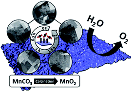
In principle, porous β-MnO2 structure can be prepared if a low density nanocrystalline precursor is abruptly converted to β-MnO2 phase. MnO2 nanostructures can be prepared from MnCO3 by calcination.18 With MnCO3 nanoparticles with a suitable structural feature to enable a fast CO2 release, it should be feasible to form highly porous manganese oxide nanomaterials. In this work, we report a facile synthetic route to single crystalline MnCO3 nanoplates (MCP) and their transformation to highly porous β-MnO2 nanoplates (PMOP) via calcination as shown in Scheme 1. Thus prepared PMOP exhibits a superior catalytic activity toward OER.


image file: c6ra01091a-s1.tif
Scheme 1 Synthetic routes to porous MnO2 nanoplate and Au deposition.

In a typical synthesis of MCP, a slurry of Mn(OAc)2 (2.50 mmol), sodium oleate (NaOA, 2.50 mmol), and oleylamine (5.00 mmol) was prepared in a 100 mL Schlenk tube equipped with a bubbler. The tube containing the slurry was evacuated for 10 min at room temperature, and then the reaction mixture was maintained at 290 °C under CO2 flow with magnetic stirring for 1 h. The precipitated white product was washed with copious amount of hexane, ethanol and water to remove any excess reagents. Finally, the white precipitate was treated with dilute acetic acid (5% v/v) for 30 min to remove surface bound surfactants, and washed several times with ethanol. The final product was dried in an oven at 45 °C. The X-ray diffraction (XRD) pattern of as-synthesized MCP indicates hexagonal close packing system with R[3 with combining macron]c space group (JCPDS #01-086-0172. See Fig. S1). As shown in the transmission electron microscope (TEM) images in Fig. 1a and b, MCP exhibits a flat rhombohedral morphology; the width of the nanoplates ranges from 100 nm to 200 nm and the average thickness is measured to be 13.1 ± 3.4 nm. Fig. 1d shows a highly magnified side-view of MCP, and the high-resolution transmission electron microscope (HRTEM) image and fast Fourier transformation (FFT) analysis of them reveal that nanoplates are single-crystalline with the wide top and bottom faces of the nanoplates being {012} facets.


image file: c6ra01091a-f1.tif
Fig. 1 Structural analysis of MnCO3 nanoplate (MCP) and porous β-MnO2 nanoplate (PMOP). (a and b) TEM images of MCP and (c) side-view of stacked nanoplates. (d) HRTEM image of stacked MCP showing that the wide top and bottom faces of a nanoplate has {012} facets; the FFT pattern of the area enclosed by a dotted rectangle is shown in (i). (e) TEM images of PMOP and (f) side-view of stacked PMOP. (g) TEM image showing the rough topography of PMOP surface. (h) HRTEM image of PMOP showing different zone axes of [111] and [113] with corresponding (ii) and (iii) FFT patterns, respectively.

The formation of MnCO3 phase and the nanoplate morphology are critically dependent on the presence of both NaOA and CO2. In the absence of either NaOA or CO2, MnO phase, instead of MnCO3, was produced from the decomposition of Mn(OAc)2 (Table S1). Obviously, the MnCO3 phase is stabilized in the presence of CO2, which shifts the chemical reaction equilibrium from MnO to MnCO3. The basic nature of NaOA might have contributed to the stability of MnCO3 phase, which can be easily destabilized in an acidic condition. NaOA seems to act as a structure directing agent as well, because the thickness of the nanoplate can be controlled by varying the amount of NaOA. (Fig. S2) The thickness of the nanoplate gradually decreases as the amount NaOA is increased, which may indicate that NaOA prefers {012} facets of MnCO3 crystal, stabilizing {012} facets and preventing the growth along the [012] zone axis.

When other Mn precursors with oxygen-containing ligands such as formate, nitrate, or acetylacetonate were used, the formation of MnCO3 phase was also observed. On the other hand, the decomposition of MnCl2 in the presence of NaOA and CO2 produced only MnO phase. Interestingly, the addition of sodium acetate to the latter reaction mixture led to the formation of plate-like MnCO3 nanostructures, indicating the role of acetate ligands in both stabilizing the MnCO3 phase and forming the nanoplate morphology. Nanoplate morphology, however, could not be obtained from decomposition of Mn precursors with formate, nitrate, or acetylacetonate ligands, even when sodium acetate was added to the reaction mixture. The acetate species might not compete effectively with these oxygen-containing ligands for binding to Mn centres to give the nanoplate morphology.

MCP can be converted into PMOP through calcination (Scheme 1). Thermogravimetric analysis (Fig. S5) of MCP under O2 reveals that the conversion into MnO2 begins around 400 °C, which is accompanied by a drastic decrease in weight due to the rapid discharge of CO2. The TEM image of PMOP in Fig. 1e and f show that the original plate morphology remains mostly intact and the surface of PMOP is very rough due to a rapid CO2 discharge at high temperature. The XRD pattern of the resulting PMOP shows very broad peaks (Fig. S6), suggesting very small grain sizes. The HRTEM analysis in Fig. 1h shows that PMOP consists of many nanosized single crystalline domains whose zone axes are randomly distributed. In addition, owing to the high porosity and rough surface (Fig. 1f and g), PMOP shows a very high Brunauer–Emmett–Teller (BET) surface area of 96.7 m2 g−1 and pore volume of 0.77 cm3 g−1 (Fig. S7).

Hetero-metal doping or deposition has been previously employed to enhance the water oxidation ability of metal oxide catalysts.19–21 Au deposition via various chemical and physical methods has been reported to enhance the catalytic activity of manganese oxide nanomaterials.22–24 In our study, three Au-deposited samples, namely, low Au/PMOP (L-Au/PMOP), medium Au/PMOP (M-Au/PMOP) and high Au/PMOP (H-Au/PMOP), were obtained via urea-assisted formation of Au nanoparticles and their deposition. The Au nanoparticle coverage in these samples and the oxidation states of constituting atoms were analysed by TEM, HRTEM and X-ray photoelectron spectroscopy (XPS) (Fig. S8 and S9). The TEM and HRTEM images of H-Au/PMOP are shown in Fig. 2 as an example, and Au nanoparticles with an average size of 2.54 nm are uniformly distributed on PMOP and, in addition, line profile analysis was performed to further confirm the presence of Au nanoparticles. (Fig. S10) On the contrary, Au nanoparticles are barely observed for L-Au/PMOP. Before Au deposition, the Mn valence state of the as-synthesized PMOP was measured to be 3.84 by XPS analysis, which may indicate the presence of Mn vacancies in MnO2 lattices.25 As presented in Table 1, the Mn valence state gradually decreases as the deposition of Au increases. Chen's group reported a similar result with Au doped MnO2 film, and the oxidation state of Mn decreases because of the gain of electrons from Au, consistent with the first principle calculation and XPS measurements.26


image file: c6ra01091a-f2.tif
Fig. 2 Structural analysis of Au deposited PMOP (H-Au/PMOP). (a) TEM image and (b) STEM image of H-Au/PMOP showing a uniform coverage of Au nanoparticles. (c and d) HRTEM images showing Au nanoparticles with 0.235 nm d-spacing which corresponds to {111} crystal planes of Au. The FFT patterns are shown in (i) and (ii). Deconvoluted XPS spectra of (e) Mn 2p and (f) Au 4f of H-Au/PMOP.
Table 1 Changes in valence states of Mn and Au depending on Au deposition
Catalyst Mn valence statea Au valence statea Au atomic % by ICP-AES
a Valence states are calculated by deconvoluting the XPS spectra of each sample.
PMOP 3.84
L-Au/PMOP 3.78 2.6
M-Au/PMOP 3.75 0.71 4.6
H-Au/PMOP 3.72 1.05 6.0


Catalytic activities of PMOP, L-Au/PMOP, M-Au/PMOP and H-Au/PMOP nanoplates toward OER were investigated by cyclic voltammetry (CV) in 1 M KOH aqueous solution. Prepared catalysts were deposited on carbon paper by drop-casting method with a loading amount of 0.4 mg cm−2. The OER activity of 20 wt% Ir/C (Premetek) was also measured to compare with those of our catalysts. The OER activity curves were obtained by averaging the forward and the reverse curves for capacitive current. As shown in Fig. 3a, PMOP exhibited an onset potential of water oxidation at an overpotential of 300 mV and a low overpotential of 450 mV at a current density of 10 mA cm−2. The Tafel slope of PMOP was measured to be 78.2 mV dec−1. It is notable that the OER activity of PMOP matches that of the commercial Ir/C which has an overpotential of 350 mV at 10 mA cm−2 and Tafel slope of 76.7 mV dec−1. The superior catalytic activity of PMOP may be attributed to the highly porous nature which can provide reactant molecules high surface area and numerous passages to catalytically active sites. Suib's group recently reported the structure–property relationship of MnO2 as well as a summary of OER catalytic activities of nanostructured MnO2 with various crystal structures.27 Notably, the catalytic performance of PMOP of our study is comparable to that of the best performing α-MnO2 catalysts and better than the recently reported screen-printed β-MnO2 film.27,28 Although the overpotential of PMOP is higher than that of the commercial Ir/C, PMOP has the lowest overpotential without any conductive support compared to those of the MnO2 catalysts reported to date. We believe that the low overpotential of POMP results from the increased contact surface with the electrode and the short travelling distance for electrons owing to its thin plate morphology. While PMOP showed excellent catalytic performance, interestingly against our expectations, the OER activities of the three Au-deposited samples deteriorated with increasing Au loading contents as can be seen in Fig. 3. The overpotentials of L-Au/PMOP, M-Au/PMOP and H-Au/PMOP were 460, 520 and 580 mV, respectively, at a current density of 10 mA cm−2 and the Tafel slopes of these Au deposited PMOP increased to 81.7, 130.2 and 141.7 mV dec−1, respectively, as shown in Fig. 3b. Furthermore, the chronopotentiometry of PMOP and Au deposited PMOP shows that bare PMOP exhibits the best stability among the MnO2 catalysts in our study (Fig. S11).


image file: c6ra01091a-f3.tif
Fig. 3 Electrochemical water oxidation measurements of Ir/C (20 wt%), PMOP, L-Au/PMOP, M-Au/PMOP and H-Au/PMOP. (a) Capacitive background corrected polarization curves in 1 M KOH electrolyte. (b) The corresponding Tafel plots.

Recent studies have suggested that the catalytic activity for OER originates from the nature of the bonding between metal centres and oxygen and that the presence of di-μ2-oxo bridged metal ions is a common feature in other transition metal oxide catalysts for water oxidation.29–31 β-MnO2 prevalently possesses mono-μ2-oxo bridged Mn ions because its crystal structure is derived from corner-shared MnO6 octahedrons, resulting in inferior catalytic activity as compared to those of other MnO2 polymorphs.25,27 Moreover, the internal Mn ions, due to narrow inner channels, are not easily accessible for reactant molecules. However, the porosity of the β-MnO2 nanostructure of this study might have provided numerous pathways to previously inaccessible internal Mn ions on β-MnO2 {110} with di-μ2-oxo bridged Mn centres.25,32 Furthermore, the 2D nanoplate morphology, while shortening the travelling distance for reactant molecules, decreases the number of inaccessible Mn ions by reducing the internal volume and increasing the surface confined atoms at the same time. In short, the increased number of accessible di-μ2-oxo bridged Mn ions in the porous MnO2 nanoplate, due to the synergy between the porosity and nanoplate morphology, might have led to the excellent catalytic performance of β-MnO2 nanoplate of this study. Another contributing factor to the enhanced catalytic performance might be the presence of defect sites such as Mn vacancies.33 When MnCO3 is converted to β-MnO2 via calcination, the conversion of crystal phase must occur from hexagonal close packing (hcp) system to a completely different phase of face-centered tetragonal (fct) system. This drastic change in crystal structures might result in the formation of very small, numerous grains of MnO2 nanocrystals with a large number of Mn vacancies. These vacancies might help di-μ2-oxo bridged Mn ions become more predominant than di-μ3-oxo bridged Mn ions, thus providing a higher number of active sites for water coordination.25 Also, uncoordinated oxygen atoms due to Mn vacancies can provide proton binding sites;34 Dau's group proposed that the resulting μ-OH units may facilitate proton-coupled electron transfer (PCET), which is considered a critical factor in water oxidation.16 Since the defect sites are coordinatively unsaturated with many dangling bonds, the nucleation and growth of Au nanoparticle occurring on those defect sites can lead to the significant passivation of OER active sites. The deteriorating catalytic performance of PMOP with increased Au contents indicates that the blockage of catalytically active sites is very severe with Au-deposition. The enhanced catalytic activity with Au-loading of catalytic metal oxides, observed in other reported studies, could not be repeated in our study.

We also investigated the catalytic performance of PMOP toward oxygen reduction reaction (ORR) (Fig. S12). According to the half-wave potentials in the ORR curves of the porous β-MnO2 nanoplates (0.647 V) and 20 wt% Pt/C (0.918 V), the MnO2 sample shows much inferior catalytic property to that of Pt/C sample. However, the ORR performance of PMOP in this study, with a slightly lower half-wave potential, is similar to that of the oxygen-deficient β-MnO2 reported by Cheng et al.35

Conclusion

In conclusion, we have synthesized PMOP with a very high surface area via calcination of highly crystalline MCP and demonstrated their excellent catalytic activity toward OER. The number density of catalytically active Mn sites in PMOP is greatly increased due to their unique morphological features such as 2D morphology and porosity. In addition, numerous Mn vacancies in PMOP, generated from abrupt material transformation, might have further contributed to the enhanced catalytic performance. The important role of surface bound catalytic sites is demonstrated by the deteriorated catalytic activity with increased Au-loading on the surface. We believe that the synthetic methodology of this study might be applicable to the preparation of other catalytically useful metal oxide nanostructures. It is quite intriguing to study the catalytic activity of other porous metal oxides of different compositions toward OER, and a related study is currently under way.

Acknowledgements

This work was supported by IBS-R023-D1, KEIT-10047717 and BioNano Health-Guard Research Center funded by the Ministry of Science, Information and Communication Technology (ICT) and Future Planning of Korea as Global Frontier Project (Grant No. H-GUARD_NRF 2013M3A6B2078946). We thank KBSI for allowing us to use their HRTEM and ICP-AES instruments, and also Prof. Chang Seop Hong and Dong Won Kang in Department of Chemistry of Korea University for BET measurements.

Notes and references

  1. A. Kudo and Y. Miseki, Chem. Soc. Rev., 2009, 38, 253 RSC.
  2. Y. Tachibana, L. Vayssieres and J. R. Durrant, Nat. Photonics, 2012, 6, 511 CrossRef CAS.
  3. T. Hisatomi, J. Kubota and K. Domen, Chem. Soc. Rev., 2014, 43, 7520 RSC.
  4. J. Suntivich, K. J. May, H. A. Gasteiger, J. B. Goodenough and Y. Shao-Horn, Science, 2011, 334, 1383 CrossRef CAS PubMed.
  5. D. Galizzioli, F. Tantardini and S. Trasatti, J. Appl. Electrochem., 1974, 4, 57 CrossRef CAS.
  6. J. Horkans and M. W. Shafer, J. Electrochem. Soc., 1977, 124, 1202 CrossRef CAS.
  7. C. Felix, T. Maiyalagan, S. Pasupathi, B. Bladergroen and V. Linkov, Int. J. Electrochem. Sci., 2012, 7, 12064 CAS.
  8. Z. Gui, E. Gillette, J. Duay, J. Hu, N. Kim and S. B. Lee, Phys. Chem. Chem. Phys., 2015, 17, 15173 RSC.
  9. D. M. Robinson, Y. B. Go, M. Mui, G. Gardner, Z. Zhang, D. Mastrogiovanni, E. Garfunkel, J. Li, M. Greenblatt and G. C. Dismukes, J. Am. Chem. Soc., 2013, 135, 3494 CrossRef CAS PubMed.
  10. J. D. Blakemore, H. B. Gray, J. R. Winkler and A. M. Muller, ACS Catal., 2013, 3, 2497 CrossRef CAS.
  11. S. C. Riha, B. M. Klahr, E. C. Tyo, S. Seifert, S. Vajda, M. J. Pellin, T. W. Hamann and A. B. F. Martinson, ACS Nano, 2013, 7, 2396 CrossRef CAS PubMed.
  12. A. Ramírez, P. Hillebrand, D. Stellmach, M. M. May, P. Bogdanoff and S. Fiechter, J. Phys. Chem. C, 2014, 118, 14073 Search PubMed.
  13. A. Singh, S. L. Y. Chang, R. K. Hocking, U. Bach and L. Spiccia, Energy Environ. Sci., 2013, 6, 579 CAS.
  14. K. Zhang, X. Han, Z. Hu, X. Zhang, Z. Tao and J. Chen, Chem. Soc. Rev., 2015, 44, 699 RSC.
  15. V. B. R. Boppana and F. Jiao, Chem. Commun., 2011, 47, 8973 RSC.
  16. I. Zaharieva, M. M. Najafpour, M. Wiechen, M. Haumann, P. Kurz and H. Dau, Energy Environ. Sci., 2011, 4, 2400 CAS.
  17. X. Duan, J. Yang, H. Gao, J. Ma, L. Jiao and W. Zheng, CrystEngComm, 2012, 14, 4196 RSC.
  18. P. Pal, S. K. Pahari, A. K. Giri, S. Pal, H. C. Bajajab and A. B. Panda, J. Mater. Chem. A, 2013, 1, 10251 CAS.
  19. Y. S. Hu, A. Kleiman-Shwarsctein, A. J. Forman, D. Hazen, J. N. Park and E. W. McFarland, Chem. Mater., 2008, 20, 3803 CrossRef CAS.
  20. X. Zhang, H. Li, S. Wang, F. R. F. Fan and A. J. Bard, J. Phys. Chem. C, 2014, 118, 16842 CAS.
  21. T. Grewe, X. Deng and H. Tüysüz, Chem. Mater., 2014, 26, 3162 CrossRef CAS.
  22. R. Frydendal, M. Busch, N. B. Halck, E. A. Paoli, P. Krtil, I. Chorkendorff and J. Rossmeisl, ChemCatChem, 2015, 7, 149 CrossRef CAS.
  23. Y. Gorlin, C. J. Chung, J. D. Benck, D. Nordlund, L. Seitz, T. C. Weng, D. Sokaras, B. M. Clemens and T. F. Jaramillo, J. Am. Chem. Soc., 2014, 136, 4920 CrossRef CAS PubMed.
  24. C. H. Kuo, W. Li, L. Pahalagedara, A. M. El-Sawy, D. Kriz, N. Genz, C. Guild, T. Ressler, S. L. Suib and J. He, Angew. Chem., Int. Ed., 2015, 54, 2345 CrossRef CAS PubMed.
  25. A. Bergmann, I. Zaharieva, H. Dau and P. Strasser, Energy Environ. Sci., 2013, 6, 2745 CAS.
  26. J. Kang, A. Hirata, L. Kang, X. Zhang, Y. Hou, L. Chen, C. Li, T. Fujita, K. Akagi and M. Chen, Angew. Chem., Int. Ed., 2013, 52, 1664 CrossRef CAS PubMed.
  27. Y. Meng, W. Song, H. Huang, Z. Ren, S. Y. Chen and S. L. Suib, J. Am. Chem. Soc., 2014, 136, 11452 CrossRef CAS PubMed.
  28. M. Fekete, R. K. Hocking, S. L. Y. Chang, C. Italiano, A. F. Patti, F. Arenae and L. Spiccia, Energy Environ. Sci., 2013, 6, 2222 CAS.
  29. M. Risch, V. Khare, I. Zaharieva, L. Gerencser, P. Chernev and H. Dau, J. Am. Chem. Soc., 2009, 131, 6936 CrossRef CAS PubMed.
  30. M. Risch, K. Klingan, J. Heidkamp, D. Ehrenberg, P. Chernev, I. Zaharieva and H. Dau, Chem. Commun., 2011, 47, 11912 RSC.
  31. J. B. Gerken, J. G. McAlpin, J. Y. C. Chen, M. L. Rigsby, W. H. Casey, R. D. Britt and S. S. Stahl, J. Am. Chem. Soc., 2011, 133, 14431 CrossRef CAS PubMed.
  32. T. A. Mellan, K. P. Maenetja, P. E. Ngoepe, S. M. Woodley, C. R. A. Catlowa and R. Grau-Crespo, J. Mater. Chem. A, 2013, 1, 14879 CAS.
  33. P. Ruetschi and R. Giovanoli, J. Electrochem. Soc., 1988, 135, 2663 CrossRef CAS.
  34. A. Iyer, J. Del-Pilar, C. K. King'ondu, E. Kissel, H. F. Garces, H. Huang, A. M. El-Sawy, P. K. Dutta and S. L. Suib, J. Phys. Chem. C, 2012, 116, 6474 CAS.
  35. F. Cheng, T. Zhang, Y. Zhang, J. Du, X. Han and J. Chen, Angew. Chem., Int. Ed., 2013, 52, 2474 CrossRef CAS PubMed.

Footnotes

Electronic supplementary information (ESI) available. See DOI: 10.1039/c6ra01091a
These authors contributed equally.

This journal is © The Royal Society of Chemistry 2016
Click here to see how this site uses Cookies. View our privacy policy here.