
Evaluation of mammalian and bacterial cell activity on titanium surface coated with dicationic imidazolium-based ionic liquids†
Izabelle M. Gindria,
Kelli L. Palmerb,
Danyal A. Siddiquia,
Shant Aghyariana,
Clarissa P. Frizzoc,
Marcos A. P. Martinsc and
Danieli C. Rodrigues*a
aDepartment of Bioengineering, University of Texas at Dallas, Richardson, TX 75080, USA. E-mail: Danieli@utdallas.edu
bDepartment of Biological Sciences, University of Texas at Dallas, Richardson, TX 75080, USA
cDepartment of Chemistry, Universidade Federal de Santa Maria, Santa Maria, Brazil–97105-900
First published on 4th April 2016
Abstract
This work presents a new strategy to protect titanium surfaces against bacterial colonization and biofilm formation using dicationic imidazolium-based ionic liquid coatings. Ionic liquids (ILs) were designed as multi-functional coatings and their compatibility with human gingival fibroblasts (HGF-1) and pre-osteoblast (MC3T3-E1) cells was investigated. Results demonstrated that IL coatings were stable and present on titanium surfaces after 7 days of immersion and showed that using phenylalanine as the anionic moiety allowed for cell proliferation and differentiation on titanium surface while also providing strong antimicrobial and anti-biofilm activity against bacterial strains relevant to the oral environment (Streptococcus sp.). Strains such as Streptococcus mutans, S. sanguinis, S salivarius, S. gordonii and S. uberis are known to colonize the surface of dental implants in the early stages after implantation (early colonizers), compromising the success of these devices. The “race for the surface” between cells and bacteria was established by correlating results obtained from cell proliferation (epithelial and osteoblast) and differentiation (osteoblast) studies with that of antimicrobial activity against early bacterial colonizers.
1. Introduction
Dental implants are successful devices typically designed with commercially pure titanium (cpTi) due to its biocompatibility, good mechanical properties, and corrosion resistance.1–3 For long-term stability, dental implants need to successfully interface and integrate with soft (gingival) and hard (bone) tissues.4 Gingival cells form a protective seal between the soft tissue and the implant surface, inhibiting bacterial penetration, while osteoblasts secrete matrix and minerals responsible for bone growth and implant anchorage.4,5The increase in the number of placed implants in the last few years has correlated to an increase in the incidence of failures. Dental implant failures are caused by a number of factors; however, recent studies have shown that bacterial biofilm adhesion plays a major role during both the early and late stages of post-implantation failure.6–10 The different stages of contamination, the microorganism's phenotypic characteristics, and the previous existence of osseointegrated implant surfaces are features that distinguish early and late stage implant failures.11,12 Early failures are related to inappropriate healing after implantation due to bacterial contamination, which prevents the occurrence of osseointegration.13 Streptococcus and Actinomyces species are associated with early stage contamination.13 On the other hand, late stage failures are characterized by the loss of previously established osseointegration.14 It has been hypothesized by our group and others that the biofilm created by early colonizers will generate anaerobic conditions required for pathogenic bacterial growth (Aggregatibacter actinomycetemcomitans, Porphyromonas gingivalis and Fusobacterium nucleatum15–17), which can ultimately cause bone and implant loss.12
According to Zhao et al.,5 the success of dental implants is driven by the “race for the surface” between bacteria and the surrounding tissues.5 If epithelial fibroblast cells are able to form a seal around the upper lateral surface of the implant, this protective barrier will hinder bacterial infiltration and biofilm formation. On the other hand, if bacteria adhere first, a biofilm may be formed, which can compromise surface integration and stability.5 Therefore, due to the microbiota naturally present in the oral environment, long-term survival of implants could be achieved by preventive measures to control bacterial adhesion and growth during the early stages of healing following post-implant insertion. A series of antimicrobial coatings has been previously developed in an attempt to improve the surface properties of dental implants.18–21 Huang et al.,18 employed TaN–Ag coating for pure Ti (cpTi) and evaluated the antimicrobial activity against Staphylococcus aureus as well as biocompatibility with human gingival fibroblast (HGF) cells.18 It was observed that the coated surface provided good antibacterial activity and compatible biological response.18 However, neither the antimicrobial activity against late colonizing bacterial strains nor biocompatibility with bone cells (osteoblasts) were assessed, which limited reaching major conclusions about coating cytotoxicity and effectiveness against bacteria colonization. Lai et al. performed a study applying nanocomposite ZrCN/amorphous carbon on titanium surfaces.19 Antimicrobial activity against A. actinomycetemcomitans and biocompatibility with HGF cells were observed to be improved.19 However, as in the previous work, only one type of cell was tested. Chlorhexidine-loaded nanoparticles have also been used to provide controlled release of antibiotics from titanium surfaces.22 Although the coating demonstrated effective antimicrobial activity, cell toxicity was not tested. Moreover, previous works using chlorhexidine as an antimicrobial agent have shown a decrease in cellular proliferation and significant reduction in both collagen and non-collagen protein production of human gingival, epithelial and osteoblast cells in vitro, thereby negatively affecting wound healing.23,24
Ionic liquids (ILs) comprise a flexible class of compounds that can have their properties tuned to meet required characteristics through structural changes.25–28 The use of these compounds as antimicrobial agents has been proposed and tested against a wide range of bacterial strains in applications involving disinfectant agents to avoid hospital acquired infections, including those triggered by multidrug resistant strains such as methicillin-resistant Staphylococcus aureus (MRSA).29–32 However, the increase in antimicrobial activity of these ILs has been typically observed to induce high toxicity against host cells.33–35 Furthermore, these compounds have not been explored before as antimicrobial coatings for medical devices, such as dental implants. Recently, a series of non-toxic and multifunctional imidazolium-based ILs, which were functionalized with dicationic moieties and organic anions, were designed by our group.36 These compounds were observed to induce antimicrobial activity against bacterial strains relevant to the oral environment while maintaining compatibility with osteoblast cells.36 Moreover, a recent report demonstrated that select formulations form stable films on titanium surfaces with high adhesion strength.37 Motivated by these previous results, the goal of the current study is to investigate antimicrobial activity and cell compatibility of titanium surfaces coated with these IL compounds.
In response to the need for designing multifunctional coatings to protect implant surface, this study aims to further understand the potential use of this IL technology to coat the surface of titanium dental implants. To simulate the initial implant healing period, IL-coated surfaces were tested in regards to their antimicrobial activity against early colonizers (Streptococcus mutans, S. sanguinis, S. salivarius, S. gordonii and S. uberis) and their compatibility with soft (human gingival fibroblasts, HGF-1) and hard tissue cells (pre-osteoblast, MC3T3-E1) in vitro. The main goal of this study was to assess the “race for the surface” by correlating results obtained for cell proliferation (epithelial and osteoblast) and differentiation (osteoblast) with antimicrobial activity against early colonizers (Streptococcus sp.).
2. Experimental
2.1. Materials
Pure titanium (cpTi) rods were acquired from McMaster-Carr (Elmhurst, IL, USA). The chemicals were received as follows: 1,10-dibromodecane, L-methionine, AMBERLITE IRN-78 OH-form and diethyl ether (Acros Organics, NJ, USA); L-phenylalanine (MP Biomedicals, Santa Ana, CA, USA); 1-methylimidazole, acetonitrile and ethanol (Fisher Scientific, Waltham, MA, USA). The reactants and solvents were of high-grade purity and were used without purification. ILs were synthesized according to the method proposed by Fukumoto et al. with slight modifications.38 1,10-bis(3-methylimidazolium-1-yl)decane dihydroxide was synthesized from 1,10-bis(3-methylimidazolium-1-yl)decane dibromide ethanolic solutions, through a reaction employing anion exchange resin. IL1 and IL2 (Fig. 1) were prepared by reacting 10 mM of 1,10-bis(3-methylimidazolium-1-yl)decane dihydroxide with 20 mM of phenylalanine and methionine, respectively. 1,10-Bis(3-methylimidazolium-1-yl)decane diphenylalanine (IL1) and 1,10-bis(3-methylimidazolium-1-yl)decane dimethionine (IL2) were characterized using nuclear magnetic resonance spectroscopy, mass spectroscopy and differential scanning calorimetry, and the data were in accordance with the literature.36 Chemical structures of the ILs investigated in this work are illustrated in Fig. 1. | ||
| Fig. 1 Chemical structure of ILs. | ||
Pre-osteoblast cells (MC3T3-E1) and human gingival fibroblasts (HGF-1) as well as Dulbecco's Modified Eagle's Medium, Alpha Minimum Essential Medium and trypsin were purchased from the American Type Culture Collection (Manassas, VA, USA). Brain heart infusion (BHI) and agar were obtained from Fisher Scientific (Waltham, MA, USA).
2.2. Coating procedure and characterization
Pure titanium (cpTi) rods were cut into disks (1 cm × 0.4 cm) and then polished with silicon carbide paper sequentially up to 1200 grit and subsequently fine-polished with 1 μm polycrystalline diamond and 0.05 μm nanometer alumina using an automatic polisher and polishing head (NANO 1000T and FEMTO 1100, Pace Technologies). Polished disks were sonicated prior to coating in acetone, ethanol, and deionized water. 100 μL of each IL solution was deposited onto each disk by pipetting to obtain homogeneous, thin films. The procedure consisted of applying 10 μL of solution every 30 minutes (repeated 10 times), resulting in 2 mM solution of IL on the polished disk surface.Coating morphology was assessed using optical microscopy (VHX-5000 Digital Microscope, Keyence) while the elemental composition was obtained using X-ray photoelectron spectroscopy (XPS). XPS experiments were carried out with a PHI 5000 Versa Probe II X-ray Photoelectron Spectrometer. Measurements were taken at an angle of 45° in relation to the sample surface, and a monochromatic Al Kα source of 1486.6 eV was used. The survey spectra were acquired using 187.850 eV pass energy and 0.8 eV step size, and high resolution spectra were obtained using 23.5 eV pass energy and 0.200 eV step size. The pressure in the analysis chamber was maintained below 10−8 Torr.
To evaluate how much IL was released from the surface after 1, 3 and 7 days of continuous immersion, ultraviolet-visible spectroscopy (UV-vis) was performed to assess the amount of IL in solution. 1× phosphate buffer saline (PBS, pH 7.4 (ref. 39)) and artificial saliva (Fusayama, pH 5.5 (ref. 40)) solutions were used as immersion media to simulate the pH of cells and bacteria experiments. Aliquots on the 1st, 3rd and 7th days were obtained from each immersion medium, and the absorbance was recorded. The IL concentration in each aliquot was obtained by employing a calibration curve for IL1 and IL2 in each medium (PBS and saliva).
2.3. Evaluation of mammalian cell growth and differentiation on IL-coated surfaces
![[thin space (1/6-em)]](https://www.rsc.org/images/entities/char_2009.gif) 000 (7 days experiments), 200000 (3 days experiments), and 350000 (1 day experiment) cells per cm2. Plates were then incubated for 1, 3 or 7 days. For long-term experiments, testing medium was changed every 2 days.
000 (7 days experiments), 200000 (3 days experiments), and 350000 (1 day experiment) cells per cm2. Plates were then incubated for 1, 3 or 7 days. For long-term experiments, testing medium was changed every 2 days.Cell anchorage, morphology and differentiation on control Ti and IL-coated Ti disks was evaluated using optical microscopy, which enabled identification of the differentiated cells due to the purple color characteristic of stained osteoblast cells. As mentioned previously, ALP is a feasible marker for osteoblasts and can be detected using 5-bromo-4-chloro-3-indolyl phosphate/nitro blue tetrazolium (MyBioSource, San Diego, CA, USA) as a substrate, which stains cells when ALP is present. Each well was rinsed with PBS; after aspiration of the PBS, 2 mL of neutral buffered formalin (10%) was added to the well. After 60 seconds, formalin was aspirated, and each well was washed with buffer solution (0.05% Tween 20 to Dulbecco's PBS, w/o Ca2+/Mg2+). Washing buffer was aspirated and 2 mL of BCIP/NBT was added to each well. After 5 minutes of incubation at 37 °C, the solution was aspirated and 2 mL of washing buffer was added to each well. Subsequently, washing buffer was removed and 2 mL of PBS solution was added. One IL-coated sample and one control sample were evaluated using optical microscopy. Epithelial fibroblast cells were not evaluated at this stage because they do not differentiate.
2.4. Anti-biofilm activity of IL-coated surfaces
Bacterial species (Streptococcus mutans UA159, Streptococcus sanguinis 10556, Streptococcus salivarius 13419, Streptococcus gordonii DL1.1 and Streptococcus uberis 13419) were tested on IL-coated and control Ti surfaces to simulate the bacterial load during the initial healing period. Strains were streaked onto brain heart infusion (BHI) agar plates and incubated at 37 °C in microaerophilic atmosphere. Broth cultures of each bacterial strain were inoculated, incubated similarly and diluted to a concentration of 105 colony forming units (CFU) per mL in artificial saliva/BHI (1.9:0.1) medium. Initially, the anti-biofilm activity of IL-coated samples was tested using monocultures, where control Ti and IL-coated Ti disks were immersed in cell density-normalized bacterial cultures for 24 h. To quantify bacterial biofilm growth, disks were retrieved from bacterial cultures, gently washed three times with buffer for removal of non-adherent cells and then vortexed vigorously in buffer and scraped with a sterile mini-spatula to remove adherent bacteria. CFU in the immersion fluid and in the buffer solution were quantified by plating on BHI agar plates. To evaluate the long-term stability of the IL coatings, control Ti and IL-coated Ti disks were immersed in saliva/BHI medium for a period of 7 days to simulate the first week of contact of the surface with oral bacteria. Bacteria were inoculated 24 h before the final immersion period and quantified as described above. Three controls and three IL-coated samples were tested in each experiment.
2.5. Statistical analysis
Statistical analysis was performed using one-way analysis of variance (ANOVA) with Tukey post hoc analysis using Origin Pro 8 Software, and the difference was considered significant when the p-value was 0.05 or less (95% confidence level).3. Results and discussion
Coating morphology and composition were investigated using optical microscopy and XPS while coating release was studied by employing UV-vis to determine the amount of IL in fluid, respectively. The interaction of control Ti and IL-coated Ti samples with epithelial gingival fibroblasts and osteoblast cells as well as bacterial strains relevant to the oral environment was evaluated subsequently.3.1 Coating morphology, composition and release profile
Fig. 2 shows the coating morphology of (a and b) control Ti, (c and d) IL1-coated Ti and (e and f) IL2-coated Ti. Pipette coating was observed to result in a homogeneous coating for IL1-coated Ti as can be seen both on the disk image (Fig. 2c) as well as on the optical microscopy image (Fig. 2d). The coating maintained the liquid/viscous feature of IL, and no solidification/crystallization was observed after coating. On the other hand, agglomeration/crystallization of IL2 was observed in some areas on the surface of the IL2-coated Ti disk (Fig. 2c and d). The ILs had the same cationic moiety and differed in terms of their anion, which was phenylalanine in IL1 and methionine in IL2, the former of which is more hydrophobic.44 | ||
| Fig. 2 Coating morphology of (a and b) control Ti, (c and d) IL1-coated Ti and (e and f) IL2-coated Ti before immersion. Sample surface after 7 days of immersion in PBS and saliva of (g and h) control Ti, (i and j) IL1-coated Ti, and (k and l) IL2-coated Ti. | ||
XPS experiments were performed to confirm the elements present on coated disks. Carbon, nitrogen and oxygen were found on surfaces of coated samples in addition to sulfur for IL2-coated Ti. Spectra obtained from both samples are demonstrated in Fig. S1 and S2 in ESI.† Titanium was not detected during the analysis and according to Lockett et al., this reveal that the coating had a thickness greater than 4–7 nm.45
The release of ILs from coated disks into the immersion media (PBS and saliva) was investigated with UV-vis, and the results are summarized in Fig. 3. Concentration of ILs in different media was calculated based on calibration curves for IL1 and IL2 in PBS and saliva (Fig. S3†). The percentage of release relative to the concentration of IL that was deposited on Ti surface is demonstrated in Fig. S4 in ESI.† The medium composition was observed to influence the release, and higher concentrations of IL were detected in PBS (pH 7.4) than in saliva (pH 5.5) after 1, 3 and 7 days for IL1 and IL2. After 7 days of immersion, the released concentrations of IL1 and IL2 in PBS were 38.2 ± 1.7% and 78.2 ± 18.3%, respectively. On the other hand, 10.2 ± 4.1% of IL1 and 19.0 ± 1.6% of IL2 were detected in saliva after 7 days. Differences in pH of the media are suggested to be associated with this behavior. According to Schmidt et al., amino acids tend to have higher affinity for titanium surfaces in acidic conditions and that could have caused the decrease of IL release in saliva.46 Optical microscopy and pictures that were taken of samples after immersion further support these results (Fig. 2g–l). After 7 days of immersion in PBS the surface of IL2-coated disks was observed to be very similar to the surface of control Ti while the yellow color characteristic of IL was observed on the IL1-coated Ti surface. This corroborates with the UV-vis results, in which most of the IL2 coating was observed to be released in PBS while IL1 demonstrated higher stability with significantly lower concentration released. In saliva, the surface of IL1-coated Ti was similar to that verified after immersion in PBS, which was evident by the yellow coloration of the disk. The more intense coloration of these samples can be associated with the lower IL release in saliva. Fig. 2 shows small crystals that were observed on the surface of IL2-coated Ti after 7 days of immersion in saliva. Images prior to immersion demonstrated that this composition tends to solidify on Ti surface.
 | ||
| Fig. 3 Release concentration of IL1 and IL2 from coated samples in PBS and saliva. *Significantly different (p < 0.05). | ||
In general, IL release monotonically increased with time. However, IL2 release was observed to actually decrease at 3 days when compared to the amount released at day 1 in PBS. IL2 release in PBS increased again after 7 days but was still lower than the amount released after 1 day. This behavior can be explained by the method used to coat samples and the chemical nature of the ILs. In contrast to IL1, IL2 is less hydrophobic, and therefore its molecules can be assumed to possess some level of mobility in solution greater than that of IL1. Furthermore, the IL coating was only applied on the polished surface area of the disks and not on the lateral surface area or underside of the disks. As a result, IL molecules in solution may have adsorbed on these previously non-coated regions, reducing the amount of IL in solution. Assuming that the adsorption kinetics is slower than the release kinetics for IL2 in PBS, it is possible that the drop in IL2 concentration seen at day 3 is a result of IL2 adsorbing to non-coated regions of the Ti disk. Furthermore, it is expected that equilibrium between the adsorption and release of IL molecules will eventually be established, which may be slower for IL2 than for IL1. IL1 may exhibit a similar initial decrease in release profile prior to immersion after 1 day. However, measurements taken at earlier time points would need to have been taken in order to confirm this trend. As shown in our previous study, IL1 was discussed to have stronger affinity for Ti surfaces than IL2. Therefore, it is reasonable to assume that IL1 may reach equilibrium between adsorption and release rates much faster than IL2.
3.2 Cell and bacterial activity
Cell viability was investigated to evaluate the ability of cells to survive and proliferate after exposure to IL-coated Ti surfaces. The viability was calculated for treated samples relative to the control. Human gingival fibroblast (HGF-1) and pre-osteoblast (MC3T3-E1) cells were cultured on control Ti and IL-coated Ti to simulate the interaction of these surfaces with soft and hard tissues. The results are summarized in Fig. 4 for IL1- and IL2-coated Ti after 1, 3 and 7 days. | ||
| Fig. 4 (a) Cell viability (%) relative to control Ti for HGF-1 after 1 and 7 days and for MC3T3-E1 cells after 1, 3 and 7 days, and (b) ALP activity for MC3T3-E1 cells after 1, 3 and 7 days. *Reduction was statistically significant (p < 0.05). | ||
Viability values obtained for HGF-1 cells after 1 day for Ti samples coated with IL1 were similar to that of the control, while lower values were observed for cells exposed to IL2-coated Ti after 1 day, and decreased for both IL1 and IL2-coated Ti after 7 days. An extra time point was evaluated for MC3T3-E1 cells due to its higher doubling time period (32 h). Pre-osteoblast cells were sensitive to the presence of both IL compositions on titanium surfaces, and the viability values were lower than 100% for IL1 and IL2-coated Ti (Fig. 4a) after 1 and 3 days. Nevertheless, after longer incubation periods (7 days), pre-osteoblast cells were able to recover and growth was observed for IL1-coated Ti, while the toxic effect persisted for IL2-coated Ti. These results corroborate with the release profile study in which high concentrations of IL1 and IL2 were released in PBS after 1 day of exposure. Considering that the cell medium was replaced every two days, we assumed that the toxic effect initially experienced by the osteoblasts was decreased due to the replacement of IL-containing medium by fresh medium. However, due to the greater amount of IL2 released in the first day, it is suggested that a higher number of cells were killed, making the recovering process more difficult for this composition, which resulted in lower viability after 7 days for samples exposed to IL2-coated samples. The ILs evaluated in this study differ by the amino acids selected as the anionic moiety. Therefore, the anion is hypothesized to play an important role on cell toxicity. Previous studies with animals and humans have shown that methionine, which was present in IL2, is the most toxic amino acid. High dosages of methionine are associated with liver damage, depression of growth and cardiovascular disease.47 On the other hand, studies have demonstrated that phenylalanine is safe in humans as they can metabolize this amino acid.47 Although the experiments in the present work only evaluated in vitro toxicity, the findings support the results that IL2 demonstrated higher toxicity than IL1 at the cellular level.
In addition to viability, differentiation of progenitor cells into osteoblasts was also tested for pre-osteoblast cells. Although the viability of IL1- and IL2-coated Ti was lower than 100% after 1 and 3 days, the degree of differentiation of pre-osteoblast into osteoblasts was not affected, as demonstrated in Fig. 4b, in which the values of ALP activity for IL1- and IL2-coated Ti were the same as those of the control. After 7 days of exposure, IL1-coated Ti continued to demonstrate similar values in comparison to the control Ti. On the other hand, the toxic effect of IL2-coated Ti after 7 days, reflected previously in the viability results, became apparent and negatively affected cell differentiation, as demonstrated in Fig. 4b.
Due to the toxic effect of IL2-coated Ti after 1 and 7 days and limited antimicrobial action (as will be discussed in Section 3.2), further morphological investigation with pre-osteoblast cells was performed only for IL1-coated Ti specimens. Attachment of pre-osteoblast cells and differentiation into osteoblasts on control Ti and IL1-coated Ti were verified using optical microscopy. All samples were stained for detection of alkaline phosphatase enzyme, and differentiated cells were distinguished due to the purple color of the staining. Fig. 5a and b show the surface of control Ti after 1 and 7 days of cell culture, and Fig. 5c and d show the same conditions for IL1-coated Ti. After 1 day, it is possible to observe that both surfaces were covered with a layer of cells, and the presence of osteoblasts was not observed. It is interesting to observe that even with lower viability after 1 day (Fig. 4a), the surface of the IL1-coated disk was entirely covered with cells. After 7 days, both control Ti and IL1-coated Ti showed differentiated cells (osteoblasts) that are indicated by black arrows. This result demonstrated that even though some degree of cytotoxicity is observed after 24 h of exposure to IL1-coated Ti, pre-osteoblast cells were still able to attach, proliferate, and differentiate into osteoblasts on IL1-coated Ti surfaces.
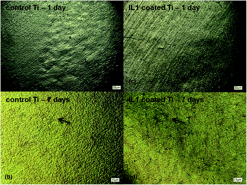 | ||
| Fig. 5 MC3T3-E1 cell attachment on Ti surfaces of (a and b) control Ti and (c and d) IL1-coated Ti after 1 and 7 days. Osteoblasts are identified by black arrows. | ||
Zhao et al. discussed that in the oral environment, the “race for the surface” is a difficult one for cells to win because it entails cell attachment and proliferation on the surface before bacterial colonization.5 The ability of cells to proliferate (soft and bone tissue cells) and differentiate (bone tissue cells) on IL-coated surfaces was observed in vitro. However, the effect against bacteria was still essential to determine coating presence and effectiveness. Antimicrobial activity was evaluated both in the fluid medium where the samples were immersed as well as on titanium disks surfaces. For this experiment, a BHI-containing saliva solution was employed in order to simulate the oral environment. Ideally, it has been established that the coating had to remain on the surface of titanium for a minimum of 7 days, which would represent the early period post-implantation, to ensure antimicrobial activity during the cell attachment process.48 Streptococcus sp. are known to be early colonizers, and the formation of biofilm on the titanium surface by these organisms can create specific conditions for colonization of other pathogens (late colonizers), which are responsible for triggering bone loss later in the process of implantation.13,49 Therefore, protection against bacterial attachment and biofilm formation early in the process post-implantation is essential to promote cell integration and protection against biofilm adhesion.5,50 Initial antimicrobial results for IL1- and IL2-coated Ti against S. sanguinis showed that IL2 did not trigger antimicrobial activity after 1 day of exposure (Fig. S5 in ESI†). This result can be attributed to the formation of crystals between the IL molecules and components found in the artificial saliva. The crystallization was suggested to inactivate the IL molecules inhibiting the antimicrobial activity of this compound. On the other hand, promising results were observed for IL1-coated Ti in reducing bacterial load. The logarithm of colony-forming units per mL (logCFU mL−1) values was quantified in the immersion fluid and on samples surfaces for control Ti and IL1-coated Ti after 1 and 7 days of immersion and are summarized in Fig. 6.
 | ||
| Fig. 6 logCFU mL−1 in fluid and on surface of control Ti and IL1-coated Ti after exposure to bacteria. *Reduction was statistically significant (p < 0.05). | ||
It was verified that, relative to control Ti, the concentration of bacteria was significantly reduced both in the fluid in which the samples were immersed in and on the surface of IL1-coated Ti samples after 1 day of immersion. Previous UV-vis measurements demonstrated that the concentration of IL1 released to the immersion fluid did not change with the time. After 1 day of immersion in saliva, 8.5 ± 0.9% of the coating was released to the fluid. The lower value of logCFU mL−1 on IL-coated sample surfaces relative to the control proved that the remaining IL on the surface acted as anti-adhesive, protecting the surface against biofilm formation. Furthermore, even with a small amount of IL released, this quantity was enough to inhibit bacterial growth (Fig. 6), which resulted in lower CFU mL−1 in the immersion fluid in comparison to the control Ti. The total amounts of S. salivarius, S. gordonii, and S. uberis cells (in suspension and adherent to the implant surface) were found to be less than the number of cells initially inoculated. This result was not observed for the control Ti and suggests that the coating conferred a bactericidal effect on these species. On the other hand, for S. mutans and S. sanguinis, the effect was anti-adhesive and bacteriostatic, as cell growth was inhibited on day 1.
Analysis of bacterial concentration in fluid and on the surface (biofilm) of control and IL1-coated Ti after 7 days of immersion was performed to evaluate the long-term effectiveness of the coating (Fig. 6). In general, there was a significant decrease of the bacterial load in the immersion fluid and on IL1-coated Ti samples in comparison to control Ti. IL1-coated Ti samples exposed to S. mutans, S. salivarius and S. gordonii had lower bacterial concentrations than control Ti after 7 days. However, bacterial load in both the immersion fluid and on the surface slightly increased in comparison to the results obtained after 1 day of immersion. After 7 days of immersion in S. sanguinis and S. uberis IL1-coated samples had the same or lower bacterial load than observed after 1 day of immersion, showing even higher antimicrobial activity against these strains.
Fig. 7 depicts images of control Ti and IL1-coated Ti disks after 1 and 7 days of exposure to S. salivarius. Bacterial biofilm (opaque white) was observed on the surface of control Ti specimens but was absent on the surface of IL1-coated Ti. This result demonstrated the antimicrobial activity of surfaces treated with this IL composition even after long periods of immersion. Although this thick, opaque white biofilm was not observed for samples exposed to the other bacterial strains tested, the bacterial concentration values were similar as shown in Fig. 6. This result suggests that different strains may form biofilms with distinct morphological features.
 | ||
| Fig. 7 Surface of (a) control Ti and (b) IL1-coated Ti after 1 day, and (c) control Ti and (d) IL1-coated Ti after 7 days of exposure to S. salivarius. Red arrows indicate biofilm formation on surface of control samples. | ||
In summary, the results of this study demonstrated that medium composition affected the release of ILs in solution, resulting in distinct toxicity against mammalian and bacterial cells. The IL coatings were, in general, more soluble in PBS, which resulted in higher toxicity against osteoblast cells although cells were able to recover after 7 days on IL1-coated samples. On the other hand, lower release of IL coatings was verified in saliva, conferring good anti-adhesive and antimicrobial activities against strains relevant to the oral environment, even after 7 days of immersion. Solidification of IL2 with artificial saliva components was observed, and this behavior was associated with the inactivation of this compound as an antimicrobial agent. Besides the evaluation of medium effect on IL release, it was possible to establish the relationship between chemical structure of ILs and cell toxicity. The selective toxicity against bacterial cells makes these compounds emerge as powerful candidates to functionalize the dental implant surface with antimicrobial activity.
4. Conclusion
After combining the results of the mammalian and bacterial cell experiments, IL coatings were able to provide antimicrobial activity while maintaining conditions for cell survival and differentiation. Cells and bacterial strains associated with the oral environment were tested to simulate the environment that would interface with IL-coated Ti samples in vivo. Initial concentrations of mammalian and bacterial cells were kept the same (about 105 cells), showing that there was a greater level of toxicity of IL1 towards bacteria. ILs are emerging as powerful candidates to assist mammalian cells in winning the race for the surface of dental implants against bacterial cells, which may improve the surface and hence success rates of these devices.Acknowledgements
The authors acknowledge the University of Texas at Dallas (UTD) for providing facilities and financial support for this study (startup funds DCR and KLP), fellowships from Coordenacao de Aperfeicoamento de Pessoal de Nivel Superior (CAPES) (IMG), the Vascular Mechanobiology laboratory at the University of Texas at Dallas for the cell culture facilities, Conselho Nacional de Desenvolvimento Cientifıco e Tecnologico (CNPq), Univerdal Proc/Proc. 475556/2012-7, and 474895/2013-0 (MAP and CPF) and the Rio Grande do Sul Foundation for Research Support (FAPERGS)—Proc. 2262-2551/14-1 and 2290-2551/14-1 (MAP and CPF).References
- D. C. Rodrigues, R. M. Urban, J. J. Jacobs and J. L. Gilbert, J. Biomed. Mater. Res., Part B, 2009, 88, 206–219 CrossRef PubMed.
- X. Liu, P. Chu and C. Ding, Mater. Sci. Eng., R, 2004, 47, 49–121 CrossRef.
- H. Hermawan, D. Ramdan and J. R. P. Djuansjah, in Biomedical Engineering – From Theory to Applications, ed. P. R. Fazel, 2011 Search PubMed.
- F. Y. Teng, C. L. Ko, H. N. Kuo, J. J. Hu, J. H. Lin, C. W. Lou, C. C. Hung, Y. L. Wang, C. Y. Cheng and W. C. Chen, Bioinorg. Chem. Appl., 2012, 2012, 1–9 CrossRef PubMed.
- B. Zhao, H. C. van der Mei, G. Subbiahdoss, J. de Vries, M. Rustema-Abbing, R. Kuijer, H. J. Busscher and Y. Ren, Dent. Mater., 2014, 30, 716–727 CrossRef CAS PubMed.
- A. Mombelli and F. Décaillet, J. Clin. Periodontol., 2011, 38(1), 203–213 CrossRef PubMed.
- J. Ata-ali, M. E. Candel-marti, A. J. Flichy-fernández, D. Peñarrocha-oltra, J. F. Balaguer-martinez and M. P. Diago, 2011, 16, e937–e943.
- A. Mombelli, N. Müller and N. Cionca, Clin. Oral Implants Res., 2012, 23(6), 67–76 CrossRef PubMed.
- T. G. Wilson, P. Valderrama and D. B. C. Rodrigues, J. Periodontol., 2014, 85, 657–660 CrossRef PubMed.
- D. C. Rodrigues, P. Valderrama, T. Wilson, K. Palmer, A. Thomas, S. Sridhar, A. Adapalli, M. Burbano and C. Wadhwani, Materials, 2013, 6, 5258–5274 CrossRef.
- Y. Manor, S. Oubaid, O. Mardinger, G. Chaushu and J. Nissan, J. Oral Surg., 2009, 67, 2649–2652 Search PubMed.
- M. Esposito, J. M. Hirsch, U. Lekholm and P. Thomsen, Eur. J. Oral Sci., 1998, 106, 527–551 CAS.
- M. C. L. G. Santos, Braz. J. Oral Sci., 2002, 1, 103–111 Search PubMed.
- M. Esposito, P. Thomsen, L. E. Ericson, L. Sennerby and U. Lekholm, Clinical Implant Dentistry and Related Research, 2000, 2, 18–32 CrossRef CAS PubMed.
- a J. van Winkelhoff, R. J. Goené, C. Benschop and T. Folmer, Clin. Oral Implants Res., 2000, 11, 511–520 CAS.
- I. S. Yeo, H. Y. Kim, K. S. Lim and J. S. Han, Int. J. Artif. Organs, 2012, 35, 762–772 CrossRef CAS PubMed.
- K. Subramani and D. Wismeijer, Int. J. Oral Maxillofac. Implants, 2012, 27, 1043–1054 Search PubMed.
- H. L. Huang, Y. Y. Chang, M. C. Lai, C. R. Lin, C. H. Lai and T. M. Shieh, Surf. Coat. Technol., 2010, 205, 1636–1641 CrossRef CAS.
- C. H. Lai, Y. Y. Chang, H. L. Huang and H. Y. Kao, Thin Solid Films, 2011, 520, 1525–1531 CrossRef CAS.
- L. Zhao, P. K. Chu, Y. Zhang and Z. Wu, J. Biomed. Mater. Res., Part B, 2009, 91, 470–480 CrossRef PubMed.
- S. Svensson, F. Suska, L. Emanuelsson, A. Palmquist, B. Norlindh, M. Trobos, H. Bäckros, L. Persson, G. Rydja, M. Ohrlander, B. Lyvén, J. Lausmaa and P. Thomsen, Nanomedicine, 2013, 9, 1048–1056 CAS.
- N. J. Wood, H. F. Jenkinson, S. a. Davis, S. Mann, D. J. O'Sullivan and M. E. Barbour, J. Mater. Sci.: Mater. Med., 2015, 26, 201 CrossRef PubMed.
- a J. Mariotti and D. a Rumpf, J. Periodontol., 1999, 70, 1443–1448 CrossRef CAS PubMed.
- T. H. Lee, C. C. Hu, S. S. Lee, M. Y. Chou and Y. C. Chang, Int. Endod. J., 2010, 43, 430–435 CrossRef PubMed.
- P. Wasserscheid and W. Keim, Angew. Chem., Int. Ed. Engl., 2000, 39, 3772–3789 CrossRef CAS.
- M. A. P. Martins, C. P. Frizzo, D. N. Moreira, A. Z. Tier, N. Zanatta and H. G. Bonacorso, Chem. Rev., 2008, 108, 2015–2050 CrossRef CAS PubMed.
- R. Caminiti and L. Gontrani, Soft and Biological Matter, 2014, p. 202 Search PubMed.
- H. Shirota, T. Mandai, H. Fukazawa and T. Kato, J. Chem. Eng. Data, 2011, 56, 2453–2459 CrossRef CAS.
- L. Carson, P. K. W. Chau, M. J. Earle, M. a. Gilea, B. F. Gilmore, S. P. Gorman, M. T. McCann and K. R. Seddon, Green Chem., 2009, 11, 492 RSC.
- M. T. Garcia, I. Ribosa, L. Perez, A. Manresa and F. Comelles, Langmuir, 2013, 29, 2536–2545 CrossRef CAS PubMed.
- R. G. Gore, L. Myles, M. Spulak, I. Beadham, T. M. Garcia, S. J. Connon and N. Gathergood, Green Chem., 2013, 15, 2747 RSC.
- P. Rajakumar, R. Raja, S. Selvam, R. Rengasamy and S. Nagaraj, Bioorg. Med. Chem. Lett., 2009, 19, 3466–3470 CrossRef CAS PubMed.
- K. Radošević, M. Cvjetko, N. Kopjar, R. Novak, J. Dumić and V. G. Srček, Ecotoxicol. Environ. Saf., 2013, 92, 112–118 CrossRef PubMed.
- K. M. Docherty and C. F. Kulpa Jr, Green Chem., 2005, 7, 185 RSC.
- S. Steudte, S. Bemowsky, M. Mahrova, U. Bottin-Weber, E. Tojo-Suarez, P. Stepnowski and S. Stolte, RSC Adv., 2014, 4, 5198 RSC.
- I. M. Gindri, D. A. Siddiqui, P. Bhardwaj, L. C. Rodriguez, K. Palmer, C. P. Frizzo, M. A. P. Martins and D. Rodrigues, RSC Adv., 2014, 4, 62594–62602 RSC.
- I. M. Gindri, C. P. Frizzo, M. A. P. Martins, A. Z. Tier, D. Siddiqui and C. Rodrigues, ACS Appl. Mater. Interfaces, 2015, 7, 27421–27431 CAS.
- K. Fukumoto, M. Yoshizawa and H. Ohno, J. Am. Chem. Soc., 2005, 127, 2398–2399 CrossRef CAS PubMed.
- M. A. Khan, R. L. Williams and D. F. Williams, Biomaterials, 1996, 17, 2117–2126 CrossRef CAS PubMed.
- N. T. Kadowaki, G. A. S. Martinez and A. Robin, Mater. Res., 2009, 12, 363–366 CrossRef CAS.
- M. Cvjetko, K. Radošević, A. Tomica, I. Slivac, J. Vorkapić-Furač and V. G. Srček, Arh. Hig. Rada Toksikol., 2012, 63, 15–20 CAS.
- B. Wang, J. Sun, S. Qian, X. Liu, S. Zhang, F. Liu, S. Dong and G. Zha, Biomed. Pharmacother., 2012, 66, 633–641 CrossRef CAS PubMed.
- Z.-Z. Shang, X. Li, H.-Q. Sun, G.-N. Xiao, C.-W. Wang and Q. Gong, Int. J. Oral Sci., 2014, 1–8 CAS.
- O. D. Monera, T. J. Sereda, N. E. Zhou, C. M. Kay and R. S. Hodges, J. Pept. Sci., 1995, 1, 319–329 CrossRef CAS PubMed.
- V. Lockett, R. Sedev, C. Bassell and J. Ralston, Phys. Chem. Chem. Phys., 2008, 10, 1330–1335 RSC.
- M. Schmidt, Arch. Orthop. Unfall-Chir., 2001, 121, 403–410 CAS.
- P. J. Garlick, J. Nutr., 2004, 134, 1633S–1639S CAS ; discussion 1664S–1666S, 1667S–1672S.
- A. Büchter, U. Joos, H.-P. Wiesmann, L. Seper and U. Meyer, Head Face Med., 2006, 2, 5 Search PubMed.
- M. C. Sánchez, A. Llama-Palacios, E. Fernández, E. Figuero, M. J. Marín, R. León, V. Blanc, D. Herrera and M. Sanz, Dent. Mater., 2014, 30, 1161–1171 CrossRef PubMed.
- M. Alhag, S. Renvert, I. Polyzois and N. Claffey, Clin. Oral Implants Res., 2008, 19, 182–187 CrossRef PubMed.
Footnote |
| † Electronic supplementary information (ESI) available: XPS spectra of IL1 and IL2, UV-vis calibration curves, release profile in percentage and antimicrobial activity of IL2. See DOI: 10.1039/c6ra01003b |
| This journal is © The Royal Society of Chemistry 2016 |