DOI:
10.1039/C6RA00673F
(Paper)
RSC Adv., 2016,
6, 29149-29158
pH-Triggered copolymer micelles as drug nanocarriers for intracellular delivery†
Received
9th January 2016
, Accepted 6th March 2016
First published on 8th March 2016
Abstract
One growing issue is how to use the weakly acidic conditions in endosomes/lysosomes for designing highly pH-sensitive drug carriers to deliver chemotherapeutic drugs accurately. In this paper, pH-sensitive amphiphilic triblock copolymers PEG8-PDPAn-PEG8 (n = 30, 50 and 100) were synthesized. Micelles composed of the copolymers were found to enter into lysosomes using a lysosome tracker method. The pH-sensitivity makes the micelles stable at pH 7.4 but swell and become looser at pH 6.0 due to protonation. Then, doxorubicin (DOX) was efficiently encapsulated in the hydrophobic cores of the micelles and released continuously in a weakly acidic environment. Cell toxicity assays were carried out using 2 human cell lines (HEK293 and Huh7) and showed good cyto-compatibility. In vitro cell viability tests proved that the DOX-loaded micelles could be more efficiently taken up by Huh7 tumor cells than free DOX, as well as actively trigger intracellular DOX release. Furthermore, in addition to small molecules (DOX), the copolymer micelles could also deliver macromolecules (such as transferrin, which could not enter into cells by itself) into lysosomes/endosomes. These nontoxic and multifunctional micelles can serve as a promising treatment candidate for efficient intracellular drug delivery and real-time monitoring.
1. Introduction
In the most recent decade, drug delivery systems based on nanotechnology have shown promising results in cancer therapy,1,2 because of their improved drug efficiency, and enhanced permeation and retention (EPR) effect.3,4 Various nanoparticles for drug delivery to tumors or for nano-therapeutics have been considered for entering clinical trials.5,6 Compared with dendrimers,7 micelles,8,9 liposomes10 and other organic/inorganic nanoparticles,11,12 polymeric micelles are much more versatile for delivering a broad spectrum of therapeutics13 because of their enormous chemical variation,8 relatively easy preparation, multiple functionality14 and high drug loading capacity. However, there were also several problems, including poor cellular internalization15 and inefficient intracellular16 drug release. To overcome these limitations, some strategies were proposed.17,18 For example, to construct an actively targeted19 drug delivery system, or to endow stimuli-responsibility,20,21 such as temperature,22 light,23 magnetic fields,24 electric fields,25 pH,26,27 redox potential28 and enzymes.29
Among them, the pH response strategy has been exploited widely to overcome various extra- and intra-cellular barriers to improve tumor-targeted nanoscale delivery systems. Till now, several strategies have been employed for designing pH-sensitive nanoparticles. It was reported that ammonium salt polymers, such as poly(2-(methacryloyloxy)ethyl phosphorylcholine) PMPC,30 poly(propylacrylic acid) PPAA31 and poly(dimethylaminoethyl methacrylate) PDMA,32 had been selected for use as pH-sensitive segments for amphipathic diblock or triblock copolymers. According to their pKa, the copolymers could provide pH-sensitivity with various triggering pH values. The drug carriers composed of these copolymers could release the drug at a specific spot, such as releasing only in the intestine33 or even inside or outside of tumor cells.34 There have been previous reports on diblock copolymers based on 2-(methacryloyloxy)ethyl phosphorylcholine (MPC) and 2-(diisopropylamino)ethyl methacrylate (DPA)35,36 that could assemble to form vesicles under neutral conditions and then dissociate to stimulate drug release when the pH decreased. However, these research reports also have several limitations such as the lack of study on intracellular drug delivery/release and the exact whereabouts of the internalized carriers. And, few reports have discussed in detail the effect of the block size of copolymers on biofunctions. In fact, the block size of copolymers affected not only the pH-sensitivity, but also the cell transfection and intracellular drug release. In addition, since many drugs and biomacromolecules, such as DOX, proteins and also DNA, could not be efficiently taken up by the cell membrane directly, efficient intercellular delivery/release systems are necessary.
We have focused on the design and preparation of pH-responsive carriers for intracellular drug delivery.37 The internalized nanoparticles generally pass through the endosome or lysosomes for degradation or deformation. Since the matrix in the endosome/lysosomes of most cells is weakly acidic (pH = 4.0–6.5),38,39 the cargo loaded in the pH-responsive carrier is expected to be released into the endosome/lysosome matrix and then target the nucleus directly.40
In this paper, amphipathic triblock copolymers, composed of poly(2-(diisopropylamino)ethylmethacrylate) (PDPA) and methoxy-poly(ethyleneglycol) (mPEG) segments (PEG8-PDPAn-PEG8 (n: 30–100)), were synthesized. PDPA was used as a pH-sensitive segment, which was hydrophobic under physiological pH conditions (pH > 7.4) and became hydrophilic at a decreased pH due to the protonation of di-isopropylamine.41 A series of micelles composed of these synthesized copolymers has been prepared through molecular self-assembly and they are expected to accomplish the task of effective cellular internalization and intracellular release. Here, the lengths of PDPAn should play an important role in the characteristics of the micelles. The pH-sensitivity as well as the drug release profile of the copolymer micelles have been investigated in vitro. Furthermore, normal human embryonic kidney cells (HEK293) and human liver cancer cells (Huh7) were incubated with the micelles to evaluate the cytotoxicity. The anti-tumor effects and intracellular delivery efficiency of the DOX-loaded micelles were evaluated. In addition to DOX, we also managed to deliver a biomacromolecule (transferrin, which could not enter into cells by itself) into lysosomes. The copolymer micelles were expected to achieve efficient intracellular drug delivery and release.
2. Experimental section
2.1 Materials
mPEG8 (Mn = 350 g mol−1) and DPA (99.5%) were purchased from Aldrich Scientific Polymer Products (USA). Copper(I) bromide (CuBr, 99.99%), diethyl-2,5-dibromohexanedioate (Br-ME-Br), N′′-pentamethyl-diethylenetriamine (PMDETA), pyrene (99%) and sodium hydride (NaH, 60%) were purchased from Aldrich and were used as received. Methylene chloride (CH2Cl2, AR), methanol (AR) and tetrahydrofuran (THF, AR) were from the Shanghai Tianlian Company. Doxorubicin hydrochloride (DOX·HCl, 98%) was purchased from J&K CHEMICAL. The two kinds of cells (HEK293T and Huh7) were supplied by the American Type Culture Collection (ATCC). Cell Counting Kit-8 (CCK-8), Dulbecco’s modified eagle medium (DMEM) and Fetal Bovine Serum (FBS) were purchased from Biotech Company of Shanghai. LysoTracker Green DND-26 (1 mM L−1, DMSO) and LysoTracker red (1 mM L−1, DMSO) were purchased from Thermo Fisher Scientific. Green fluorescently labeled transferrin, calcein acetoxy-methylester (calcein-AM, green) and propidium iodide (PI, red) were purchased from Beyotime Biotechnology Co. Ltd. (Nantong, China).
2.2 Synthesis of PDPAn blocks
PDPA50 block, as an example, was synthesized using an ATRP method. The DPA monomer (2.13 g, 10 mmol), Br-ME-Br initiator (0.072 g, 0.201 mmol), PMDETA ligand (45.6 μL, 0.222 mmol) and dry CH2Cl2 (4.0 mL) were mixed into a Schlenk flask with a magnetic stir bar. This solution was deoxygenated by freezing with liquid nitrogen 3 times, for 30 min each time, before adding CuBr catalyst (0.0290 g, 0.200 mmol). The [DPA]
:
[Br-ME-Br]
:
[CuBr]
:
[PMDETA] molar ratio was 50
:
1
:
1
:
1.1. The reaction was carried out under an argon atmosphere for 10 h at 25 °C. Finally, the reaction solution was diluted with THF (about 200 mL) and was then passed through an alumina column to remove the catalyst. The copolymer was then washed with methanol at least 3 times and the solvent was removed on a rotatory evaporator at 45 °C. The 1H NMR spectra were recorded on a Bruker AV400 (400 MHz) spectrometer at ambient temperature using CCl3D as the solvent.
2.3 Synthesis of PEG8-PDPAn-PEG8 triblock copolymers
A 50 mL flask with a magnetic stir bar and a rubber septum was charged with dry mPEG8 (0.25 g, 0.714 mmol) and dry argon. Dry THF (10 mL) was injected and NaH (0.06 g, 2.5 mmol) was poured into the flask and kept stirring in an ice bath. The reaction was carried out under an argon atmosphere at 20 °C for 3 h. After adding the PDPA50 (1.0 g, 0.0938 mmol) solution dropwise, the reaction was carried out for 3 h. The solvent was removed using a rotatory evaporator. The copolymer was then washed with methanol at least 3 times and the solvent was removed on a rotatory evaporator at 45 °C. The Mn and Mw/Mn values of the PEG8-PDPAn-PEG8 triblock copolymers were assessed using gel permeation chromatography (GPC) at 25 °C in combination with a refractive index detector. The GPC setup involves a polymer laboratories (PL) gel column. The eluent was dry THF with a flow rate of 1.0 mL min−1.
2.4 Characterization of PEG8-PDPAn-PEG8 triblock copolymers
The pH-sensitivities of PEG8-PDPAn-PEG8 were investigated using DLS (Malvern Instruments) and UV-vis spectroscopy (UV-2450, Shimadzu). The transmittance of the copolymer solutions (0.1 mg mL−1) at 400 nm was recorded at different pH values and the diameter and zeta-potential were measured using dynamic light scattering (DLS) (Zem4228, Malvern Instruments) at room temperature. The critical micelle concentration (CMC) values of the triblock copolymers were measured using a fluorescence spectrometer (F-4500, HITACHI, Japan). Pyrene was used as a fluorescent probe and the concentration of pyrene solution was 1 × 10⁻⁶ mol L−1. The pyrene excitation wavelength was set to 333 nm and the fluorescence intensity was measured at 372 nm and 383 nm. The intensity ratio of the peaks excited at 372 nm to those at 383 nm (I372/I383) was plotted against the logarithm of the concentration to determine the CMC.
2.5 Preparation of polymeric micelles
The blank micelles were prepared using a solvent evaporation method. Briefly, 3 mg of copolymer was dissolved in 1 mL of THF. Then phosphate buffered saline (PBS, pH 7.4, 1 mmol L−1) was added dropwise under high-speed stirring. The polymeric micelles then formed after the complete evaporation of THF. The final concentration of copolymer was 0.3 mg mL−1. For the DOX-loaded micelles, they were prepared by dissolving DOX in THF. DOX was treated with a drop of THF to remove hydrochloric acid. The final concentration of DOX was 0.05 mg mL−1. The DOX-loaded micelles were transferred into a dialysis bag (cutoff Mn = 3500 g mol−1) and dialyzed against deionized water to remove the unloaded DOX. The size and morphology of the micelles were studied using DLS and transmission electron microscopy (TEM, JEM-1400). To prepare the TEM samples, 0.02 mL (0.3 mg mL−1) of micelle solution was dropped onto a carbon-coated copper grid and then a drop of phosphotungstic acid solution (1% w/w) was added to stain the micelles.
2.6 The drug loading and release of micelles
The DOX-loading content (DLC) and encapsulation efficiency (DEE) were determined using UV-vis spectroscopy at a wavelength of 485 nm. The values of the DLC and DEE are defined by the following equations: |
 | (1) |
|
 | (2) |
The concentration of the copolymer was 0.3 mg mL−1. Then the pH-dependent release rates of DOX from the micelles were investigated. Briefly, 2 mL of DOX-loaded micelles was sealed in a dialysis tube (cutoff Mn = 3500 g mol−1), which was placed in a beaker containing 50 mL of PBS (10 mmol L−1, pH = 3.0, 6.0, 7.4 and 10.0) at 37 °C. The concentrations of DOX in PBS were measured after different time intervals using eqn (3).
|
 | (3) |
A1 is the absorption intensity of DOX at time
t and
A2 is the intensity of the total amount of DOX (including free and encapsulated DOX).
2.7 Cytotoxicity
The cytotoxicity of the blank micelles, toward HEK 293 cells or Huh7 cells, was measured using CCK-8 assays. Briefly, cells were cultured in a formulated DMEM nutrient solution supplemented with 10% FBS, 100 U mL−1 of penicillin G and 100 μg mL−1 of streptomycin (37 °C, humidified 5% CO2). The cells were seeded in 96 well plates at an initial density of 2000 cells per well for 24 h prior to treatment and then treated with blank micelle solutions with different concentrations (0–300 μg mL−1). The micelle solution was removed 24 h later and the cells were rinsed three times with PBS. Then, 20 μL of CCK-8 was added for a further 1 h of incubation and read using an automated microplate spectrophotometer (ELX800 Biotek, USA) at 450 nm. The results were determined from an average of four separate wells.
2.8 Cellular uptake
The Huh7 cells were incubated with free DOX, DOX-loaded micelles or transferrin-loaded micelles in DMEM at an equivalent dose (5 μg mL−1) for 1 and 5 h. The cells were imaged using a confocal laser scanning microscope (CLSM) with various differential interference contrast channels, including a blue channel (DAPI), red channel (DOX, LysoTracker red), and green channel (transferrin, LysoTracker green) excited at 358, 543 and 488 nm, respectively.
2.9 Anti-tumor cell effect
The tumor cell killing activity was investigated for different formulations of free DOX and micelles loaded with DOX. Huh7 cells were incubated in 24-well plates containing DMEM without FBS (about pH 7.4) for 5 h with free DOX and DOX-loaded micelles (the relative concentrations of DOX were fixed as 0, 2.5, 5, 10, 25 and 50 μg mL−1). Then, fresh nutrient solution without FBS was added and the cells were cultured for a further 19 h. The cells were treated with 2 μM calcein-AM and 4.5 μM PI for 30 min. Live cells were stained green and dead cells were stained red when visualized using fluorescence microscopy (EVOS, AMG). The quantitative viability of the Huh7 cells was determined using CCK-8. The cells were rinsed after 5 h and cultured in normal DMEM for 19 h.
3. Results and discussion
3.1 The characteristics of the copolymer
The triblock copolymers with different block lengths (PEG8-PDPAn-PEG8) were synthesized through two steps: an ATRP method and then a substitution reaction for the synthesis of the PDPAn segment and PEG8-PDPAn-PEG8, respectively. The 1H NMR spectra of mPEG8, PDPA50 and PEG8-PDPA50-PEG8 are shown in Fig. 1. As shown in Fig. 1A, the characteristic peaks at δ 3.67, δ 3.28 and δ 3.06 ppm represent the –CH2–CH2–, –OCH3 and –OH groups of the mPEG8 block. Fig. 1B shows the characteristic peaks of PDPA. The characteristic peaks of PDPA also appeared in the spectrum of PEG8-PDPA50-PEG8 (Fig. 1C), and the peak at δ 3.06 ppm (which represents the –OH groups) vanished, indicating that the –OH groups were displaced by the PDPA segments. These 1H NMR results confirmed that all residual monomers and impurities had been removed. The molecular weight of PEG8-PDPA50-PEG8 estimated from the 1H NMR results was 11
152 g mol−1 and its Mn value was found to be 13
679 g mol−1 with a polydispersity index (Mw/Mn) of 1.20 according to the GPC measurement. The composition and molecular weights of PEG8-PDPA50-PEG8 are listed in Table 1 with the data of the other two copolymers. These results showed that the desired molecular structures with a narrow molecular weight distribution were successfully synthesized. And furthermore, their CMC values were also measured using a fluorescent probe method. The fluorescence intensity ratios (I372/I383) under various concentrations are shown in the ESI (S1†). From the results shown in Table 1, the CMC values were found to decrease with the increase in PDPA length.
 |
| Fig. 1 The 1H NMR spectra of (A) mPEG8, (B) PDPA50 and (C) PEG8-PDPA50-PEG8 in CDCl3. | |
Table 1 The chemical compositions, molecular weights, polydispersity indexes and triggering pH values of the synthesized copolymers
Copolymer |
Mn,NMR (g mol−1) |
Mn,GPC (g mol−1) |
Mw/Mn |
CMC (mmol L−1) |
Triggering pH |
PEG8-PDPA30-PEG8 |
7093 |
9275 |
1.22 |
1.02 × 10⁻³ |
6.37 |
PEG8-PDPA50-PEG8 |
11 152 |
13 679 |
1.20 |
3.31 × 10⁻⁴ |
6.66 |
PEG8-PDPA100-PEG8 |
22 032 |
34 863 |
1.34 |
1.54 × 10⁻⁴ |
6.95 |
3.2 The properties of PEG8-PDPAn-PEG8
The triggering pH values of the synthesized copolymers were determined from the transmittance of the copolymer solutions at various pH values. Their transmittance was measured at an increasing pH and the results are shown in Fig. 2a. The configurations of the copolymers in solution were sensitive to the addition of NaOH. The solutions of the copolymers were transparent below pH 5.0 and became turbid suddenly from pH 6.0. The triggering pH values were found to be related to the PDPA length. An increase in PDPA length meant there were more tertiary amine groups to deprotonate resulting in a slight increase in pH. The synthesized copolymers were positively charged because the PDPA residues were protonated at a lower pH. Fig. 2b shows that their zeta potentials decreased to negative values when the solutions were titrated from acid to alkali. The results suggested that the synthesized copolymers had a triggering pH in the range of 6.37–6.95 depending on PDPA size.
 |
| Fig. 2 The pH-sensitivity of the synthesized copolymers (0.1 mg mL−1). (a) The transmittance of the copolymer solutions at various pH values. (b) The zeta potentials of the copolymers at various pH values. | |
3.3 The properties of the micelles
Micelles were prepared through the solvent volatilization method and THF was used as the solvent. The physico-chemical properties of the micelles composed of the synthesized copolymers are summarized in Table 2. Firstly, as shown in Table 2, the concentration of the copolymer seemed to have little influence on the size of the micelle. However, the diameters of the micelles increased obviously when the PDPA segments were lengthened. From the TEM images (Fig. 3a), it can be seen that the micelles have core–shell structures, which were thought to be composed of extended PEG shells and compacted PDPA cores, respectively. Then, it was easy to understand that with a longer PDPA length a larger core could be formed. Their size distribution graphs (Fig. 3b) and PDI values obtained using DLS indicated that the micelles were uniform in size, which could also be verified from the TEM images. And it was also found, at pH 7.4, that the micelles were negatively charged and their zeta potentials were almost unchanged by the PDPA length.
Table 2 Properties of the micelles composed of copolymers in PBS (pH 7.4)
Micelle composition |
Concentration (mg mL−1) |
Diameter (nm) |
PDI (±0.015) |
Zeta (±1.2 mV) |
DLC (±1%) |
DEE (±5%) |
PEG8-PDPA30-PEG8 |
0.15 |
122 ± 13 |
0.081 |
−9.5 |
— |
— |
0.20 |
124 ± 15 |
0.067 |
−10.2 |
— |
— |
0.30 |
131 ± 13 |
0.098 |
−11.5 |
12 |
72 |
PEG8-PDPA50-PEG8 |
0.15 |
134 ± 14 |
0.061 |
−8.5 |
— |
— |
0.20 |
136 ± 15 |
0.063 |
−9.3 |
— |
— |
0.30 |
149 ± 10 |
0.039 |
−10.6 |
14 |
82 |
PEG8-PDPA100-PEG8 |
0.15 |
163 ± 11 |
0.095 |
−7.1 |
— |
— |
0.20 |
156 ± 13 |
0.078 |
−7.6 |
— |
— |
0.30 |
188 ± 15 |
0.095 |
−8.8 |
15 |
90 |
 |
| Fig. 3 (a) From left to right: TEM images of PEG8-PDPA30-PEG8, PEG8-PDPA50-PEG8 and PEG8-PDPA100-PEG8; (b) the size distribution graphs of the micelles determined using DLS. The concentration of the micelles was 0.3 mg mL−1 (pH 7.4). | |
3.4 The DOX release of micelles
The values of the DLC and DEE of the micelles are also listed in Table 2 with the micelle properties. The DEE and DLC values of the micelles indicated the high encapsulation efficiencies of these micelles and were found to increase with PDPA length. DOX was thought to be incorporated in the hydrophobic PDPA core. Micelles with larger cores seemed to be able to bind and encapsulate more DOX. The micelles composed of PEG8-PDPA100-PEG8 had a maximum DEE value of 90%. The cumulative release of DOX from the micelles was monitored at various pH values at 37 °C in vitro. As shown in Fig. 4, the release profiles of the three micelles showed that the DOX accumulated release increased quickly with decreasing pH. As shown in Fig. 4a, at pH 3.0, nearly 100% of the DOX was released from the micelles (PEG8-PDPA30-PEG8) after 15 h, while only 10% of the DOX was released at pH 10.0. As shown in Fig. 4b and c, the results for the other two micelles showed similar release profiles to those shown in Fig. 4a.
 |
| Fig. 4 DOX accumulated release from the micelles at various pH values. The concentration of the micelles was 0.3 mg mL−1. | |
Comparing the release rates of the three micelles at pH 7.4 or 10.0, they were found to be very low. However, at pH 3.0 and 6.0, the DOX release rates increased by different degrees depending on the length of the PDPA chain. After 24 h, the micelles composed of PEG8-PDPA100-PEG8 had accumulated releases of about 75% and 60% at pH 3.0 and 6.0, respectively. The DOX molecules were encapsulated into the micelles through hydrophobic force under neutral/basic conditions and released due to the protonation of PDPA when the pH decreased. The micelles composed of PEG8-PDPA100-PEG8 showed the best sustained release effect.
These results suggested that the hydrophobic PDPA core might be protonated gradually from the outside into the centre, which could also be demonstrated by the 1H NMR spectra. The 1H NMR spectra of the micelles composed of PEG8-PDPA50-PEG8 in a solution of pH 6.0 were obtained after various intervals (Fig. 5a). Peak a (δ = 1.3 ppm) corresponding to the –N–H groups did not exist in the beginning and became more and more obvious with time, showing the evolution of the protonation degree.42 The peaks at δ 1.75 and δ 3.67 ppm, corresponding to the –CH2– and –CH2–CH2– groups, also increased with time, suggesting that the alkyl chain of PDPA was exposed gradually due to the loosened cores. The TEM and DLS results (Fig. 5b and c) also showed that the diameter of the micelles increased with time at pH 6.0. It could be speculated that the tightly tangled structure of the PDPA core became looser and looser as shown in the inset of Fig. 5a. The large values of the diameter further suggested that the micelles did not collapse even after 24 h but just swelled.
 |
| Fig. 5 The evolution process with time for micelles composed of PEG8-PDPA50-PEG8 (0.3 mg mL−1) at pH 6.0 studied using (a) 1H NMR (D2O/DCl), (b) TEM and (c) DLS. | |
The results shown in Fig. 4 indicate that the DOX accumulated release rate decreases with an increase in PDPA length. Under acidic conditions, PEG8-PDPA100-PEG8 with the longest PDPA chain might form micelles with the most compact hydrophobic core and then induce the slowest protonation and drug release. In this experiment, a sustained release effect of DOX from the micelles was achieved under weakly acidic conditions and the release kinetics depended on the length of PDPA.
3.5 The cell toxicity of micelles in vitro
To evaluate the cyto-compatibility of these micelles (PEG8-PDPA30-PEG8, PEG8-PDPA50-PEG8 and PEG8-PDPA100-PEG8), human embryonic kidney cells HEK 293 as well as human liver cancer cells Huh7 were incubated with different concentrations of the three micelles individually for 24 h. The CCK-8 assay demonstrated that the micelles had high cell viability for both normal and tumor cells in the concentration range of 10–300 μg mL−1 (Fig. 6a and b). All of the cell viabilities were nearly 100%. It was shown that the polymeric micelles possessed excellent cyto-compatibility.
 |
| Fig. 6 Cell viability of HEK 293T and Huh7 cells cultured with different concentrations of micelles, 0, 10, 25, 50, 75, 100, 200 and 300 μg mL−1, for 24 h. | |
3.6 Cellular uptake efficiency
The cellular uptake efficiency of free DOX and DOX-loaded micelles was observed using CLSM. The micelles composed of PEG8-PDPA50-PEG8 were used in the following cell experiments. As shown in Fig. 7a, we compared the results of free DOX and DOX-loaded micelles cultured with liver tumor cells Huh7 for 1 h and 5 h. After 1 h of incubation, both free DOX and DOX-loaded micelles were seldom taken up by the cells (Fig. 7aI and II). However, 5 h later, DOX-loaded micelles could be better ingested by Huh7 cells and internalized into the cytoplasm more effectively compared with free DOX as shown in Fig. 7aIII and IV. In Fig. 7aIV, it can also be seen that there was a phenomenon of apoptosis from the disappearance of some blue stained cell nuclei.
 |
| Fig. 7 (a) The CLSM images of Huh7 cells incubated with free DOX or DOX-loaded micelles (DOX formulation was 5 μg mL−1) for 1 h (I and II) and 5 h (III and IV). DOX is red and the cellular nuclei stained with DAPI are blue. (b) The CLSM images of Huh7 cells treated with DOX-loaded micelles (red fluorescence) for 5 h and then stained with a lysosome tracker (green fluorescence). (c) The CLSM images of Huh7 cells treated with transferrin-loaded micelles (green fluorescence) for 5 h and then stained with a lysosome tracker (red fluorescence). The bars are 10 μm. | |
In order to investigate if the DOX-loaded micelles could enter into lysosomes, a lysosome tracker (green) was used to stain the Huh7 cells. The Huh7 cells were cultured with the DOX-loaded micelles for 5 h and then stained immediately. Their CLSM images are shown in Fig. 7b. As shown in Fig. 7b, the red fluorescence indicated that the DOX-loaded micelles had entered into the cells. The green domains represented a lysosome site, which partly overlapped with the red domains, suggesting that the DOX-loaded micelles had entered into the lysosome.
We also tried to verify whether these copolymer micelles could deliver macromolecules into cells or not. So green fluorescently labeled transferrin has been used instead of DOX. Here, to distinguish the red fluorescence of DOX, a lysosome tracker labeled with a green fluorescent marker was selected. The results in Fig. 7c also show the overlap of the red and green fluorescence domains, indicating that transferrin has been delivered to the lysosomes. These results suggested that the pH-sensitive micelles could deliver both small molecules (DOX) and macromolecules (transferrin) into lysosomes/endosomes. Since the pH of the lysosome matrix is usually weakly acidic, the cargo loaded in these copolymer micelles is expected to be released easily and quickly in situ.
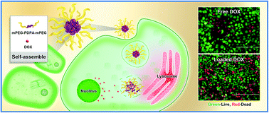
Furthermore, the cell viability was investigated for cancer cells cultured with DOX in different formulations. The fluorescence images of Huh7 cells treated with free DOX and DOX-loaded micelles are shown in Fig. 8a. The cells survived better with free DOX than with DOX-loaded micelles at certain drug concentrations in the range of 2.5–50 μg mL−1 (Fig. 8a). The quantitative data showed that about 20% of the cells were killed by DOX-loaded micelles as the drug concentration was increased to 50 μg mL−1, while only 5% of the cells were killed by free DOX (Fig. 8b). The results showed that DOX-loaded micelles were easier for cancer cells to take up than free DOX. After the micelles were internalized by Huh7 cells, intracellular DOX release was successfully triggered by the lower pH of the lysosome matrix and then the therapy efficacy was enhanced (Fig. 8c). These results indicated that the copolymer micelles prepared here could be used as a good carrier for intracellular delivery and release.
 |
| Fig. 8 (a) The visual images of Huh7 cells after treatment with different DOX formulations (0–50 μg mL−1) were recorded using fluorescence imaging. The micelles were composed of PEG8-PDPA50-PEG8. Green calcein-AM fluorescence and red PI fluorescence indicated live and dead cells, respectively. The bar is 100 μm. (b) The quantitative viability of Huh7 cells after being incubated with different DOX formulations. (c) The transport process of pH-sensitive micelles for intracellular drug delivery and release. | |
From these results, we demonstrated that the micelles could enter into lysosomes/endosomes after being incubated with Huh7 cells for 5 h. The cargo could be released in the lysosomes/endosomes because of the lower pH (pH 4.0–6.5). However, the release kinetics of DOX and transferrin could not be measured. If we estimated using the results shown in Fig. 4(b), the DOX release percentage should be 30–40% after 5 h and 80–90% after 24 h. Furthermore, the cell anti-tumor effect experiments were carried out using a process of 5 h of incubation with DOX-loaded micelles and then were cultured following the general route for 19 h, which gave enough time for DOX release. The good anti-tumor effect shown in Fig. 8 indicated that DOX had been released from the micelles, escaped from the lysosomes/endosomes and reached the cell nucleus ultimately.
4. Conclusion
In conclusion, we prepared a new pH-sensitive polymeric micelle for intracellular drug delivery and release. The micelles, which were used as nano-carriers, exhibit a high loading capacity and pH-triggered release of DOX. The micelles exhibit no cell toxicity both to normal human cells and tumor cells. Compared with free DOX, the pH-sensitive micelles loaded with DOX were more easily internalized by cancer cells and got to the lysosome site quickly. Due to the pH-sensitivity, the micelles achieved the release of DOX at the weakly acidic site (lysosome) before it entered into nuclei and then induced cell apoptosis. The research results confirmed the DOX release and cell uptake mechanism, the final fate of the novel pH-sensitive micelles inside a cell, and even the transfection of biomacromolecules such as transferrin, which will provide a better understanding of the mechanisms and in turn allow for the optimization of their function as drug delivery vehicles. This approach of designing and tailoring polymeric micelles may provide a useful means for the development of various delivery vehicles suitable for cancer therapy.
Conflict of interest
The authors declare no conflict of interest.
Acknowledgements
Financial support for this work was provided by the National Natural Science Foundation of China (No. 21276074), the 111 Project (No. B08021) of China and the Fundamental Research Funds for the Centre Universities of China.
References
- R. A. Petros and J. M. DeSimone, Nat. Rev. Drug Discovery, 2010, 9, 615–627 CrossRef CAS PubMed.
- J. M. Gamboa and K. W. Leong, Adv. Drug Delivery Rev., 2013, 65, 800–810 CrossRef CAS PubMed.
- H. Maeda, H. Nakamura and J. Fang, Adv. Drug Delivery Rev., 2013, 65, 71–79 CrossRef CAS PubMed.
- J. Fang, H. Nakamura and H. Maeda, Adv. Drug Delivery Rev., 2011, 63, 136–151 CrossRef CAS PubMed.
- S. Tang, X. Huang, X. Chen and N. Zheng, Adv. Funct. Mater., 2010, 20, 2442–2447 CrossRef CAS.
- H. C. Yuki Mochida, Y. Miura, F. Albertini, S. Fukushima, N. N. Kensuke Osada and K. Kataoka, ACS Nano, 2014, 8, 6724–6738 CrossRef PubMed.
- H. C. L. Hanxiang Zeng, T. N. Tiambeng, G. A. Williams and Z. Guan, J. Am. Chem. Soc., 2013, 135, 4962–4965 CrossRef PubMed.
- E. Jin, B. Zhang, X. Sun, Z. Zhou, X. Ma, Q. Sun, J. Tang, Y. Shen, E. Van Kirk, W. J. Murdoch and M. Radosz, J. Am. Chem. Soc., 2013, 135, 933–940 CrossRef CAS PubMed.
- X. Duan, J. Xiao, Q. Yin, Z. Zhang, H. Yu, S. Mao and Y. Li, ACS Nano, 2013, 7, 5858–5869 CrossRef CAS PubMed.
- N. Lozano, W. T. Al-Jamal, A. Taruttis, N. Beziere, N. C. Burton, J. Van den Bossche, M. Mazza, E. Herzog, V. Ntziachristos and K. Kostarelos, J. Am. Chem. Soc., 2012, 134, 13256–13258 CrossRef CAS PubMed.
- Y. Zhang, M. Wang, Y.-G. Zheng, H. Tan, B. Y.-W. Hsu, Z.-C. Yang, S. Y. Wong, A. Y.-C. Chang, M. Choolani, X. Li and J. Wang, Chem. Mater., 2013, 25, 2976–2985 CrossRef CAS.
- K. Kim, J. H. Kim, H. Park, Y. S. Kim, K. Park, H. Nam, S. Lee, J. H. Park, R. W. Park, I. S. Kim, K. Choi, S. Y. Kim and I. C. Kwon, J. Controlled Release, 2010, 146, 219–227 CrossRef CAS PubMed.
- R. Tan, Z. She, M. Wang, Z. Fang, Y. Liu and Q. Feng, Carbohydr. Polym., 2012, 87, 1515–1521 CrossRef CAS.
- Y. Wang, L. Dou, H. He, Y. Zhang and Q. Shen, Mol. Pharmaceutics, 2014, 11, 885–894 CrossRef CAS PubMed.
- Y. Brudno, E. A. Silva, C. J. Kearney, S. A. Lewin, A. Miller, K. D. Martinick, M. Aizenberg and D. J. Mooney, Proc. Natl. Acad. Sci. U. S. A., 2014, 111, 12722–12727 CrossRef CAS PubMed.
- X. Guo, D. Li, G. Yang, C. Shi, Z. Tang, J. Wang and S. Zhou, ACS Appl. Mater. Interfaces, 2014, 6, 8549–8559 CAS.
- V. Stepankova, S. Bidmanova, T. Koudelakova, Z. Prokop, R. Chaloupkova and J. Damborsky, ACS Catal., 2013, 3, 2823–2836 CrossRef CAS.
- I. Yildiz, S. Impellizzeri, E. Deniz, B. McCaughan, J. F. Callan and F. M. Raymo, J. Am. Chem. Soc., 2011, 133, 871–879 CrossRef CAS PubMed.
- J. H. Na, S. Y. Lee, S. Lee, H. Koo, K. H. Min, S. Y. Jeong, S. H. Yuk, K. Kim and I. C. Kwon, J. Controlled Release, 2012, 163, 2–9 CrossRef CAS PubMed.
- D. Roy, J. N. Cambre and B. S. Sumerlin, Prog. Polym. Sci., 2010, 35, 278–301 CrossRef CAS.
- R. P. Dumitriu, G. R. Mitchell and C. Vasile, Polym. Int., 2011, 60, 222–233 CrossRef CAS.
- M. M. Soledad Lencina, Z. Iatridi, M. A. Villar and C. Tsitsilianis, Eur. Polym. J., 2014, 61, 33–44 CrossRef CAS.
- N. C. Fan, F. Y. Cheng, J. A. Ho and C. S. Yeh, Angew. Chem., Int. Ed. Engl., 2012, 51, 8806–8810 CrossRef PubMed.
- J. H. Lee, K. J. Chen, S. H. Noh, M. A. Garcia, H. Wang, W. Y. Lin, H. Jeong, B. J. Kong, D. B. Stout, J. Cheon and H. R. Tseng, Angew. Chem., Int. Ed. Engl., 2013, 52, 4384–4388 CrossRef CAS PubMed.
- X. Shan, Y. Fang, S. Wang, Y. Guan, H. Y. Chen and N. Tao, Nano Lett., 2014, 14, 4151–4157 CrossRef CAS PubMed.
- Y. Huang, Z. Tang, X. Zhang, H. Yu, H. Sun, X. Pang and X. Chen, Biomacromolecules, 2013, 14, 2023–2032 CrossRef CAS PubMed.
- Y. H. Liang, C. H. Liu, S. H. Liao, Y. Y. Lin, H. W. Tang, S. Y. Liu, I. R. Lai and K. C. Wu, ACS Appl. Mater. Interfaces, 2012, 4, 6720–6727 CAS.
- H. N. Abdelhamid and H.-F. Wu, TrAC, Trends Anal. Chem., 2015, 65, 30–46 CrossRef CAS.
- E. H. Hill, Y. Zhang, D. G. Evans and D. G. Whitten, ACS Appl. Mater. Interfaces, 2015, 7, 5550–5560 CAS.
- R. T. Pearson, N. J. Warren, A. L. Lewis, S. P. Armes and G. Battaglia, Macromolecules, 2013, 46, 1400–1407 CrossRef CAS.
- S. Mishra, L. Y. Peddada, D. I. Devore and C. M. Roth, Acc. Chem. Res., 2012, 45, 1057–1066 CrossRef CAS PubMed.
- H. Yu, Y. Zou, Y. Wang, X. Huang, G. Huang, B. D. Sumer, D. A. Boothman and J. Gao, ACS Nano, 2011, 5, 9246–9255 CrossRef CAS PubMed.
- M. Drozdzik, C. Groer, J. Penski, J. Lapczuk, M. Ostrowski, Y. Lai, B. Prasad, J. D. Unadkat, W. Siegmund and S. Oswald, Mol. Pharm., 2014, 11, 3547–3555 CrossRef CAS PubMed.
- L. M. Kaminskas, V. M. McLeod, B. D. Kelly, C. Cullinane, G. Sberna, M. Williamson, B. J. Boyd, D. J. Owen and C. J. Porter, Mol. Pharm., 2012, 9, 422–432 CrossRef CAS PubMed.
- B. Yuan, Q. Chen, W. Q. Ding, P. S. Liu, S. S. Wu, S. C. Lin, J. Shen and Y. Gai, ACS Appl. Mater. Interfaces, 2012, 4, 4031–4039 CAS.
- M. Ciftci, M. A. Tasdelen, W. Li, K. Matyjaszewski and Y. Yagci, Macromolecules, 2013, 46, 9537–9543 CrossRef CAS.
- J. C. Yu, Y. L. Chen, Y. Q. Zhang, X. K. Yao, C. G. Qian, J. Huang, S. Zhu, X. Q. Jiang, Q. D. Shen and Z. Gu, Chem. Commun., 2014, 50, 4699–4702 RSC.
- F. Wang, Y.-C. Wang, S. Dou, M.-H. Xiong, T.-M. Sun and J. Wang, ACS Nano, 2011, 5, 3679–3692 CrossRef CAS PubMed.
- Y. Li, J. Yang, B. Xu, F. Gao, W. Wang and W. Liu, ACS Appl. Mater. Interfaces, 2015, 7, 8114–8124 CAS.
- H. Li, Y. Cui, J. Sui, S. Bian, Y. Sun, J. Liang, Y. Fan and X. Zhang, ACS Appl. Mater. Interfaces, 2015, 7, 15855–15865 CAS.
- K.-I. Shimizu, N. Imaiida, K. Kon, S. M. A. Hakim Siddiki and A. Satsuma, ACS Catal., 2013, 3, 998–1005 CrossRef CAS.
- W. Hao, X. Han, Y. Shang, S. Xu and H. Liu, Colloids Surf., B, 2015, 136, 809–816 CrossRef CAS PubMed.
Footnote |
† Electronic supplementary information (ESI) available. See DOI: 10.1039/c6ra00673f |
|
This journal is © The Royal Society of Chemistry 2016 |
Click here to see how this site uses Cookies. View our privacy policy here.