Methanation of carbon monoxide on ordered mesoporous NiO–TiO2–Al2O3 composite oxides

Qing Liu*a, Yuanyu Tianab and Hongmei Aic
aKey Laboratory of Low Carbon Energy and Chemical Engineering, Shandong University of Science and Technology, Qingdao, Shandong 266590, P. R. China. E-mail: sdsslq@163.com; Tel: +86-532-86057766
bState Key Laboratory of Heavy Oil Processing, China University of Petroleum (East China), Qingdao, Shandong 266580, P. R. China
cDepartment of Economy & Management, Shandong University of Science and Technology, Tai’an 271021, P. R. China

Received 6th January 2016 , Accepted 11th February 2016

First published on 12th February 2016


Abstract

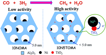
Simultaneous inhibition of the sintering of Ni particles and coke formation, while maintaining high catalytic activity, is greatly challenging for supported Ni catalysts in high-temperature applications. To address this problem, a series of ternary ordered mesoporous NiO–TiO2–Al2O3 composite oxides were synthesized via the evaporation-induced self-assembly (EISA) method and applied in the CO methanation reaction. The addition of TiO2 could significantly promote the catalytic activity, and the optimal ordered mesoporous NiO–TiO2–Al2O3 catalyst with a composition of 10 wt% NiO and 5 wt% TiO2 (10N5TOMA) achieved the maximum CH4 yield of 93% at 380 °C, 0.1 MPa and a weight hourly space velocity of 60[thin space (1/6-em)]000 mL g−1 h−1. Our characterization results showed that the promotive effect of TiO2 appeared to be two-fold: on the one hand, the Ti species could decrease the Ni particle size and improve the reducibility of Ni particles, leading to increased H2 uptake and the dispersion of the metallic Ni; on the other hand, the electron cloud density of Ni was increased by electron transfer from Ti4+/Ti3+ redox, which might facilitate CO dissociation from the catalyst surface. In addition, the ordered mesoporous 10N5TOMA catalyst showed superior anti-sintering and anti-coking properties in a 550 °C-139 h-lifetime test, mainly because the Ni particles were anchored in the alumina matrix with a strong interaction between the Ni species and the ordered mesoporous alumina (OMA) framework.


1. Introduction

Natural gas is a highly efficient form of energy and is considered to be the cleanest fossil fuel.1,2 However, due to its poor reserves in some regions of the world, conversion of syngas from coal or biomass to synthetic natural gas (SNG) via the methanation reaction is attracting great attention. Considering the exothermic nature of CO methanation (CO + 3H2 → CH4 + H2O, ΔH298 K = −206.1 kJ mol−1), thermodynamically, this reaction is favored at low temperature due to LeChatelier’s principle; but kinetically, this reaction is favored at high temperatures, because the higher the temperature is, the faster reaction rate is. High operation temperatures are usually employed in industry due to economic considerations, for example being above 600 °C in the first methanation reactor of the TREMP™ process.3,4 Ni/Al2O3 is the most commonly used catalyst for CO methanation due to its high catalytic activity and relatively low cost.5,6 However, it often suffers from sintering of the Ni particles and coking during the methanation process.2,7 Thus, the development of catalysts retaining high stability against sintering and carbon deposition while maintaining high catalytic activity at high temperatures is desirable.

Generally, formation of an alloy is an effective method to stabilize metal nanoparticles, but the broad distribution of compositions can result in a lower-than-expected catalytic performance.8,9 Encapsulation of metal nanoparticles in well-defined cavities or channels, such as core–shell, core–sheath, ordered mesoporous or lamellar structures, is a more straightforward way of stabilizing metal nanoparticles.8,9 For the materials with core–shell and lamellar structures, the thickness of the encapsulation shell is often poorly controlled, which leads to a decrease in the catalytic activity resulting from mass transfer resistance due to the thick shells.8–10 For the materials with a core–sheath structure, it is difficult to accurately control the location of the metal nanoparticles inside the sheath, and the migration of metal particles is hard to completely inhibit along the longitudinal direction.11 In contrast, ordered mesoporous supports can not only confine metal nanoparticles in a fixed space to hamper their sintering and coke formation, but also offer a large surface area for the high dispersion of active metal atoms, thus promising to enhance both the activity and stability of the catalyst simultaneously.12–14 By comprehensively considering the catalytic effect, production scalability, operability and economic cost issues, ordered mesoporous structures are the most feasible for industrial production among the above-mentioned structures. Recently, ordered mesoporous Ni–V–Al,15 Ni–Zr–Al16 and Ni–Al17 catalysts have exhibited high anti-sintering and anti-coking properties for high-temperature CO methanation due to the confinement effect of ordered mesoporous structures; however, their activities are still poor, and high reaction temperatures (400 or 450 °C) are required to achieve the maximum CO conversion and CH4 yield at atmospheric pressure. Therefore, considering the industrial application, the catalytic activity of ordered mesoporous catalysts should be further enhanced.

Promoters play an important role in the catalytic performance of a heterogeneous catalyst and usually affect the electron mobility, crystal texture, metal dispersion and thermal stability of the catalyst.18 Doping with an appropriate promoter is a commonly used strategy to improve the catalytic performance. Various promoters, such as MgO,1 ZrO2,7 CeO2[thin space (1/6-em)]5 and TiO2,19 have been employed to enhance Ni/Al2O3 catalysts for CO methanation. Among them, TiO2 has been found to effectively restrict the formation of the NiAl2O4 spinel phase and weaken the NiO–Al2O3 interaction,19,20 leading to a greater exposure of Ni species and improved carbon monoxide adsorption capacity.19 In addition, Escobar et al.20 found that both the Ni dispersion and H2 chemisorption could be improved via the addition of an appropriate amount of TiO2 in the Ni/Al2O3–TiO2 catalyst. As we know, the hydrogenation of *CHx species and the dissociation of CO are the rate-controlling steps in the CO methanation reaction,21,22 thus, the enhanced H2 and CO chemisorption by addition of TiO2 can facilitate CO methanation. It is expected that TiO2 can play a similar role in improving the catalytic activity of the ordered mesoporous NiO–TiO2–Al2O3 catalysts for CO methanation. Furthermore, Morris et al.12 found that the ordered mesoporous structure still remained after the incorporation of TiO2 species into the ordered mesoporous alumina (OMA) framework, indicating that it is feasible to synthesize the NiO–TiO2–Al2O3 composite oxides with an ordered mesoporous structure.

Herein, to enhance the catalytic performance as well as the anti-coking and anti-sintering properties of Ni catalysts for the CO methanation reaction, as well as to investigate the influence of the TiO2 promoter, a group of ternary NiO–TiO2–Al2O3 composite oxides with ordered mesoporous structures were synthetized via a one-pot evaporation-induced self-assembly (EISA) strategy. To our knowledge, the design and synthesis of ordered mesoporous NiO–TiO2–Al2O3 composite oxides for CO methanation have not yet been formally reported in the literature. In this work, in order to gain insight into the structure–catalytic performance relationship and to determine how the TiO2 promoter impacts on the activity and stability, various techniques, including N2 adsorption, transmission electron microscopy (TEM), scanning electron microscopy (SEM), X-ray diffraction (XRD), H2 temperature-programmed reduction (H2-TPR), H2 temperature-programmed desorption (H2-TPD), and X-ray photoelectron spectroscopy (XPS), were used to characterize the catalysts before and after the catalytic reaction.

2. Experimental

2.1. Catalyst preparation

All chemicals are of analytical grade and used as received. Aluminum isopropoxide (Al(OPri)3), nickel(II) nitrate hexahydrate (Ni(NO3)2·6H2O), 67 wt% nitric acid (HNO3) and ethanol were purchased from Sinopharm Chemical Reagent Co. Ltd., China, titanium(IV) isopropoxide (Ti(OPri)4) from Alfa Aesar, and (EO)20(PO)70(EO)20 triblock copolymer (Pluronic P123, typical Mn = 5800) from Sigma-Aldrich. All the gases, with purity of >99.999%, were purchased from Qingdao Hengxiang Gas Company Ltd., China and used as received.

The ordered mesoporous NiO–TiO2–Al2O3 composite oxides were prepared using a one-pot EISA method following our previous procedure.15,16 Typically, 2.2 g P123 was dissolved in 40.0 mL of anhydrous ethanol, followed by addition of 3.3 mL 67 wt% nitric acid, 4.08 g Al(OPri)3 and stoichiometric quantities of Ni(NO3)2·6H2O and Ti(OPri)4 in sequence under vigorous stirring. The obtained mixture was covered with PE film and stirred at room temperature for at least 5 h. Finally, the mixture was transferred to a Petri dish, which was covered with PE film with holes, and placed into an oven to undergo the slow EISA process at 60 °C for 48 h. The final gel was calcined with a first step to 400 °C for 4 h with a heating rate of 1 °C min−1 and a second step to 550 °C for 2 h with a heating rate of 5 °C min−1. The obtained samples were denoted as 10NxTOMA (x = 0, 2, 5 and 7), in which x and OMA represent the mass percentage of TiO2 and the ordered mesoporous Al2O3, respectively; the NiO content was fixed at 10 wt% in all the catalysts in this work.

2.2. Catalyst characterization

N2 adsorption was measured at −196 °C using a Quantachrome surface area & pore size analyzer NOVA 3200e. Prior to the measurement, the sample was degassed at 300 °C for 2 h under vacuum. The specific surface area was determined according to the Brunauer–Emmett–Teller (BET) method. The pore size distribution (PSD) was calculated based on the Barrett–Joyner–Halenda (BJH) method using the adsorption isotherm branch.

The morphology of the samples was observed by field emission scanning electron microscopy (FE-SEM) (JSM-6700F, JEOL, Japan) and bright field transmission electron microscopy (BF-TEM) (JEM-2010F, JEOL, Japan) under a working voltage of 200 kV, equipped with a liquid nitrogen-cooled energy-dispersive X-ray spectroscopy (EDS) detector for elemental analysis. The dark field transmission electron microscopy (DF-TEM) and high-angle annular dark field scanning transmission electron microscopy (HAADF-STEM) were performed on a Tecnai G2 F20 U-TWIN transmission electron microscope (FEI, USA) at an acceleration voltage of 200 kV.

X-ray diffraction (XRD) patterns in the range of 10.0 to 90.0° (wide angle) or 0.5 to 5.0° (small angle) were recorded on a PANalytical X’Pert PRO MPD using Cu Kα radiation (λ = 1.5418 Å) at 40 kV and 40 mA. The crystal size of the sample was estimated using the Debye–Scherrer equation.

H2 temperature-programmed reduction (H2-TPR) and H2 temperature-programmed desorption (H2-TPD) experiments were conducted on a Quantachrome automated chemisorption analyzer (chemBET pulsar TPR/TPD) following the procedures reported previously.6 For H2-TPR, prior to the measurement, the sample was pretreated at 200 °C for 1 h in a He flow. Then, the sample was cooled to room temperature, followed by heating to 1000 °C with a heating rate of 10 °C min−1 in a 10 vol% H2/Ar flow (30 mL min−1). For H2-TPD, after reduction in situ in a H2/Ar flow at 700 °C for 1 h, the sample was cooled down to room temperature and saturated with H2/Ar for 2 h. After removing the physically adsorbed H2 by flushing in Ar (30 mL min−1) for 1 h, the sample was heated to 650 °C at 10 °C min−1 in Ar. The consumed or desorbed H2 was detected continuously as a function of temperature using a thermal conductivity detector (TCD). The dispersion of Ni was calculated from the volume of chemisorbed H2 based on the formula described in our previous work.15

The X-ray photoelectron spectroscopy (XPS) was conducted on a VG ESCALAB 250 spectrometer (Thermo Electron, U.K.) with a non-monochromatized Al Kα X-ray source (1486 eV).

Thermogravimetric (TG) analysis was conducted on a STA449F3 thermal gravimetric analysis system (NETZSCH, Germany). A 10 mg sample was used and was heated in air (200 mL min−1) from room temperature up to 1000 °C (10 °C min−1).

2.3. Catalytic measurements

The catalytic measurements are similar to our previous work.16 Typically, the CO methanation reactions were carried out in a fixed bed reactor at 0.1 MPa with a quartz tube (720 mm length by 8 mm inner diameter). A 0.1 g catalyst sample (20–40 mesh) diluted with 5.0 g quartz sand (20–40 mesh) was reduced at 700 °C in pure H2 (100 mL min−1) for 1 h and then cooled to the starting reaction temperature in H2. The mixed H2 and CO with N2 (as an internal standard) (H2/CO/N2 = 3/1/1, molar ratio) was introduced into the reactor, and the outlet gas stream from the reactor was cooled using a cold trap. The inlet and outlet gases were analyzed on-line using a Micro GC 3000A (Agilent Technologies). The concentrations of H2, N2, CH4 and CO in the gas products were detected by a TCD detector connected to a molecular sieve column, while the concentrations of CO2, C2H4, C2H6, C3H6 and C3H8 were analyzed by another TCD connected to a Plot Q column. A lifetime test of the CO methanation was performed at 550 °C and 0.1 MPa. The CO conversion, CH4 selectivity and yield are defined as follows:
image file: c6ra00392c-t1.tif

image file: c6ra00392c-t2.tif

image file: c6ra00392c-t3.tif
here, X is the conversion of CO, S is the selectivity for CH4, Y is the yield of CH4, and Fi,in and Fi,out are the volume flow rates of species i (i = CO or CH4) at the inlet and outlet, respectively.

3. Results and discussion

3.1. Characterization of the catalysts

The nitrogen physisorption isotherms of the calcined 10NxTOMA are shown in Fig. 1a. All the isotherms belong to type IV with H1-type hysteresis loops, which indicate the presence of uniform cylindrically shaped channels.15,23 The shapes of the isotherms and hysteresis loops of 10NxTOMA (x = 2, 5 and 7) are similar to those of 10NOMA, indicating that the ordered mesoporous structure still remains after the doping with titanium species. In addition, the PSD curves of the 10NxTOMA samples have only one narrow peak around 10 nm, confirming the high uniformity of the channels. The textural properties of the calcined samples are summarized in Table 1. All the 10NxTOMA samples possess high specific surface areas up to 315 m2 g−1, large pore volumes up to 0.70 cm3 g−1 and average pore diameters around 9 nm. Furthermore, the specific surface areas, pore volumes and average pore diameters of 10NxTOMA all show little change, indicating the similar geometrical structure of 10NxTOMA after the addition of a small amount of TiO2 species. Overall, the ordered mesoporous 10NxTOMA samples have uniform mesopores, high specific surface areas and large pore volumes, which indicate promise in their potential applications as catalysts.
image file: c6ra00392c-f1.tif
Fig. 1 N2 adsorption isotherms (a) and PSD curves (b) of the calcined catalysts.
Table 1 Physical and chemical properties of the samples
Sample SBETa (m2 g−1) Vpb (cm3 g−1) Dpc (nm) Ni particled size (nm) H2 uptake (μmol g−1) De (%)
a SBET, surface area of the calcined sample derived from the BET equation.b VP, pore volume of the calcined sample obtained from the volume of nitrogen adsorbed at the relative pressure of 0.97.c DP, average pore diameter of the calcined sample derived from the BJH method using the following equation: image file: c6ra00392c-t4.tif.d Estimated from the XRD diffraction peak (2θ = 44.6) using the Debye–Scherrer equation. The Ni particle size was denoted as ‘<5.0’ if the calculated value was smaller than 5.0 nm, considering the XRD detection limit.e D, Ni dispersion calculated based on the H2-TPR and H2-TPD results.
10NOMA 315.8 0.75 9.5 5.0 58.6 8.9
10N2TOMA 331.1 0.77 9.3 <5.0 82.5 12.5
10N5TOMA 322.4 0.71 8.8 <5.0 92.9 14.1
10N7TOMA 325.6 0.78 9.6 <5.0 72.4 11.0


Evidence of the presence of hexagonally ordered mesopores for the 700°C-reduced 10NxTOMA is provided by the TEM images in Fig. 2. As seen in the BF-TEM images in Fig. 2a–d, the typical ordered mesostructure is obvious, with a regular alignment of cylindrical pores along the (100) direction,12,15,23 and the ordered mesostructure can be retained after incorporation of different amounts of Ti species, which is in good agreement with the results of N2 adsorption. In addition, the Ni particles in all the 10NxTOMA catalysts are located in the OMA channels, being anchored on the OMA channel wall, and have uniform sizes without any obvious agglomeration. Further characterization of the reduced 10N5TOMA was carried out by EDS, HRTEM, DF-TEM and HAADF-STEM (Fig. 2d–k). The EDS spectrum (inset of Fig. 2d) shows clear peaks of O, Ni, Al and Ti, implying that all these elements have been successfully introduced into the ordered mesoporous 10N5TOMA. In the HRTEM image (Fig. 2e), the lattice spacing of ca. 0.20 nm corresponding to the Ni (111) plane can be clearly observed, while there is no lattice corresponding to TiO2, which may be related to its poor crystallinity. In the DF-TEM images, the cylindrical pores are regularly aligned and the bright (Fig. 2f and g) spots representing Ni particles are homogeneously embedded in the OMA framework. However, the TiO2 particles are hardly observed in either the BF-TEM and DF-TEM images due to the high dispersion and poor contrast compared with the Ni particles and OMA framework. Interestingly, in the HAADF-STEM images, the corresponding elemental maps of Al, O, Ni and Ti demonstrate the actual distributions of Al, O, Ni and Ti elements separately (Fig. 2h–k). It is seen that both Ni and Ti elements are highly dispersed in the catalyst, and some Ti species can be located in the vicinity of Ni particles in both the channels and skeleton of the OMA framework, which is feasible for the interaction between Ti and Ni species during the reaction.


image file: c6ra00392c-f2.tif
Fig. 2 BF-TEM images of the reduced samples: (a) 10NOMA, (b) 10N2TOMA, (c) 10N7TOMA, and (d) 10N5TOMA; the EDS spectrum of 10N5TOMA is provided in (d) inset, units for y axis: counts [a.u.]; HRTEM image of the reduced 10N5TOMA (e); DF-TEM image of the reduced 10N5TOMA (f); HAADF-STEM image of the reduced 10N5TOMA catalyst (g), and elemental mapping images of Al (h), O (i), Ni (j) and Ti (k).

As we know, small angle X-ray diffraction (SXRD) patterns can provide further evidence for the formation of an ordered mesostructure. As shown in Fig. 3a, the SXRD patterns of the calcined 10NxTOMA exhibit two peaks at around 0.85 and 1.4°, corresponding to the (100) and (110) planes respectively, which further confirms the formation of a hexagonally ordered mesoporous structure (p6mm symmetry),12,23 coinciding with the above N2 adsorption and TEM results. Moreover, there are no apparent diffraction peaks of NiO, TiO2 and Al2O3 in the wide angle X-ray diffraction (WXRD) patterns of the calcined 10NxTOMA due to their incorporation into the OMA skeleton, poor crystallinity, or high dispersion (Fig. 3b).15 Meanwhile, the WXRD patterns of the 700 °C-reduced catalysts are shown in Fig. 3c. Apparently, even after reduction at 700 °C for 1 h, the OMA framework is still amorphous, while there are three new peaks at around 44.5, 51.8 and 75.9°, corresponding to the (111), (200) and (220) planes of metallic Ni (JCPDS 01-070-1849), respectively. The estimated Ni nanoparticle sizes from the WXRD patterns using the Debye–Scherrer equation are listed in Table 1. The Ni particle size of 10NOMA is 5.0 nm; after the incorporation of Ti species, the Ni particles of 10NxTOMA (x = 2, 5 and 7) are all smaller than 5.0 nm, indicating that the addition of Ti species can decrease the Ni particle size and improve Ni dispersion. The decrease in Ni particle size after the addition of TiO2 may be because the TiO2 species can act as a physical barrier which restricts the migration and agglomeration of Ni particles during the reduction process. Furthermore, there is still no observation of any diffraction peaks of TiO2 even after the incorporation of 7 wt% TiO2 in 10N7TOMA, indicating their high dispersion, or poor crystallinity after 700 °C-reduction treatment. In all, the ordered mesoporous NiO–TiO2–Al2O3 catalyst preparation using the EISA method can successfully restrict the Ni particle size, especially after incorporation of Ti species.


image file: c6ra00392c-f3.tif
Fig. 3 Small-angle XRD (a) and wide-angle XRD (b) patterns of the calcined samples and wide-angle XRD patterns of the reduced samples (c).

The H2-TPR profiles of 10NxTOMA are displayed in Fig. 4a. For 10NOMA, there is only one reduction peak at around 647 °C, corresponding to the reduction of NiO species in the OMA skeleton with a strong interaction with the support.1,24 Compared with 10NOMA, there is also one reduction peak in the H2-TPR profiles of 10NxTOMA (x = 2, 5 and 7), while the peaks shift to lower temperatures with increasing TiO2 content, and the lowest peak temperature is observed at about 607 °C for 10N7TOMA, indicating that the addition of Ti species could decrease the reduction temperature and improve the reducibility of the Ni particles in the 10NxTOMA catalysts. Herein, the Ti species plays a similar role to the Ti promoter in Ni/TiO2–Al2O3 catalysts19 and the V promoter in Ni/V2O3–Al2O3 catalysts,15 decreasing the interaction between the metal and the support. For 10N7TOMA, it is of note that there is a small new peak around 487 °C. Considering its small integral area and low peak value, this peak may correspond to the reduction of a small amount of the NiO–TiO2–Al2O3 composite with a slight decrease in the mesoporous uniformity in 10N7TOMA after a further increase in the mass fraction of the Ti species.


image file: c6ra00392c-f4.tif
Fig. 4 H2-TPR profiles (a) and H2-TPD profiles (b) of the catalysts.

The H2-TPD profiles of 10NxTOMA are displayed in Fig. 4b. As described in the literature,25 there is no obvious H2 adsorption peak for TiO2, and all the hydrogen desorption peaks in the H2-TPD profiles are attributed to the Ni particles in the Ni–TiO2–Al2O3 catalysts. For 10NOMA, there are two main H2 desorption peaks at around 149 and 453 °C. The peak at 149 °C is attributed to the chemisorbed hydrogen on the highly dispersed Ni nanoparticles;25 and the peak located at 453 °C can be derived from the H2 adsorbed in the surface bulk or poorly-dispersed Ni particles.25,26 After the TiO2 addition, there is almost no change in either the peak value or integrated area of the first desorption peak; however, the second desorption peak of 10NxTOMA shifts to low temperatures with increasing TiO2 content, and there is an obvious increment in the integrated area of the second desorption peak (except for 10N7TOMA), because of the improved the reducibility of the Ni particles and the presence of more exposed metallic Ni atoms in 10N2TOMA and 10N5TOMA. For 10N7TOMA, the second desorption peak shifts to the lowest temperature of around 377 °C, while there is a new third desorption peak at around 453 °C, which should correspond to the Ni particles covered by the excess TiO2, resulting in more difficult desorption of H2. Interestingly, Escobar et al.20 also found that the H2 chemisorption suppression effect was evident for the ∼10 wt% Ni catalyst at high TiO2 content, because of the partial coverage of the metallic nickel particles by TiOx moieties.

The calculated hydrogen uptakes and the Ni dispersion of the 10NxTOMA are listed in Table 1. It is seen that the addition of an appropriate amount of TiO2 can increase the H2 uptake and the dispersion of metallic Ni in the 10NxTOMA catalysts, while an excess of Ti species has an adverse effect due to the coverage of the exposed Ni atom. Among them, 10N5TOMA has the highest total H2 uptake of 92.9 mmol g−1 and Ni dispersion of 14.1%.

XPS analysis of the 700 °C-reduced 10N5TOMA and 10NOMA was employed to further study the oxidative state of the Ni species, and the results are shown in Fig. 5a. For 10NOMA, the peaks of Ni 2p3/2 at 855.7 and 861.9 eV belong to Ni2+, and the peak at 852.6 eV is attributed to the metallic Ni.15 The formation of Ni2+ is because of the partial oxidation of surface metallic Ni during the sample transfer before the XPS test. In addition, there is a shift of 0.5 eV for Ni0 from 852.6 eV for 10NOMA to 852.1 eV for 10N5TOMA; meanwhile, the binding energy of Ni2+ for 10N5TOMA decreases 0.2 eV compared to that of 10NOMA. This indicates that an interaction may occur between the Ni species and TiO2 in 10N5TOMA, which is similar to the results in the literature.19,25 The decrease in binding energy may be due to the increase in electron cloud density of the Ni atoms (Ni0 → Niδ−) by electron transfer from Ti4+/Ti3+ redox. Zeng et al.19 found that the increase in electron cloud density of Ni atoms could promote CO dissociation on the catalyst surface, leading to a relatively high catalytic performance. In order to confirm the existence of Ti3+ species, the Ti 2p spectra of the reduced 10N5TOMA catalyst are fitted and shown in Fig. 5b. The two main peaks at around 464.3 and 458.6 eV correspond to Ti 2p3/2 and Ti 2p1/2 of TiO2, respectively;27 meanwhile, there is a weak peak at around 456.9 eV, attributed to Ti 2p3/2 of Ti2O3.28 Escobar et al.20 found that titania could be stabilized to a partial reduction state as TiOx (x < 2) in alumina-rich TiO2–Al2O3 binary oxides, treated in a H2 flow at 500 °C. Hence, the minority Ti3+ may be formed through the reduction of Ti4+ in H2 at 700 °C. Combined with the above results, it is inferred that there is an occurrence of Ti4+/Ti3+ redox, which can transfer electrons to Ni species and is related to the enhanced catalytic performances of 10NxTOMA.


image file: c6ra00392c-f5.tif
Fig. 5 XPS spectra of the reduced 10N5TOMA and 10NOMA: (a) Ni 2p and (b) Ti 2p.

3.2. Catalytic performances of the catalysts

The CO methanation reaction over 10NxTOMA was carried out in the temperature range of 300−500 °C at 0.1 MPa using a weight hourly space velocity (WHSV) of 60[thin space (1/6-em)]000 mL g−1 h−1, and the results are displayed in Fig. 6a–c. For 10NOMA, an increased catalytic activity is observed with the increase in the temperature, and its CO conversion can reach the thermodynamic equilibrium calculated using the Gibbs free energy minimization method at 450 °C.29 After the Ti addition, the CO conversion and CH4 yield over 10NxTOMA (x = 2, 5 and 7) are obviously improved, and can reach the thermodynamic equilibrium at 400 °C; however, the CH4 selectivity of the various catalysts (except for 10N7TOMA at below 420 °C and 10N2TOMA at 360 °C) is similar, and the by-product detected by GC is CO2, which may be the product of the CO Boudouard reaction (2CO → CO2 + C), reverse methane reforming reaction by CO2 (2CO + 2H2 → CO2 + CH4), and water-gas shift reaction (CO + H2O → CO2 + H2).7 Overall, the similar CH4 selectivity indicates the simultaneous promotion of both CO methanation and the side reactions by the ordered mesoporous NiO–TiO2–Al2O3 catalysts.30,31 Among them, 10N5TOMA is the best due to its largest H2 uptake and Ni dispersion, and it can reach the maximum CH4 yield of 93% at only 380 °C. In addition, compared to the ordered mesoporous Ni–V–Al,15 Ni–Zr–Al16 and Ni–Al17 catalysts in the previous literature, 10N5TOMA is even more active, and can achieve higher maximum CO conversion and CH4 yield at lower temperatures. In short, TiO2 is an efficient promoter and 10N5TOMA can be a promising catalyst for CO methanation.
image file: c6ra00392c-f6.tif
Fig. 6 Catalytic properties of the catalysts at 0.1 MPa, 60[thin space (1/6-em)]000 mL g−1 h−1: (a) CO conversion, (b) CH4 selectivity, and (c) CH4 yield; (d) lifetime test of 10N5TOMA at 550 °C, 0.1 MPa.

The lifetime test is essential for evaluation of a heterogeneous catalyst. In order to carry out the lifetime test for the optimal 10N5TOMA catalyst in a relatively short time in the laboratory, the harsh reaction condition of high temperature (550 °C) was employed. As seen from the results in Fig. 6d, both CO conversion and CH4 selectivity can remain stable at 60[thin space (1/6-em)]000 mL g−1 h−1, 0.1 MPa, 550 °C through the whole lifetime test of 139 h, indicating that 10N5TOMA has high stability at high temperature.

After the lifetime test, the spent 10N5TOMA was further characterized by XRD, TEM and SEM, and the results are displayed in Fig. 7. As seen in Fig. 7a, the XRD pattern of the spent 10N5TOMA is similar to that of the reduced one, except for a new peak at around 26.7°, which is the characteristic diffraction peak of rutile-type TiO2 (JCPDS 00-021-1276), indicating the formation of well-crystallized TiO2 in 10N5TOMA under the hydrothermal conditions (H2O is one of the products in CO methanation) at high temperature during the 139 h-lifetime test. Interestingly, it seems that the crystal phase transformation of TiO2 has little influence on the catalytic performance and structure of 10N5TOMA. The OMA is still amorphous and the calculated Ni particle size is still smaller than 5.0 nm after the lifetime test, indicating that 10N5TOMA has high stability and sintering-resistance of the Ni particles. Moreover, as seen from the TEM image of the spent 10N5TOMA, the ordered mesostructure remains almost unchanged and the Ni particles are embedded in the framework with an average size of ca. 4.0 ± 1.2 nm without any obvious agglomeration (Fig. 7b), which further confirms the high stability and anti-sintering property of 10N5TOMA. Furthermore, there is no diffraction peak corresponding to graphitic carbon in the XRD pattern (Fig. 7a), and no carbon filaments are observed in either the TEM (Fig. 7b) or SEM (Fig. 7c) images, which indicates that the deposited carbon should be quite small in quantity and most probably is amorphous. The amount of deposited carbon on the spent catalyst is determined by TG analysis, and the carbon content in the spent 10N5TOMA catalyst is estimated to be only 0.2 wt% (Fig. 7d), suggesting its excellent anti-coking capacity. The superior anti-sintering property may be because the Ni particles are anchored in the alumina matrix and partially exposed in the OMA channels, and this confinement effect of the OMA framework can effectively reduce Ni sintering.32 Simultaneously, the strong interaction between Ni and the OMA support can also suppress the growth of carbon filaments which can lift the Ni particles from the support,33 resulting in the high anti-coking property of 10N5TOMA. In short, the high catalytic activity and stability of the 10N5TOMA catalyst can be attributed to the incorporation of the TiO2 promoter and the special location of the Ni particles in the mesoporous framework.


image file: c6ra00392c-f7.tif
Fig. 7 XRD patterns of the reduced and spent 10N5TOMA (a), TEM (b) and SEM (c) images of the spent 10N5TOMA, and TG curves of the reduced and spent 10N5TOMA (d).

4. Conclusions

A series of ordered mesoporous NiO–TiO2–Al2O3 composite oxides with TiO2 contents ranging from 0 wt% to 7 wt% have been successfully synthesized using the facile one-pot EISA method and are used in the CO methanation reaction to produce SNG. The addition of TiO2 can promote the catalytic activity of 10NxTOMA, mainly because of two reasons. On the one hand, TiO2 can decrease the Ni particle size and improve the reducibility of the Ni particles leading to enhanced H2 uptake and Ni dispersion; on the other hand, the electron cloud density of Ni was increased by electron transfer from Ti4+/Ti3+ redox, leading to a relatively high catalytic performance of the 10NxTOMA catalyst. In addition, the optimal ordered mesoporous 10N5TOMA catalyst shows superior anti-sintering and anti-coking properties in a 550 °C-139 h-lifetime test, mainly because the Ni particles are anchored in the alumina matrix with a strong interaction between Ni and the OMA framework. This work shows that the ordered mesoporous NiO–TiO2–Al2O3 catalyst is a promising candidate for CO methanation that can simultaneously prevent sintering of Ni particles and coking, while maintaining high catalytic activity at high temperatures.

References

  1. D. C. Hu, J. J. Gao, Y. Ping, L. H. Jia, P. Gunawan, Z. Y. Zhong, G. W. Xu, F. N. Gu and F. B. Su, Ind. Eng. Chem. Res., 2012, 51, 4875–4886 CrossRef CAS.
  2. V. M. Shinde and G. Madras, AIChE J., 2014, 60, 1027–1035 CrossRef CAS.
  3. J. Kopyscinski, T. J. Schildhauer and S. M. A. Biollaz, Fuel, 2010, 89, 1763–1783 CrossRef CAS.
  4. J. R. Rostrup-Nielsen, K. Pedersen and J. Sehested, Appl. Catal., A, 2007, 330, 134–138 CrossRef CAS.
  5. Q. Liu, J. J. Gao, M. J. Zhang, H. F. Li, F. N. Gu, G. W. Xu, Z. Y. Zhong and F. B. Su, RSC Adv., 2014, 4, 16094–16103 RSC.
  6. Q. Liu, F. N. Gu, X. P. Lu, Y. J. Liu, H. F. Li, Z. Y. Zhong, G. W. Xu and F. B. Su, Appl. Catal., A, 2014, 488, 37–47 CrossRef CAS.
  7. Q. Liu, F. N. Gu, J. J. Gao, H. F. Li, G. W. Xu and F. B. Su, J. Energy Chem., 2014, 23, 761–770 CrossRef.
  8. A. M. Cao, R. W. Lu and G. Veser, Phys. Chem. Chem. Phys., 2010, 12, 13499–13510 RSC.
  9. S. R. Li and J. L. Gong, Chem. Soc. Rev., 2014, 43, 7245–7256 RSC.
  10. J. Lu, B. Fu, M. C. Kung, G. Xiao, J. W. Elam, H. H. Kung and P. C. Stair, Science, 2012, 335, 1205–1208 CrossRef CAS PubMed.
  11. L. M. Chew, P. Kangvansura, H. Ruland, H. J. Schulte, C. Somsen, W. Xia, G. Eggeler, A. Worayingyong and M. Muhler, Appl. Catal., A, 2014, 482, 163–170 CrossRef CAS.
  12. S. M. Morris, P. F. Fulvio and M. Jaroniec, J. Am. Chem. Soc., 2008, 130, 15210–15216 CrossRef CAS PubMed.
  13. J. Y. Zhang, Z. Xin, X. Meng, Y. Lv and M. Tao, Fuel, 2014, 116, 25–33 CrossRef CAS.
  14. J. Y. Zhang, Z. Xin, X. Meng and M. Tao, Fuel, 2013, 109, 693–701 CrossRef CAS.
  15. Q. Liu, J. J. Gao, F. N. Gu, X. P. Lu, Y. J. Liu, H. F. Li, Z. Y. Zhong, B. Liu, G. W. Xu and F. B. Su, J. Catal., 2015, 326, 127–138 CrossRef CAS.
  16. Q. Liu, F. N. Gu, X. Y. Wang, G. J. Jin, H. F. Li, F. Gao, Z. Y. Zhong, G. W. Xu and F. B. Su, RSC Adv., 2015, 5, 84186–84194 RSC.
  17. H. Tian, S. R. Li, L. Zeng, H. Y. Ma and J. L. Gong, Science China Materials, 2015, 58, 9–15 CrossRef CAS.
  18. J. J. Gao, Q. Liu, F. N. Gu, B. Liu, Z. Y. Zhong and F. B. Su, RSC Adv., 2015, 5, 22759–22776 RSC.
  19. Y. Zeng, H. F. Ma, H. T. Zhang, W. Y. Ying and D. Y. Fang, Fuel, 2014, 137, 155–163 CrossRef CAS.
  20. J. Escobar, J. Antonio De Los Reyes and T. Viveros, Appl. Catal., A, 2003, 253, 151–163 CrossRef CAS.
  21. J. Happel, I. Suzuki, P. Kokayeff and V. Fthenakis, J. Catal., 1980, 65, 59–77 CrossRef CAS.
  22. D. M. Stockwell, J. S. Chung and C. O. Bennett, J. Catal., 1988, 112, 135–144 CrossRef CAS.
  23. Q. Yuan, A. X. Yin, C. Luo, L. D. Sun, Y. W. Zhang, W. T. Duan, H. C. Liu and C. H. Yan, J. Am. Chem. Soc., 2008, 130, 3465–3472 CrossRef CAS PubMed.
  24. J. J. Gao, C. M. Jia, J. Li, F. N. Gu, G. W. Xu, Z. Y. Zhong and F. B. Su, Ind. Eng. Chem. Res., 2012, 51, 10345–10353 CrossRef CAS.
  25. J. Liu, C. M. Li, F. Wang, S. He, H. Chen, Y. F. Zhao, M. Wei, D. G. Evans and X. Duan, Catal. Sci. Technol., 2013, 3, 2627–2633 CAS.
  26. S. Velu and S. Gangwal, Solid State Ionics, 2006, 177, 803–811 CrossRef CAS.
  27. H. Y. Min, X. Q. Ran, J. W. Fan, Y. Sun, J. P. Yang, W. Teng, W. X. Zhang, G. M. Li and D. Y. Zhao, J. Mater. Chem. A, 2015, 3, 7399–7405 CAS.
  28. D. Gonbeau, C. Guimon, G. Pfister-Guillouzo, A. Levasseur, G. Meunier and R. Dormoy, Surf. Sci., 1991, 254, 81–89 CrossRef CAS.
  29. J. J. Gao, Y. L. Wang, Y. Ping, D. C. Hu, G. W. Xu, F. N. Gu and F. B. Su, RSC Adv., 2012, 2, 2358–2368 RSC.
  30. G. A. Somorjai and M. C. Yang, Top. Catal., 2003, 24, 61–72 CrossRef CAS.
  31. G. A. Somorjai and Y. G. Borodko, Catal. Lett., 2001, 76, 1–5 CrossRef CAS.
  32. N. Wang, K. Shen, L. H. Huang, X. P. Yu, W. Z. Qian and W. Chu, ACS Catal., 2013, 3, 1638–1651 CrossRef CAS.
  33. H. Y. Ma, L. Zeng, H. Tian, D. Li, X. Wang, X. Y. Li and J. L. Gong, Appl. Catal., B, 2016, 181, 321–331 CrossRef CAS.

This journal is © The Royal Society of Chemistry 2016
Click here to see how this site uses Cookies. View our privacy policy here.