Q. Luo†
ab,
L. Zhang†*a,
X. Chena,
O. K. Tan*a and
K. C. Leongb
aNanomaterials Lab, School of Electrical and Electronic Engineering, Nanyang Technological University, 50 Nanyang Avenue, 639798, Singapore. E-mail: zh0012li@e.ntu.edu.sg; EOKTAN@ntu.edu.sg
bGLOBALFOUNDRIES Singapore Pte Ltd, 738406, Singapore
First published on 1st February 2016
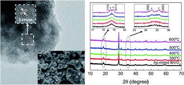
In this work, a mechanochemical high energy ball milling approach was used to synthesize monoclinic BiVO4 (m-BiVO4) nanoparticles in an attempt to simultaneously reduce the particle size and improve the throughput for practical photocatalytic applications. The effect of annealing to eliminate the induced defects and thus enhance the reactivity was studied on the mechanochemically synthesized BiVO4 nanoparticles. Besides using the conventional characterization tools of XRD, Raman, FE-SEM, HRTEM, XPS and UV-vis diffuse reflectance to examine the crystalline structure, morphology, chemical states and visible light absorption, a customized Kelvin probe coupled with an LED light source was developed as a non-contact tool to study the surface photovoltage (SPV) response for understanding charge generation and separation. The photocatalytic performance was finally evaluated for the degradation of Rhodamine B (RhB) under visible light irradiation to correlate with these physicochemical properties.
Among a number of non-titania based visible light driven photocatalysts under consideration, bismuth vanadate (BiVO4) emerges as one of the most promising candidates since its first demonstration for O2 evolution under visible light irradiation by Kudo et al.5 In nature, BiVO4 exists in three crystalline phase forms: tetragonal zircon (z-t), tetragonal scheelite (s-t) and monoclinic sheetlite (m).6 Under different thermal or preparation conditions, these phases can undergo phase transformations.7 As compared to the tetragonal counterparts, m-BiVO4 with a narrow bandgap of ca. 2.4 eV demonstrates superior photocatalytic effect under visible light irradiation7 and has been therefore investigated and synthesized via various approaches, including solid state reaction,8 co-precipitation process,9 hydrothermal route,10,11 chemical bath deposition,12 flame spray pyrolysis,13 sonochemical route14 and microwave-assisted methods.15,16 It is well known that the photocatalytic activity intimately relates to the particle size and surface area of the powders. However, many of the abovementioned methods usually lead to the formation of gross particles with small surface area, either due to the high calcination temperature in solid state reaction or because of the tiny solubility product of BiVO4 in the aqueous solution.17 In this regard, it is essential to develop methods to synthesize nano-sized m-BiVO4 with high surface area so as to improve the photocatalytic efficiency.
There have been some reported works on preparation of nano-sized m-BiVO4 in literature.10,11,15,18,19 Our group has previously synthesized m-BiVO4 octahedral nano-crystals using hydrothermal route in the presence of sodium dodecyl benzene sulfonate (SDBS).10 Controllable morphologies and uniform sizes could be obtained. Yet, the BET surface area remains unsatisfactory only up to 4 m2 g−1. Sun et al. has reported modified hydrothermal method to synthesize nano-BiVO4 particles in the presence of ethylene diamine tetraacetic acid (EDTA).11 Instead of using EDTA as a chelating agent as in the conventional hydrothermal process,18 EDTA was introduced prior to the precipitation process so the growth of BiVO4 crystallite was well controlled to achieve a large surface area up to 10 m2 g−1. Reverse-microemulsion process has been reported by Chung and Lu to produce nano-scaled m-BiVO4 at a heating temperature of 400 °C to obtain average particle size down to 35 nm.18 More recently, a novel microwave-assisted approach based on in situ twin polymerization has been reported by Hofmann et al. for the preparation of nano-sized m-BiVO4 with BET surface areas of 7–16 m2 g−1.15 Conventional high energy ball milling process has also been explored by Venkatesan et al. to obtain BiVO4 nanoparticles with spherical-like morphology and average sizes about 20 nm by optimizing milling times and ball to powder ratio (BPR).19
The last mechanochemical process represents a simple and effective mean with relative high yield to obtain fine particles with large surface area as compared to other complex approaches involving many steps or with a low throughput, and was thus investigated in the present work for the synthesis of m-BiVO4 nanoparticles for photocatalytic degradation of organic pollutants. It is noteworthy that along with decreasing the particle size, intensive milling often produces defects and microstress due to the meta-stable high energy state. Thus, post-annealing to eliminate the induced defects and enhance the photo-reactivity20,21 of the mechanochemically synthesized m-BiVO4 nanoparticles was also of our particular interests and was investigated and optimized in our present study. The physicochemical properties of the mechanochemically synthesized m-BiVO4 annealed at different temperatures were characterized, and their photocatalytic performance was evaluated and compared for the degradation of Rhodamine B (RhB) dye under visible light irradiation. In particular, surface photovoltage (SPV) in dark and upon irradiation were recorded for the first time to correlate with the photocatalytic activities, which is also a surface phenomenon.
:
BiVO4 powder
:
ethanol = 20
:
1
:
0.5 in an YSZ bowl, and a milling speed of 550 rpm for 8 h. The powders were finally separated from YSZ balls using 325# sieve and subject to annealing at different temperatures from 300 to 600 °C in dry air for 1 h to eliminate the defects induced during the ball milling process.
The surface photovoltage spectroscopy (SPS) of the powders was characterized in vacuum by measuring the change of contact potential difference (ΔCPD) in dark and under illumination using a commercial UHV Kelvin probe unit (KP Technology Ltd) incorporated with high-power LED sources (Mightex LED) with switchable wavelength from UV (365 nm) to near infrared (850 nm) through a quartz window. The maximum power at its output for λ = 455 nm is 350 mW. The particulate films for KP measurements were prepared by dispersing the powders in propylene carbonate binder (QPAC@40) and coated onto FTO glass by doctor-blade method. 1.5 g of QPAC@40 was dissolved in 10 g acetone. After overnight stirring, 0.5 ml of the mixture solvent was extracted and added in 0.1 g powder to prepare the paste that was used in doctor blade.
In SPS measurement, the contact potential difference (CPD) is defined as:
| eCPD = ∅TIP − ∅S | (1) |
| eSPV = eΔ∅S = eΔCPD | (2) |
| SPV = ΔCPD = CPDdark − CPDlight | (3) |
| Annealing T (°C) | Crystallite sizea (nm) | BET SSA (m2 g−1) | Particle sizeb (nm) | Bandgap (eV) | SPV (mV) | Reaction constant k (h−1) |
|---|---|---|---|---|---|---|
a Estimated with Scherrer formula D = 0.9λ/β cos θ, where λ is the wavelength of the X-ray (1.54 Å), and β is the full width at half maximum (FWHM) in radians for peak centered at 2θ = 28.8° and θ is the Bragg angle in the diffraction pattern.b dBET = 6/SBETρ assuming the particles are roughly spherical, ρBiVO4 = 6.95 g cm−3. |
||||||
| — | 20 | 17 | 51 | 2.66 | — | — |
| 300 | 21 | 16 | 53 | 2.64 | 160 | 0.056 |
| 400 | 26 | 10 | 91 | 2.61 | 210 | 0.107 |
| 500 | 32 | 3 | 276 | 2.60 | 100 | 0.046 |
| 600 | 36 | 1 | 639 | 2.59 | 80 | 0.031 |
The local crystal structure was further examined by Raman study (Fig. 2). The five characteristic bands of monoclinic BiVO4 were clearly identified in all the samples, specifically, at 210 cm−1 for external mode, 327 and 368 cm−1 for asymmetric and symmetric bending of VO43− tetrahedron, 710 and 827 cm−1 for stretching mode of V–O bond and shorter V–O bond in VO43− tetrahedron.23 Raman band positions are very sensitive to the short range order.24 No obvious shift in band positions was noticeable at the first glance. However, close inspection of the strongest and sharpest peak (inset) revealed a blue shift towards higher frequency with annealing. The band position of the as-milled BiVO4 at 823 cm−1 was lower than the commonly reported value of 826–827 cm−1 as of typical BiVO4 particles25 as well as those with annealing in the present study. This could imply longer bond lengths of VO43− tetrahedron based on the established functional relationships between the Raman stretching frequencies and the metal–oxygen bond length.24,25 It could also be attributed to its nanoscale crystal grains in analogy to quantum confinement effect, which was theoretically predicted by Richter et al.26 whereby natural Lorentzian phonon band characteristics of infinite bulk solids evolve into an asymmetric Raman band when the physical size decreases and experimentally observed by Li et al. on mesoporous BiVO4 with particle size of ca. 8 nm.27 With elevated annealing temperature, the peak intensity increased significantly, confirming the improved crystallinity with post-annealing.
Fig. 3(a)–(e) presents the FESEM images of the as-milled BiVO4 and those with post-annealing at 300–600 °C. The as-milled BiVO4 sample was composed of spherical particles with average size of 50 nm. Annealing at 300 °C did not change the morphology and size distribution significantly. At an annealing temperature of 400 °C, despite with a significant segregation, the average particle size was still well below 100 nm. A significant increase in particle size was observed at elevated annealing temperatures above 500 °C as expected. The observation was in good agreement with the change of particle sizes estimated from the measured BET surface area (Table 1). It was also noteworthy that BiVO4 samples annealed up to 400 °C exhibited much higher BET surface area (10–17 m2 g−1) than most of the reported works in literature, affirming the effectiveness of mechanochemical high-energy ball milling process in producing nano-sized particles. The obtained surface areas were also in accordance with those of the nanosized BiVO4 reported by Sun et al. (10 m2 g−1)11 and Hofmann et al. (7–16 m2 g−1)15 or the nanoplatelets reported by Ressnig et al. (7 m2 g−1).28 TEM image in Fig. 3(f) re-confirmed the nano-crystalline nature of BiVO4 annealed at 400 °C. Corresponding HRTEM images in Fig. 3(g) and (h) exhibited well-crystallized grains with lattice spacing of 0.291 nm and 0.286 nm, which can be identified as (040) plane of monoclinic BiVO4,8 in good agreement with XRD analysis. In short, BiVO4 nanoparticles with monoclinic sheetlite structure were successfully synthesized by mechanochemical ball milling approach followed by post-annealing process. Good crystallinity and small particle size need to be compromised at optimum annealing temperature to achieve the desirable photoactivity.
O) and adsorbed water (OHads) bonds due to contamination from environment during characterization.30 Overall, the binding energies of the different elements were originated from the typical monoclinic scheelite BiVO4, consistent with the XRD analysis.
![]() | ||
| Fig. 4 XPS spectra of Bi 4f (a), V 2p (b), O 1s (c) and VB scan (d) of mechanochemically synthesized BiVO4 annealed at 400 °C. | ||
The VB scan as shown in Fig. 4(d) allows us to understand the VB electronic structure of the m-BiVO4. It was noted that the VB edge was 2.0 eV below the Fermi energy level from the photoemission onset, suggesting it was an n-type semiconductor considering the overall bandgap energy of ca. 2.4 eV. The peak positions were consistent with those calculated and demonstrated by Cooper et al.31 Specifically, an isolated photoemission peak centered at ca. 12 eV was assigned to the partial DOS of Bi 6s states. The broad photoemission between 9 and 2 eV was ascribed to a combined DOS of unhybridized O 2pπ mixed with Bi 6s states at ca. 2.65 eV, O 2pπ state at ca. 3.67 eV, the hybridized O sp2/V 3d state at ca. 5.63 eV, and the hybridized O sp2/Bi 6p state near 7.45 eV with respect to the Fermi level in the VB.
![]() | ||
| Fig. 5 UV-vis absorption spectra of mechanochemically synthesized BiVO4 annealed at different temperatures (a) and their extrapolated bandgap (b). | ||
BiVO4 was commonly reported as a direct bandgap semiconductor material,32 with the conduction band minimum and valence band maximum comprised primarily of unoccupied V 3d orbital and hybrid orbitals of Bi 6s and O 2p, respectively.31 Thus, the bandgap of BiVO4 can be extrapolated from (αhν)2 versus photon energy plot (Tauc plot) based on the relation:
| α(hν) ∝ (hν − Eg)1/2 | (4) |
Kelvin probe based SPV measures the contact potential change as a result of illumination. Different from diffuse reflectance absorption spectra, the magnitude of the SPV does not only reply on the number of electron–hole pairs generated by photon absorption, but also the diffusion of these carriers to surface states which in turn, also depends on the charge separation efficiency.30 A larger surface photovoltage signal therefore indicates that more charge carriers can diffuse to the particle surface to cause a larger contact potential difference.33 As shown in Fig. 6, BiVO4 annealed at 400–600 °C showed a typical n-type semiconductor behavior with positive SPV values upon illumination, consistent with the conclusion drawn from XPS VB scan. The sample annealed at 400 °C had the largest SPV value of 210 mV as compared to those annealed at 500–600 °C as illustrated in Table 1, implying a better charge separation. With post-annealing, the good crystallization with fewer structural defects effectively reduced the recombination and promoted charge transportation. Simultaneously, the relative small size shortened the carrier transportation path to increase the charge separation efficiency, which might lead to a faster photodegradation rate.
![]() | ||
| Fig. 7 RhB degradation in the presence of annealed BiVO4 under white LED irradiation of 1.5 mW cm−2 with the inset showing the first order reaction kinetics. | ||
Footnote |
| † These authors contributed equally to this work. |
| This journal is © The Royal Society of Chemistry 2016 |