3D flexible hydrogen evolution electrodes with Se-promoted molybdenum sulfide nanosheet arrays

Zonghua Pua, Shiyong Weib, Zhibao Chenb and Shichun Mu*a
aState Key Laboratory of Advanced Technology for Materials Synthesis and Processing, Wuhan University of Technology, Wuhan 430070, China. E-mail: msc@whut.edu.cn
bJiangxi Key Laboratory for Advanced Copper and Tungsten Materials, Jiangxi Academy of Sciences, Nanchang 330029, China

Received 30th December 2015 , Accepted 19th January 2016

First published on 21st January 2016


Abstract

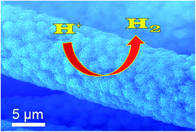
Developing high-efficiency and stable catalysts from earth-abundant elements for the hydrogen evolution reaction (HER) is essential for renewable energy conversion. In this work, Se-promoted molybdenum sulfide nanosheet arrays supported on flexible carbon cloth (Se–MoS2/CC) have been successfully synthesized and explored for the first time as a 3D hydrogen evolution cathode. Without any active process, the Se–MoS2/CC electrode exhibits greatly enhanced activity with a smaller Tafel slope (63 mV dec−1) and a higher exchange current density (0.16 mA cm−2) than the pristine MoS2/CC one (79 mV dec−1 and 0.1 mA cm−2). The overpotentials needed to attain the current densities of 10 and 100 mA cm−2 are merely 127 and 218 mV, respectively. In addition, Se–MoS2/CC maintains its high electrocatalytic activity for at least 25 h in acidic media.


Hydrogen is considered as a clean and renewable energy resource in the hydrogen-economy paradigm.1 The electrolysis of water is one of the easiest and cleanest routes to produce highly pure hydrogen via hydrogen evolution reaction (HER).2,3 However, the use of Pt, as the best HER electrocatalyst, has been prohibited due to its high expense and scarcity.4–6 Such a limitation has motivated great efforts to design and fabricate Pt-free catalysts from earth-abundant elements with high catalytic activity for HER.

In recent years, both density functional theory (DFT) and experimental results have confirmed that MoS2 is a promising Pt-free HER electrocatalyst and its activity predominately originates from the S-edges.7,8 In order to maximally exposed S-edges and strikingly enhanced the catalytic activity of MoS2, great efforts have been focused on the growth of MoS2 nanostructures.9–11 For instance, defective MoS2 nanosheets decorated with Au nanoparticles,12 defect-rich MoS2 nanosheets,13 mesoporous MoS2 architecture,14 vertically aligned layers of MoS2,15 ultrathin MoS2 nanoplates,16 low crystalline MoS2 nanosheet,17 as well as MoS2 quantum dots are used to obtain good catalytic performance.18 It is confirmed that utilizing precious-metal-free elements can also improve the HER catalytic activity of MoS2. Such as Ni, Co, Fe and V can significantly promote the catalytic performance of MoS2.19,20 Se atom, as the same group to S, is more metallic nature than S, thus the Se doping into MoS2 can improve the electrical conductivity and further promote the HER catalytic activity of MoS2.21

Generally, the preparation of catalysts for HER measurements requires multi-step procedures, including mixing the catalysts with polymer binders (such as Nafion), and then immobilizing the mixture on electrode surfaces. Unfortunately, the utilization of such electrical insulating polymer binders might decrease the electrical conductivity and block catalytic active sites, all of which significantly reduce the effective catalytic activity.22–24 Such problems could be avoided by developing hydrogen evolution cathodes via growing the active materials onto current collectors directly.25 Carbon cloth (CC) is a cheap, conductive and flexible carbon microfiber, not only provides a 3D substrate, but also has poor electrocatalytic activity and thus could provide a clean background for exhibiting the HER activity of supported catalysts.26

Herein, differently from the previous report, we have rationally designed and synthesized novel Se-promoted molybdenum sulfide nanosheet arrays supported on flexible carbon cloth (Se–MoS2/CC) through a two-step strategy: to hydrothermally growing MoS2 nanosheet arrays on CC (MoS2/CC) first, and then hydrothermally converting the MoS2/CC precursor into Se–MoS2/CC using NaHSe as the Se source. As a flexible electrode for HER, Se–MoS2/CC reveals enhanced electrocatalytic activity with a lower onset overpotential and smaller Tafel slops as well as a larger exchange current density (j0) than that of pure MoS2/CC.

Fig. 1a presents the X-ray diffraction (XRD) patterns of Se–MoS2/CC, MoS2/CC and blank CC. For MoS2/CC, four diffraction peaks around 2θ = 14.2°, 32.8°, 39.7° and 58.3° are assigned to the (002), (100), (103) and (110) planes of MoS2, respectively,27 in good agreement with the standard pattern of hexagonal MoS2 (JPCDS no. 37-1492). After hydrothermal selenization treatments, the diffraction peaks slightly shift toward smaller diffraction angles, suggesting that the addition of Se increases the interplanar distance.27 In addition, as shown in Fig. 1b, the peaks of Se–MoS2/CC (002) are weaker and broader, suggesting that the reduced grain size (according to Scherrer equation).28 For both samples, the peaks at 2θ = 26° and 43° can be assigned to the (002) and (101) planes of the CC substrate,29 respectively. Scanning electron microscopy (SEM) images of blank CC (Fig. S1) indicate it consists of smooth microfibers with an average diameter of about 10 μm. SEM images of MoS2/CC (Fig. 1c) show the full coverage of all fibers with MoS2 nanosheets interconnected with each other to form a network-like array and such nanosheets have obvious ripples and corrugations. Followed by selenization, the basic morphology is still maintained (Fig. 1d). In addition, the synthesized Se–MoS2/CC represents a high flexibility and can even endure folding and twisting without destroying its construction (Fig. S2). The energy dispersive X-ray (EDX) spectrum (Fig. S3) confirms the presence of elemental C, Mo, S and Se. The Transmission electron microscopy (TEM) image (Fig. 1e) further reveals its structure is nanosheet. A representative high-resolution TEM (HRTEM) image (Fig. 1f) of Se–MoS2 clearly exhibits well-defined lattice fringes with an interplane spacing of 0.66 nm, which is larger than that of pristine MoS2 (Fig. S4, 0.61 nm), as witnessed by the XRD results. SEM image and EDX elemental mapping images (Fig. 2a–d) show that all elements (Mo, S and Se) in Se–MoS2/CC are uniformly distributed in the whole microfiber. These results suggest the successful synthesis of Se–MoS2/CC by a two-step method.


image file: c5ra28078h-f1.tif
Fig. 1 (a) XRD patterns of blank CC, MoS2/CC and Se–MoS2/CC, (b) the enlarged (002) plane of MoS2/CC and Se–MoS2/CC. SEM images of (c) MoS2/CC and (d) Se–MoS2/CC. (e) TEM and (f) HRTEM images of Se–MoS2 nanosheets.

image file: c5ra28078h-f2.tif
Fig. 2 (a) SEM image and EDX elemental mapping images of (b) Mo, (c) S and (d) Se for Se–MoS2/CC.

X-ray photoelectron spectroscopy (XPS) analyses were performed to elucidate the valence states of Mo, S and Se in Se–MoS2/CC. The XPS survey spectrum of Se–MoS2/CC (Fig. 3a) shows the peaks of Mo 3d, S 2p, Se 3d, C 1s and O 1s signals. Fig. 3b–d present the high-resolution XPS spectra of Mo 3d, S 2p and Se 3d. The binding energies of Mo 3d5/2 and Mo 3d3/2 are 229.3 and 232.5 eV, respectively, which can be assigned to Mo4+.26 The high-resolution spectrum of S 2p shows two peaks at 163.4 and 162.2 eV assigned to S 2p1/2 and S 2p3/2, respectively, confirming the formation of S2−.26 The Se 3d peak is split into well-defined 3d5/2 and 3d3/2 peaks at 54.8 and 55.5 eV,30 a well known characteristic of substitutional doping of Se to S, in comparison of S occupying the interstitial sites of MoS2.27 The quantification analysis of the Se 3d and S 2p peaks gives the Se doping concentration (∼6.6%) in MoS2.


image file: c5ra28078h-f3.tif
Fig. 3 (a) XPS survey spectrum of Se-doped MoS2/CC. XPS spectra in the (b) Mo 3d, (c) S 2p and (d) Se 3d regions for Se–MoS2/CC.

Without any active process, MoS2/CC and Se–MoS2/CC were directly investigated as the working electrodes in a typical three-electrode cell using H2-saturated 0.5 M H2SO4 solution at room temperature. As a reference, the blank CC and the 20 wt% commercial Pt/C were also tested. Fig. 4a and b show the HER polarization curves. As expected, the blank CC shows very limited HER activity, while the commercial Pt/C displays superior HER performance with a negligible overpotential (η). It is noted that MoS2/CC shows intrinsic electrocatalytic performance toward HER with an onset overpotential of near 72 mV and significant H2 evolution (j = 10 mA cm−2) can not be achieved until an overpotential of 152 mV. In contrast, the Se–MoS2/CC electrode exhibits much superior catalytic performance over MoS2/CC with a smaller onset overpotential (60 mV). Moreover, the overpotentials required to drive the current densities of 10 and 100 mA cm−2 are 127 ± 2 and 218 ± 2 mV (mean s.d.), respectively. This can be confirmed by measuring five Se–MoS2/CC electrodes under same conditions. Table S1 lists HER activity parameters of Mo-based HER catalysts in acidic solutions. Interestingly, Se–MoS2/CC shows a high catalytic activity comparable with other catalysts. We further confirmed that the selenization time of 8 h (Se doping concentration is 6.4%, which is consistent within experimental error with the XPS result (Table S2)) is optimal for HER activity of Se–MoS2/CC (Fig. S5).


image file: c5ra28078h-f4.tif
Fig. 4 (a and b) Polarization curves of Se–MoS2/CC, MoS2/CC, Pt/C and blank CC in 0.5 M H2SO4 with a scan rate of 2 mV s−1. (c) Corresponding Tafel plots. (d) Nyquist plots of Se–MoS2/CC and MoS2/CC recorded at 0 V vs. RHE in 0.5 M H2SO4.

Tafel plots are useful for quantitative kinetic analysis of HER.8 As shown in Fig. 4c, the Tafel slope for Pt/C is 30 mV dec−1.31,32 The Tafel slope of Se–MoS2/CC is 63 mV dec−1 in the region η = 80–180 mV, suggesting that HER proceeds by a Volmer–Heyrovsky mechanism.33–35 MoS2/CC has a higher Tafel slope (79 mV dec−1) than Se–MoS2/CC. The smaller Tafel slope is advantageous in practical application as it implies a faster increase in reaction rate with increasing the overpotential.36 By extrapolating the Tafel plot, the j0 value of Se–MoS2/CC (0.16 mA cm−2) is about 1.6 times of that of MoS2/CC (0.1 mA cm−2) (Fig. S6). The earlier onset overpotential and smaller Tafel slope as well as larger j0 clearly demonstrate that the catalytic activity of the Se–MoS2/CC is better than that of the MoS2/CC.

The enhanced catalytic activity of Se–MoS2/CC could be explained as follows facts: first, the incorporation of Se into MoS2 reduces the average grain size, resulting in more active edge sites,27 as witnessed by the XRD pattern. Second, Se atoms are more metallic nature than S, thus the Se doped into MoS2 can improve the electrical conductivity.37 As shown in Fig. 4d, Nyquist plots and data fittings to a simplified Randles circuit reveal that the charge transfer resistance (Rct) value of Se–MoS2/CC (143 Ω) is much lower than MoS2/CC (302 Ω). This result indicates that Se–MoS2/CC has better electronic and ionic conductivity than MoS2/CC, which contributes to the greatly enhanced HER activity.

For practical application the durability of HER electrocatalyst is another crucial factor. The accelerated stability tests of the Se–MoS2/CC electrode were probed by continuous cyclic voltammetry (CV) sweeping from −0.27 to +0.08 V vs. RHE with a scan rate of 100 mV s−1. As shown in Fig. 5a, after 2000 CV cycles, this electrode exhibits a similar polarization curve to the initial one with a negligible current loss. Moreover, to deliver a current density of 20 mA cm−2 (without iR correction), the potential is slightly decayed (∼31 mV from 0.145 to 0.176 V) over 25 h testing (Fig. 5b). It should be noted that the structure of the Se–MoS2/CC can be well preserved after 25 h durability tests (Fig. S7). These observations demonstrate the excellent stability of the 3D flexible Se–MoS2/CC electrode for the HER in acidic solutions.


image file: c5ra28078h-f5.tif
Fig. 5 (a) Polarization curves recorded for Se–MoS2/CC before and after 2000 CV cycles between −0.27 and +0.08 V vs. RHE. (b) Long-term stability test for Se–MoS2/CC carried out under a constant current density of 20 mA cm−2 (without iR correction).

In conclusion, we have developed a two-step hydrothermal strategy to prepare a highly active and stable 3D flexible Se–MoS2/CC hierarchical electrode, which is facile and easily scale-up. When used as the integrated 3D hydrogen evolution cathode, the Se–MoS2/CC shows excellent activity and durability in acidic solution. The facile, low-cost scalable fabrication process, together with its superior activity and long-term electrochemical stability of this flexible 3D electrode, promises its application toward large-scale production of hydrogen by water splitting.

Acknowledgements

This work was supported by the National Natural Science Foundation of China (51372186), and the open Foundation of Jiangxi Key Laboratory of Advanced Copper and Tungsten Materials (2013-KLP-05).

Notes and references

  1. T. R. Cook, D. K. Dogutan, S. Y. Reece, Y. Surendranath, T. S. Teets and D. G. Nocera, Chem. Rev., 2010, 110, 6474–6502 CrossRef CAS PubMed.
  2. M. Dresselhaus and I. Thomas, Nature, 2001, 414, 332–337 CrossRef CAS PubMed.
  3. M. G. Walter, E. L. Warren, J. R. McKone, S. W. Boettcher, Q. Mi, E. A. Santori and N. S. Lewis, Chem. Rev., 2010, 110, 6446–6473 CrossRef CAS PubMed.
  4. Y. Hara, N. Minami, H. Matsumoto and H. Itagaki, Appl. Catal., A, 2007, 332, 289–296 CrossRef CAS.
  5. M. Wu, P. K. Shen, Z. Wei, S. Song and M. Nie, J. Power Sources, 2007, 166, 310–316 CrossRef CAS.
  6. J. Greeley, T. F. Jaramillo, J. Bonde, I. Chorkendorff and J. K. Nørskov, Nat. Mater., 2006, 5, 909–913 CrossRef CAS PubMed.
  7. B. Hinnemann, P. G. Moses, J. Bonde, K. P. Jorgensen, J. H. Nielsen, S. Horch, I. Chorkendorff and J. K. Nørskov, J. Am. Chem. Soc., 2005, 127, 5308–5309 CrossRef CAS PubMed.
  8. T. F. Jaramillo, K. P. Jorgensen, J. Bonde, J. H. Nielsen, S. Horch and I. Chorkendorff, Science, 2007, 317, 100–102 CrossRef CAS PubMed.
  9. Z. Lu, W. Zhu, X. Yu, H. Zhang, Y. Li, X. Sun, X. Wang, H. Wang, J. Wang, J. Luo, X. Lei and L. Jiang, Adv. Mater., 2014, 26, 2683–2687 CrossRef CAS PubMed.
  10. Y. Yan, B. Xia, Z. Xu and X. Wang, ACS Catal., 2014, 4, 1693–1705 CrossRef CAS.
  11. P. D. Tran, S. S. Pramana, V. S. Kale, M. Nguyen, S. Y. Chiam, S. K. Batabyal, L. H. Wong, J. Barber and J. Loo, Chem.–Eur. J., 2012, 18, 13994–13999 CrossRef CAS PubMed.
  12. J. Kim, S. Byun, A. J. Smith, J. Yu and J. X. Huang, J. Phys. Chem. Lett., 2013, 4, 1227–1232 CrossRef CAS PubMed.
  13. J. Xie, H. Zhang, S. Li, R. Wang, X. Sun, M. Zhou, J. Zhou, X. W. Lou and Y. Xie, Adv. Mater., 2013, 25, 5807–5813 CrossRef CAS PubMed.
  14. J. Kibsgaard, Z. Chen, B. N. Reinecke and T. F. Jaramillo, Nat. Mater., 2012, 11, 963–969 CrossRef CAS PubMed.
  15. D. S. Kong, H. T. Wang, J. J. Cha, M. Pasta, K. J. Koski, J. Yao and Y. Cui, Nano Lett., 2013, 13, 1341–1347 CrossRef CAS PubMed.
  16. Y. Yan, B. Xia, X. Ge, Z. Liu, J. Wang and X. Wang, ACS Appl. Mater. Interfaces, 2013, 5, 12794–12798 CAS.
  17. Y. Yan, X. Ge, Z. Liu, J. Wang, J. Lee and X. Wang, Nanoscale, 2013, 5, 7768–7771 RSC.
  18. X. Ren, L. Pang, Y. Zhang, X. Ren, H. Fan and S. F. Liu, J. Mater. Chem. A, 2015, 3, 10693–10697 CAS.
  19. J. Bonde, P. G. Moses, T. F. Jaramillo, J. K. Nørskov and I. Chorkendorff, Faraday Discuss., 2008, 140, 219–231 RSC.
  20. X. Sun, J. Dai, Y. Guo, C. Wu, F. Hu, J. Zhao, X. Zeng and Y. Xie, Nanoscale, 2014, 6, 8359–8367 RSC.
  21. X. Ren, Q. Ma, H. Fan, L. Pang, Y. Zhang, Y. Yao, X. Ren and S. Liu, Chem. Commun., 2015, 51, 15997–16000 RSC.
  22. A. I. Carim, F. H. Saadi, M. P. Soriaga and N. S. Lewis, J. Mater. Chem. A, 2014, 2, 13835–13839 CAS.
  23. D. Kong, H. Wang, Z. Lu and Y. Cui, J. Am. Chem. Soc., 2014, 136, 4897–4900 CrossRef CAS PubMed.
  24. J. D. Roy-Mayhew, G. Boschloo, A. Hagfeldt and I. A. Aksay, ACS Appl. Mater. Interfaces, 2012, 4, 2794–2800 CAS.
  25. J. Tian, Q. Liu, A. M. Asiri and X. Sun, J. Am. Chem. Soc., 2014, 136, 7587–7590 CrossRef CAS PubMed.
  26. Y. Yan, B. Xia, N. Li, Z. Xu, A. Fisherc and X. Wang, J. Mater. Chem. A, 2015, 3, 131–135 CAS.
  27. Q. Liu, J. Tian, A. M. Asiri and X. Sun, Angew Chem., Int. Ed., 2014, 53, 6710–6714 CrossRef CAS PubMed.
  28. J. I. Langford and A. J. C. Wilson, J. Appl. Crystallogr., 1978, 11, 102–113 CrossRef CAS.
  29. H. Li, X. Wang, B. Ding, G. Pang, P. Nie and L. Shen, ChemElectroChem, 2014, 1, 1118–1125 CrossRef CAS.
  30. H. Tang, K. P. Dou, C. C. Kaun, Q. Kuang and S. H. Yang, J. Mater. Chem. A, 2014, 2, 360–364 CAS.
  31. K. Chang, D. Geng, X. Li, J. Yang, Y. Tang, M. Cai, R. Li and X. Sun, Adv. Energy Mater., 2013, 3, 839–844 CrossRef CAS.
  32. E. J. Popczun, J. R. McKone, C. G. Read, A. J. Biacchi, A. M. Wiltrout, N. S. Lewis and R. E. Schaak, J. Am. Chem. Soc., 2013, 135, 9267–9270 CrossRef CAS PubMed.
  33. Y. F. Xu, M. R. Gao, Y. R. Zheng, J. Jiang and S. H. Yu, Angew. Chem., Int. Ed., 2013, 52, 8546–8550 CrossRef CAS PubMed.
  34. Z. Pu, Q. Liu, C. Tang, A. M. Asiri and X. Sun, Nanoscale, 2014, 6, 11031–11034 RSC.
  35. Z. Pu, Q. Liu, A. M. Asiri and X. Sun, ACS Appl. Mater. Interfaces, 2014, 6, 21874–21879 CAS.
  36. D. Kong, H. Wang, Z. Lu and Y. Cui, J. Am. Chem. Soc., 2014, 136, 4897–4900 CrossRef CAS PubMed.
  37. H. Fei, Y. Yang, X. Fan, G. Wang, G. Ruan and J. Tour, J. Mater. Chem. A, 2015, 3, 5798–5804 CAS.

Footnote

Electronic supplementary information (ESI) available: Experimental section and figures. See DOI: 10.1039/c5ra28078h

This journal is © The Royal Society of Chemistry 2016
Click here to see how this site uses Cookies. View our privacy policy here.