DOI:
10.1039/C5RA28037K
(Paper)
RSC Adv., 2016,
6, 21119-21126
Tuning the band-gap of h-boron nitride nanoplatelets by covalent grafting of imidazolium ionic liquids†
Received
30th December 2015
, Accepted 16th February 2016
First published on 16th February 2016
Abstract
Imidazolium ionic liquids with three different anions, viz. bis(salicylato)borate (BScB), oleate (OL) and hexafluorophosphate (PF6), are covalently grafted on h-boron nitride nanoplatelets (h-BNNPs) to probe the shifts in the band gap energy. The grafting of ionic liquids on h-BNNPs was confirmed by FTIR, XPS, TGA and EDX analyses, whereas XRD and HRTEM results suggested that the crystalline and layering structure of h-BNNPs remained intact after covalent grafting of ionic liquids. The characteristic band gap energy of h-BNNPs (5.9 eV) was reduced to 5.78, 5.25 and 5.00 eV in h-BNNPs-OL, h-BNNPs-PF6 and h-BNNPs-BScB, respectively. The band gap of h-BNNPs-ILs is controlled by charge transfer between the ionic liquids and h-BNNPs, and exhibited strong correlation with the chemical structure of the associated anion in h-BNNPs-ILs.
1. Introduction
The emergent phenomenon of graphene has inspired researchers to exploit other two-dimensional nanomaterials, including hexagonal boron nitride nanosheets (h-BNNs), for fundamental understanding to application perspectives.1 The h-BNNs exhibit remarkably higher chemical stability and resistance to oxidation compared to that of graphene.2 In h-BNNs, heat transfer mainly takes place via phonons and shows excellent thermal conductivity along the basal plane.3 These intrinsic properties, including excellent mechanical strength, promise the potential of h-BNNs for several applications such as ultraviolet nanoelectronics, luminescence, electron field emission, polymeric composites, thermo-conductive fluids, lubrication etc.4–6 Unlike graphene, h-BNNs exhibit wide band gap (∼6 eV).7 The functionalization and doping of h-BNNs is one of the most promising approach to adjust the electronic structure. Recently, Golberg et al. demonstrated that electronic structure of h-BN nanotubes can be changed by their covalent functionalization.8 Further, the first-principle calculations revealed that energy gap can be adjusted by varying concentration and changing the electro negativity of the functionalized species.
The h-BN nanosheets and nanotubes have limited avenues for their chemical functionalization compared to that of graphene. The defect and edge sites in h-BN nanotubes are considered to be most promising sites for the chemical functionalization.9 The electron-deficient boron at defect and edge sites of h-BN acts as Lewis acid. These sites are prone to interact with electron-rich Lewis bases such as amino terminated poly(ethylene glycol), trialkylamines and trialkylphosphines.10 Recently, boron sites of h-BN skeleton were oxidized using H2O2, HNO3/H2SO4, oleum, HNO3/H2SO4/KMnO4 to generate hydroxyl functionalities for grafting of organic species.6d,11 Ionic liquids have been established as green medium for preparation and stabilization of various nanomaterials.12 Excellent thermal and chemical stability, inherent polarity, negligible volatility and high conductivity of ionic liquids offers several advantages over the conventional solvents, surfactants and stabilizers.13 The h-BN nanotubes induced a new phase in imidazolium ionic liquids and form the gel structure driven by non-covalent cation–π interactions.14 Molecular dynamics studies revealed that anions in the ionic liquids have a stronger interaction with h-BN surface than imidazolium, pyridinium and ammonium cations. Furthermore, thermo-chemical analysis revealed that the free energy of adsorption of ionic liquids on h-BN surface is negative, and thus, the adsorption occurs spontaneously.15 The modification of h-BN surface with ionic liquids via covalent interaction could provides novel optical properties, which can be controlled by tailoring the chemical structure of ionic liquids on the h-BNNs surface. Herein, for the first time, we present systematic approach for covalent grafting of imidazolium ionic liquids on the h-boron nitride nanoplatelets (h-BNNPs). The changes in band gap energy and optical properties of ionic liquids-grafted h-BNNPs (h-BNNPs-ILs) surface are examined by UV-vis absorption measurements as a function of chemical structure of associated anions.
2. Experimental
2.1. Materials
1-Methylimidazole (99%, Sigma Aldrich), N-methyl-2-pyrrolidone (>99.5%, Merck Chemicals), 3-chloropropyltrimethoxysilane (CPTMS, 97%, Alfa Aesar), boric acid (99.5%, Loba Chemie), lithium carbonate (99.9%, Sigma Aldrich), salicylic acid (99.8%, Sigma Aldrich) and sodium oleate (90%, Loba Chemie) were used as received. The h-BN powder was procured from MK Impex, Canada.
2.2. Grafting of ionic liquids on h-BNNPs
2.2.1. Preparation of h-BNNPs. The h-BN powder with average particle size of 70 nm was dispersed in the N-methyl-2-pyrrolidone (NMP) and then exfoliated into nanoplatelets using ultrasonic probe (power: 500 W, intensity: 35%, ultrasonic probe diameter: 13 mm, ultrasonication time: 90 minutes). The resultant dispersion was kept overnight to settle down the non-exfoliated and bigger size contents of h-BN particles. The supernatant having nicely dispersed fine platelets/sheets of h-BN in NMP was decanted and then centrifuged at 4000 rpm for 20 minutes. The deposited h-BNNPs in the centrifuge tubes was collected and then washed with ethanol to remove the NMP traces.
2.2.2. Oxidation of h-BNNPs. The h-BNNPs was oxidized using a mixture of strong oxidizing reagents (H2SO4, NaNO3, KMnO4) to generate the oxygen functionalities on the defect and edge sites of h-BNNPs (Scheme 1a and b). In a typical exercise, 200 mg h-BNNPs powder, 200 mg NaNO3 and 9 mL H2SO4 were mixed in a reaction vessel. The 3 g KMnO4 was then added into the reaction vessel under the continuous stirring. Owing to explosive nature of this reaction, KMnO4 was added very slowly and the reaction vessel was kept on the ice bath. This was followed by stirring of reaction mixture for 24 hours at room temperature. Subsequently, 16 mL of distilled water was added gradually to the reaction mixture and then the temperature of reaction vessel was raised to 98 °C using an oil bath. The reaction mixture was cool down after 48 hours and 4 mL of 30% H2O2 was added in the reaction mixture. This was followed by washing of oxidized product (ox-h-BNNPs) with distilled water. The wet cake of ox-h-BNNPs collected by centrifugation was rinsed with 10 mL of 3.5% HCl. Then the ox-h-BNNPs was repeatedly washed with distilled water, until neutral pH was attained. The ox-h-BNNPs was finally rinsed with ethanol and dried in the oven at 70 °C.
 |
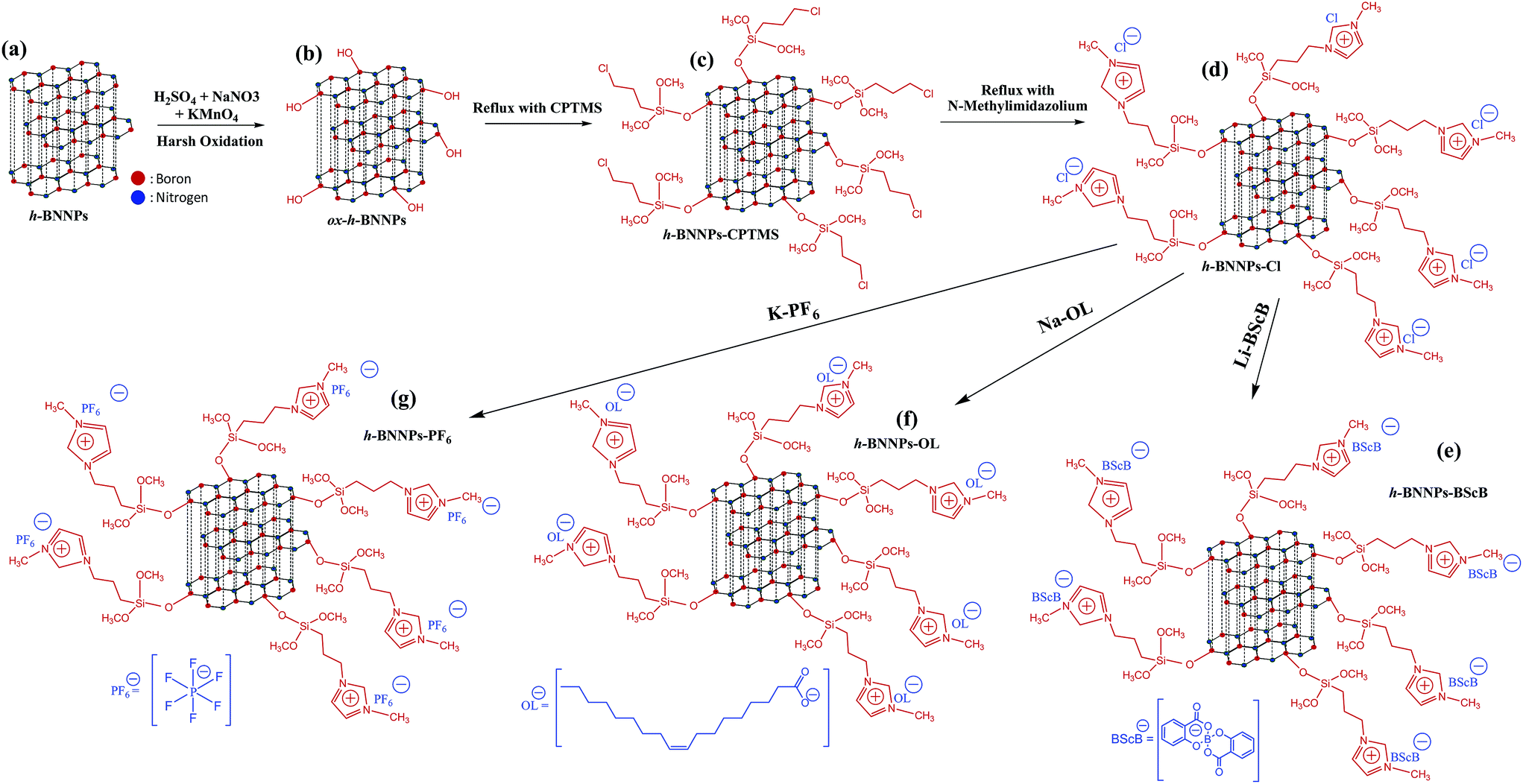
| | Scheme 1 Schematic models of (a) h-BNNPs and (b) ox-h-BNNPs having hydroxyl groups on edge and defect sites of boron. (c) CPTMS was grafted on ox-h-BNNPs by chemical interaction between methoxy groups of CPTMS and hydroxyl groups of ox-h-BNNPs. (d) 1-Methylimidazole was grafted on CPTMS-modified h-BNNPs via covalent linkage and eliminated chloro groups of CPTMS are associated to the imidazolium ring as chloride anion. In the final step, chloride anion of h-BNNPs-Cl was replaced by (e) BScB, (f) OL and (g) PF6 anions via metathesis reaction with Li-BScB, Na-OL and K-PF6 salts, respectively. | |
2.2.3. Preparation of h-BNNPs-ILs. The ox-h-BNNPs was dispersed in toluene using an ultrasonic bath and the dispersion was purged with nitrogen for 30 minutes. This was followed by addition of CPTMS as a bifunctional chemical linker. The reaction mixture was refluxed under N2 atmosphere for 24 hours. Herein, the methoxy groups of CPTMS are prone to interact with hydroxyl functionality of ox-h-BNNPs via covalent interaction as shown in Scheme 1b and c. The developed reaction product (h-BNNPs-CPTMS), having chloro group as terminal site, was washed several times with ethanol to remove the non-reacted content of CPTMS. In the subsequent step, chloro-terminal of h-BNNPs-CPTMS was targeted to prepare the imidazolium ionic liquid. In this context, the dispersion of h-BNNPs-CPTMS in toluene was refluxed with 1-methylimidazole for 36 hours under uninterrupted stirring. In this reaction, 1-methylimidazole replaced the chloro-terminal of h-BNNPs-CPTMS and attached as imidazolium cation. The eliminated chloro group retained as chloride anion as shown in the Scheme 1c and d. The developed product (h-BNNPs-Cl) was then washed with toluene and ethanol to remove the un-reacted contents. This was followed by replacement of chloride anion in h-BNNPs-Cl by oleate, bis(salicylato)borate and hexafluorophosphate anions via metathesis reaction (Scheme 1d–g) using sodium-oleate, lithium-bis(salicylato)borate, and potassium-hexafluorophosphate salts, respectively. In this context, the aqueous dispersion of h-BNNPs-Cl was mixed with aqueous solution of sodium oleate and the reaction mixture was stirred at 50 °C for 12 hours. The developed product grafted with imidazolium oleate ionic liquid (h-BNNPs-OL) was washed with water and ethanol in subsequent order and then dried in oven at 60 °C. Likewise, an aqueous dispersion of h-BNNPs-Cl was reacted with lithium-bis(salicylato)borate and potassium-hexafluorophosphate salts (individually) to obtained bis(salicylato)borate and hexafluorophosphate anion associated h-BNNPs, respectively. The imidazolium ionic liquids-grafted h-BNNPs having oleate, bis(salicylato)borate and hexafluorophosphate anions were designated as h-BNNPs-OL, h-BNNPs-BScB and h-BNNPs-PF6 respectively, throughout the manuscript.
2.3. Chemical and structural characterization
FTIR spectra of all samples were recorded using a Thermo-Nicolet 8700 research spectrometer with a spectral resolution of 4 cm−1. Each sample with known quantity was mixed with KBr to prepare the pellets for FTIR measurements. X-ray Photoelectron Spectra (XPS) of all samples were collected using ESCA 3400 spectrometer (Kratos Analytical Ltd). The peak fitting of all XPS spectra was carried out through Gaussian–Lorentzian function after performing a Shirley background correction. Diffraction patterns of all samples were collected for 2θ of 2–80° using Bruker D8 advance diffractometer operated at 40 kV and 40 mA with Cu Kα radiation of 0.15418 nm wavelength. The microstructural features and elemental distribution of all samples were probed using scanning electron microscope (SEM) coupled with energy dispersive X-ray (EDX) spectroscopy using a Quanta-200F (FEI) electron microscope. High resolution transmission electron microscopic (HR-TEM) images of h-BNNPs and h-BNNPs-ILs were collected to probe their nanoscopic features including layering structure.
2.4. Optical properties of h-BNNPs-ILs
The UV-visible absorption spectra of all samples were collected using a Shimadzu UV-2600 spectrophotometer. The band gap energy of each sample was extracted by Tauc plot of UV-visible absorption results. The extrapolating of distinct linear regime of the absorption edge to the x-axis yielded the band gap energy of the sample.
3. Results and discussion
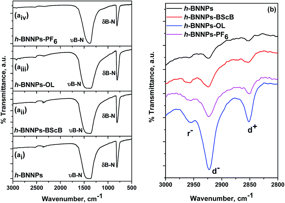
The imidazolium ionic liquids having three different anions viz. BScB, OL and PF6− were grafted on the h-BNNPs to probe their effect on optical properties of pristine h-BNNPs. The changes in the chemical features owing to grafting of imidazolium ionic liquids were examined by FTIR and XPS analyses. The vibrational spectra (Fig. 1ai–iv) of h-BNNPs and h-BNNPs-ILs exhibited two strong peaks at 809 and ∼1390 cm−1, revealing the B–N linkages of h-BNNPs skeleton. The vibrational modes in the range of 3000–2800 cm−1 (Fig. 1b) were attributed to C–H stretches of methylene and methyl units and confirmed the grafting of bifunctional CPTMS linker and imidazolium ring in the h-BNNPs-ILs (h-BNNPs-BScB, h-BNNPs-PF6 and h-BNNPs-OL). A higher intensity of C–H stretches in h-BNNPs-OL was attributed to longer chain (higher methylene units) of oleate anion.
 |
| | Fig. 1 FTIR spectra of (ai–iv) h-BNNPs and h-BNNPs-ILs samples in the range of 3000–500 cm−1, representing the characteristic modes of B–N. (b) An expanded region of 3000–2800 cm−1 shows C–H stretches attributed to the methylene units of CPTMS and imidazolium ring in the h-BNNPs-ILs. | |
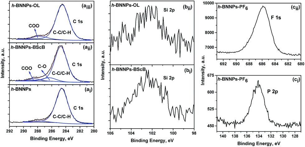
Fig. 2 shows high-resolution B 1s and N 1s XPS spectra of h-BNNPs and h-BNNPs-ILs. The strong abundance of B 1s and N 1s peaks at 190.7 and 398.3 eV, respectively, demonstrate the h-BN skeleton in all samples. The B 1s spectrum could be deconvoluted into two components at 190.7 and 192.2 eV. The peak component at 190.7 eV with very strong intensity was attributed to the B–N linkage of core skeleton, whereas peak at 192.2 eV having very low intensity was assigned to the B–O functionality in the h-BNNPs-ILs samples. The presence of B–OH groups on the edge and defect sites in the h-BN scaffold, prepared by harsh oxidation, contributes to the B–O peak component. The preparation of ionic liquids thin films on the h-BNNPs was confirmed by the appearance of C 1s and Si 2p peaks as shown in Fig. 3. The deconvoluted peak component of C 1s spectrum (Fig. 3ai) at higher binding energy having very low intensity, suggested the adsorption of environmental contamination on the h-BNNPs. The chemically shifted deconvoluted peak components of C 1s spectrum at higher binding energy revealed the presence of carboxylate groups in the h-BNNPs-OL owing to oleate anion (Fig. 3aiii) and presence of carbonyl and carboxylate functionalities in the h-BNNPs-BScB because of BScB anion (Fig. 3aii). The significant increased of % atomic concentration of carbon in h-BNNPs-OL further support the grafting of oleate anion (Table 1). The increased ratio of B/N = 1.083 in h-BNNPs-BScB (Table 1) compared to that of h-BNNPs (B/N = 1.057), revealed the grafting of boron based BScB ionic liquid. The P 2p and F 1s peaks at 134.1 and 686.0 eV, respectively (Fig. 3ci–ii), were assigned to the P–F linkage of PF6− anion of h-BNNPs-PF6. These results suggested the grafting of ionic liquids on the h-BNNPs via covalent interaction.
 |
| | Fig. 2 B 1s XPS spectra of (ai) h-BNNPs, (aii) h-BNNPs-BScB, (aiii) h-BNNPs-OL and (aiv) h-BNNPs-PF6 samples. N 1s spectra of (bi) h-BNNPs, (bii) h-BNNPs-BScB, (biii) h-BNNPs-OL and (biv) h-BNNPs-PF6 samples. | |
 |
| | Fig. 3 High resolution (ai–aiii) C 1s XPS spectra of h-BNNPs, h-BNNPs-BScB and h-BNNPs-OL, (bi and bii) Si 2p XPS spectra of h-BNNPs-BScB and h-BNNPs-OL samples. (ci) P 2p and (cii) F 1s XPS spectra of h-BNNPs-PF6. The deconvoluted components of C 1s spectra illustrate chemical linkages to various types of functionalities in the h-BNNPs and h-BNNPs-ILs. | |
Table 1 The % atomic concentration of h-BNNPs and ionic liquids-grafted h-BNNPs, based on the XPS results
| Sample description |
% atomic concentration |
B/N |
| B |
N |
C |
O |
Si |
P |
F |
| h-BNNPs |
40.5 |
38.3 |
13.6 |
7.6 |
— |
— |
— |
1.057 |
| h-BNNPs-BScB |
39.2 |
36.2 |
16.5 |
7.6 |
0.5 |
— |
— |
1.083 |
| h-BNNPs-PF6 |
38.4 |
36.2 |
11.8 |
7.8 |
0.5 |
0.9 |
4.5 |
1.061 |
| h-BNNPs-OL |
34.5 |
32.7 |
24.4 |
8.1 |
0.4 |
— |
— |
1.055 |
The thermo-gravimetric analyses of h-BNNPs and h-BNNPs-ILs were carried out to probe their thermal decomposition property (Fig. 4). The h-BNNPs showed 2.48 wt% loss up to 700 °C and was attributed to absorbed contamination from the environment. The h-BNNPs-ILs exhibited 4.7 to 6.5 wt% loss up to 700 °C and is variable with grafted anion. The higher wt% loss of material in the h-BNNPs-ILs compared to that of h-BNNPs reveals the thermal decomposition of chemically grafted ionic liquids.
 |
| | Fig. 4 Thermo-gravimetric patterns of h-BNNPs, h-BNNPs-BScB, h-BNNPs-OL and h-BNNPs-PF6 over the temperature range of 25–700 °C under nitrogen flow. Inset graph reveals the % weight loss of h-BNNPs and h-BNNPs-ILs at 700 °C. | |
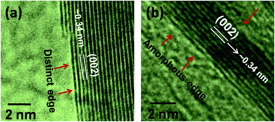
The XRD patterns (Fig. 5) of h-BNNPs and h-BNNPs-ILs depict characteristic diffraction peaks corresponding to the (002), (100), (101), (102), (004), and (110) planes. These patterns revealed that after grating of ionic liquids, crystalline layered structure of h-BNNPs remained intact. The TEM images (Fig. 6) suggested the platelets structure of h-BN with lateral thickness of 5–40 nm. These h-BNNPs are prepared by exfoliation of h-BN powder using ultrasonic probe. No changes were observed in lamellar structure of h-BNNPs after grafting of ionic liquids. The high-resolution images (Fig. 7) of h-BNNPs and h-BNNPs-ILs explicitly demonstrate the stacking of h-BN atomic lamellae with interlayer spacing of ∼0.34 nm in (002) crystal plane and is further corroborated by XRD results. Fig. 7a displays distinct boundary of h-BNNPs lamellae, whereas a representative high-resolution TEM image (Fig. 7b) of h-BNNPs-OL shows the amorphous edges and are indicated by arrows. The amorphous edges in h-BNNPs-OL could be attributed to the chemically grafted ionic liquid thin film. The elemental analysis based on EDX measurements coupled with SEM provides elemental detail of h-BNNPs-ILs. The homogeneous distribution of Si, C, O besides the core element B and N in h-BNNPs-BScB (Fig. 8 and S1, ESI†) suggested the uniform grafting of imidazolium-BScB ionic liquid on the h-BNNPs skeleton via silane linker. Likewise, regular distribution of characteristic elements Si, P, F, C, O in h-BNNPs-PF6 (Fig. 8b and S2 ESI†) and Si, C, O in h-BNNPs-OL (Fig. S3 ESI†) besides the B and N revealed the homogeneous grafting of imidazolium-PF6 and imidazolium-OL ionic liquids, respectively.
 |
| | Fig. 5 Powder XRD patterns of h-BNNPs, h-BNNPs-BScB, h-BNNPs-OL and h-BNNPs-PF6 samples. | |
 |
| | Fig. 6 Low and high resolutions TEM images of (ai–aiii) h-BNNPs, (bi–biii) h-BNNPs–BScB and (ci–ciii) h-BNNPs-OL. | |
 |
| | Fig. 7 High resolution TEM images of (a) h-BNNPs and (b) h-BNNPs-OL revealing lamellar structure with interlayer distance of ∼0.34 nm. The presence of amorphous phase at the edges of h-BNNPs-OL is plausibly attributed to the grafted ionic liquid thin film. | |
 |
| | Fig. 8 SEM micrograph and corresponding elemental mapping of (ai and aii) h-BNNPs-BScB and (bi–biv) h-BNNPs-PF6. The uniform distribution of Si in h-BNNPs-BScB and h-BNNPs-PF6 revealed the grafting of CPTMS. Further, thorough distribution of P and F in h-BNNPs-PF6 confirmed the grafting of PF6 anion. | |
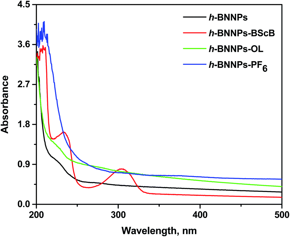
The UV-visible absorption spectra of h-BNNPs and h-BNNPs-ILs (Fig. 9) were collected to examine the electronic structure. The pristine h-BNNPs and h-BNNPs-OL showed absorption maxima below 200 nm, whereas the absorption maxima of h-BNNPs-BScB and h-BNNPs-PF6 shifted towards higher wavelength (∼215 nm). Besides the B–N characteristics absorption features, the BNNPs-BScB exhibited two additional absorbance maxima at 234 and 306 nm and were attributed to bis(salicylato)borate anion of chemically grafted ionic liquids. The band gap energy of each sample was extracted from the intersection point between the energy (x-axis) and the line extrapolated from the linear portion of the absorption edge as shown in Fig. 10. The pristine h-BNNPs exhibits strong absorption below 200 nm, which corresponds to band gap energy of 5.9 eV. The grafting of imidazolium ionic liquids thin films on the h-BNNPs shows significant changes in their UV-visible absorption spectra, as a result the band gap energy shifts to lower values, i.e., 5.78, 5.25 and 5.00 eV for h-BNNPs-OL, h-BNNPs-PF6 and h-BNNPs-BScB, respectively (Table 2).
 |
| | Fig. 9 UV-visible absorption spectra of pristine h-BNNPs and ionic liquids-functionalized h-BNNPs. | |
 |
| | Fig. 10 Tauc plots of h-BNNPs and ionic liquids-grafted h-BNNPs illustrating the band gap energy, determined from their optical absorption spectra. | |
Table 2 The band gap energy of h-BNNPs and ionic liquids-grafted h-BNNPs
| No. |
Material description |
Band gap energy, eV |
| 1 |
h-BNNPs |
5.90 ± 0.04 |
| 2 |
h-BNNPs-OL |
5.78 ± 0.03 |
| 3 |
h-BNNPs-PF6 |
5.25 ± 0.04 |
| 4 |
h-BNNPs-BScB |
5.00 ± 0.02, 3.7 ± 0.03 |
It is believed that the charge transfer between ionic liquids and h-BNNPs changed the positions of HOMO and LUMO, consequently, shifts in the band gap energy. The grafting of ionic liquids introduce additional electronic levels in the large gap and reduced the band gap energy.8 The chemical functionalization of carbon nanotubes, h-BN nanotubes and graphene transforms the electronic structure and alters the band gap.9a,16,17 The chemical nature of grafted species plays important role to control the magnitude of band gap. The h-BNNPs-ILs, having imidazolium cation and three different anions; BScB, PF6 and OL, exhibited structural dependent shifts in the band gap energy and suggested that chemical structure of anions control the band gap energy. Golberg et al. demonstrated that anionic moieties strongly interact with h-BN scaffold than the counter cations and changes electronic structure of h-BN scaffold.14 The h-BNNPs-BScB exhibited two additional absorption peak features beside the shift of main peak towards the higher wavelength (Fig. 9). These features are corresponds to band gap energies of 5.0 and 3.7 eV respectively, and are significantly lower than that of pristine h-BNNPs (5.9 eV). The BScB anion (Scheme 1) in h-BNNPs-BScB exhibits two electron rich aromatic ring, which are probably reduce the band gap and improve the charge mobility. The introduction of double bond, π-electrons and extended π-conjugated system are known to reduce the band gap energy.18 The oleate anion in h-BNNPs-OL shows no significant shift in the band gap energy and this might be attributed to the presence of long hydrocarbon chain. While the PF6− anion has shift of main peak towards the higher wavelength as shown in Fig. 9 with corresponding band gap energy of 5.25 eV. These results suggest that presence of π-electron rich system in the aromatic rings in BScB anion significantly reduces the band gap energy of h-BNNPs by functionalization with ionic liquids, as a result changes in the optical properties.
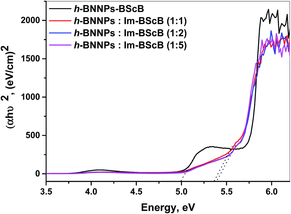
The band gap energy of ionic liquids-grafted h-BNNPs is strongly controlled by chemical structure of anion. In the h-BNNPs-ILs, the imidazolium cation is covalently attached to the h-BNNPs and anions are associates with imidazolium cation via coulombic interaction. In order to probe that covalent grafting of ionic liquids on h-BNNPs is essential to change the band gap energy, the h-BNNPs was simply mixed with corresponding ionic liquid: 1-propyl-3-methyl imidazolium-BScB (Im-BScB) in 1![[thin space (1/6-em)]](https://www.rsc.org/images/entities/char_2009.gif) :1, 1:2 and 1:5 wt% ratio. The UV-visible absorption spectra (Fig. S4, ESI†) of physical mixture of h-BNNPs and Im-BScB revealed diminished shoulder in the range of 220–240 nm owing to band gap of ∼5.4 eV (Fig. 11). However, the covalently grafted h-BNNPs-BScB showed very distinct absorptions at 234 and 306 nm with corresponding band gaps of 5.0 and 3.7 eV, which are significantly lower than the physical mixture of its constituents. Further an increasing wt% ratio of ionic liquid in physical mixture of h-BNNPs and Im-BScB shows no significant changes in the band gap energy (Fig. 11). Likewise, physical mixture of h-BNNPs and Im-PF6 (Fig. S5 and S6†) shows high band-gap compared to that of covalently grafted h-BNNPs-PF6. These results revealed that covalently grafted-ionic liquids on h-BNNPs significantly lower the band-gap compared to the physical mixtures of their constituents.
:1, 1:2 and 1:5 wt% ratio. The UV-visible absorption spectra (Fig. S4, ESI†) of physical mixture of h-BNNPs and Im-BScB revealed diminished shoulder in the range of 220–240 nm owing to band gap of ∼5.4 eV (Fig. 11). However, the covalently grafted h-BNNPs-BScB showed very distinct absorptions at 234 and 306 nm with corresponding band gaps of 5.0 and 3.7 eV, which are significantly lower than the physical mixture of its constituents. Further an increasing wt% ratio of ionic liquid in physical mixture of h-BNNPs and Im-BScB shows no significant changes in the band gap energy (Fig. 11). Likewise, physical mixture of h-BNNPs and Im-PF6 (Fig. S5 and S6†) shows high band-gap compared to that of covalently grafted h-BNNPs-PF6. These results revealed that covalently grafted-ionic liquids on h-BNNPs significantly lower the band-gap compared to the physical mixtures of their constituents.
 |
| | Fig. 11 Tauc plots of h-BNNPs-BScB and physical mixtures of h-BNNPs and 1-propyl-3-methyl imidazolium-BScB ionic liquid in variable wt% ratio. | |
The pristine h-BNNPs exhibits high band-gap, which limits its applications for designing and fabricating optoelectronic nanodevices and as energy storage electronic materials for supercapacitors.19,20 In this context, grafting of ionic liquids to alter the band gap of h-BNNPs is explored as a promising approach to reduce the band gap energy. Currently, more efforts are in progress to further reduce the band-gap of h-BNNPs and develop the materials for nanoelectronics/nanophotonic applications.
4. Conclusions
In summary, the imidazolium ionic liquids were grafted on h-BNNPs via covalent interaction and then evaluated their optical properties. The different anions viz. BScB, OL and PF6 were selected to probe their effects on changes in band gap energy of h-BNNPs. FTIR, XPS, TGA and EDX results confirmed the covalent grafting of imidazolium ionic liquids. The UV-visible absorption characteristics of h-BNNPs were changed after grafting of the ionic liquids and absorption maxima shifted towards higher wavelength. Correspondingly, the band gap energy (5.9 eV) of h-BNNPs was reduced significantly. Among three variable anions of ionic liquids, BScB showed maximum reduction in band gap energy and was attributed to presence of π-electron rich aromatic system. It is believed that charge transfer facilitated by ionic liquids, particularly anion, reduces the band gap energy of h-BNNPs. The anions exhibit stronger interaction with h-BN skeleton, hence their structure plays critical role to tune the band gap energy.
Acknowledgements
This work was generously supported by CSIR, Govt. of India through five year research program (CSC-0118/04). We kindly acknowledge the Director CSIR-IIP for his kind permission to publish these results. The ASD and RTD of CSIR-IIP Dehradun; IIT Madras, Chennai and Kyoto University, Japan are acknowledged for instrumentation support. RG thankful to CSIR India, for the fellowship support.
Notes and references
- K. S. Novoselov, V. I. Fal'ko, L. Colombo, P. R. Gellert, M. G. Schwab and K. Kim, Nature, 2012, 490, 192–200 CrossRef CAS PubMed.
-
(a) Y. Chen, J. Zou, S. J. Campbell and G. L. Caer, Appl. Phys. Lett., 2004, 84, 2430–2432 CrossRef CAS;
(b) C. Y. Zhi, Y. Bando, C. C. Tang, Q. Huang and D. Golberg, J. Mater. Chem., 2008, 18, 3900–3908 RSC.
- I. Jo, M. T. Pettes, J. Kim, K. Watanabe, T. Taniguchi, Z. Yao and L. Shi, Nano Lett., 2013, 13, 550–554 CrossRef CAS PubMed.
-
(a) A. Pakdal, Y. Bando and D. Golberg, Chem. Soc. Rev., 2014, 43, 934–959 RSC;
(b) X. Jiang, Q. Weng, X. Wang, X. Li, J. Zhang, D. Golberg and Y. Bando, J. Mater. Sci. Technol., 2015, 31, 589–598 CrossRef.
-
(a) L. Boldrin, F. Scarpa, R. Chowdhury and S. Adhikari, Nanotechnology, 2011, 22, 505702 CrossRef CAS PubMed;
(b) T. Sugino, C. Kimura and T. Yamamoto, Appl. Phys. Lett., 2002, 80, 3602–3604 CrossRef CAS;
(c) Y. Kubota, K. Watanabe, O. Tsuda and T. Taniguchi, Science, 2007, 317, 932–934 CrossRef CAS PubMed.
-
(a) C. Zhi, Y. Bando, C. Tang, H. Kuwahara and D. Golberg, Adv. Mater., 2009, 21, 2889–2893 CrossRef CAS;
(b) M. G. Silly, P. Jaffrennou, J. Barjon, J.-S. Lauret, F. Ducastelle, A. Loiseau, E. Obraztsova, B. Attal-Tretout and E. Rosencher, Phys. Rev. B: Condens. Matter Mater. Phys., 2007, 75, 085205 CrossRef;
(c) C. Zhi, Y. Xu, Y. Bando and D. Golberg, ACS Nano, 2011, 5, 6571–6577 CrossRef CAS PubMed;
(d) S. Kumari, O. P. Sharma, R. Gusain, H. P. Mungse, A. Kukrety, N. Kumar, H. Sugimura and O. P. Khatri, ACS Appl. Mater. Interfaces, 2015, 7, 3708–3716 CrossRef CAS PubMed.
- K. Watanabe, T. Taniguchi and H. Kanda, Nat. Mater., 2004, 3, 404–409 CrossRef CAS PubMed.
- C. Zhi, Y. Bando, C. Tang and D. Golberg, Phys. Rev. B: Condens. Matter Mater. Phys., 2006, 74, 153413 CrossRef.
-
(a) C. Zhi, Y. Bando, C. Tang, S. Honda, K. Sato, H. Kuwahara and D. Golberg, Angew. Chem., Int. Ed., 2005, 44, 7932–7935 CrossRef CAS PubMed;
(b) T. Sainsbury, T. Ikuno, D. Okawa, D. Pacile, J. M. J. Frechet and A. Zettl, J. Phys. Chem. C, 2007, 111, 12992–12999 CrossRef CAS.
-
(a) S.-U. Xie, W. Wang, K. A. Shiral Fernando, X. Wang, Y. Lin and Y.-P. Sun, Chem. Commun., 2005, 3670–3672 RSC;
(b) S. Pal, S. R. C. Vivekchand, A. Govindaraj and C. N. R. Rao, J. Mater. Chem., 2007, 17, 450–452 RSC;
(c) Y. Lin, T. V. Williams, W. Cao, H. E. Elsayed-Ali and J.
W. Connell, J. Phys. Chem. C, 2010, 114, 17434–17439 CrossRef CAS.
-
(a) A. S. Nazarov, V. N. Demin, E. D. Grayfer, A. I. Bulavchenko, A. T. Arymbaeva, H.-J. Shin, J.-Y. Choi and V. E. Fedorov, Chem.–Asian J., 2012, 7, 554–560 CrossRef CAS PubMed;
(b) J. Hou, G. Li, N. Yang, L. Qin, M. E. Grami, Q. Zhang, N. Wang and X. Qu, RSC Adv., 2014, 4, 44282–44290 RSC.
-
(a) J. Dupont and J. D. Scholten, Chem. Soc. Rev., 2010, 39, 1780–1804 RSC;
(b) O. P. Khatri, K. Adachi, K. Murase, K. Okazaki, T. Torimoto, N. Tanaka, S. Kuwabata and H. Sugimura, Langmuir, 2008, 24, 7785–7792 CrossRef CAS PubMed.
-
(a) N. V. Plechkova and K. R. Seddon, Chem. Soc. Rev., 2008, 37, 123–150 RSC;
(b) H. Weingartner, Angew. Chem., Int. Ed., 2008, 47, 654–670 CrossRef PubMed.
- C. Zhi, Y. Bando, W. Wang, C. Tang, H. Kuwahara and D. Golberg, J. Phys. Chem. C, 2007, 111, 18545–18549 CAS.
-
(a) M. Shakourian-Fard, G. Kamath and Z. Jamshidi, J. Phys. Chem. C, 2014, 118, 26003–26016 CrossRef CAS;
(b) G. Kamath and G. A. Baker, RSC Adv., 2013, 3, 8197–8202 RSC.
- K. S. Kim, D. J. Bae, J. R. Kim, K. A. Park, K. G. Jeon, S. C. Lim, J. J. Kim, W. B. Choi, C. Y. Park and Y. H. Lee, Curr. Appl. Phys, 2004, 4, 559–562 CrossRef.
- H. Zhang, E. Bekyarova, J. W. Huang, Z. Zhao, W. Bao, F. Wang, R. C. Haddon and C. N. Lau, Nano Lett., 2011, 11, 4047–4051 CrossRef CAS PubMed.
- W. Wu, Y. Liu and D. Zhu, Chem. Soc. Rev., 2010, 39, 1489–1502 RSC.
- Y. Xue, Q. Liu, G. He, K. Xu, L. Jiang, X. Hu and J. Hu, Nanoscale Res. Lett., 2013, 8, 49 CrossRef PubMed.
- S. Saha, M. Jana, P. Khanra, P. Samanta, H. Koo, N. C. Murmu and T. Kuila, ACS Appl. Mater. Interfaces, 2015, 7, 14211–14222 CAS.
Footnote |
| † Electronic supplementary information (ESI) available. See DOI: 10.1039/c5ra28037k |
|
| This journal is © The Royal Society of Chemistry 2016 |
Click here to see how this site uses Cookies. View our privacy policy here.