
Optical sensitivity of mussel protein-coated double-walled carbon nanotubes on the iron–DOPA conjugation bond†
Yong-Il Koa,
Cheon-Soo Kanga,
Eun-Ae Shinb,
Yong Chae Jungc,
Hiroyuki Muramatsua,
Takuya Hayashia,
Yoong Ahm Kim*b and
Mildred S. Dresselhausd
aFaculty of Engineering and Carbon Institute of Science and Technology, Shinshu University, 4-17-1 Wakasato, Nagano 380-8553, Japan
bDepartment of Polymer Engineering, Graduated School & School of Polymer Science and Engineering, Chonnam National University, 77 Yongbong-ro, Buk-gu, Gwangju, 500-757, Republic of Korea. E-mail: yak@jnu.ac.kr; Fax: +82-62-530-1779; Tel: +82-62-530-1871
cInstitute of Advanced Composite Materials, Korea Institute of Science and Technology (KIST), 92 Chundong-ro, Bongdong-eup, Wanju-gun, Jeonbuk 565-905, Republic of Korea
dDepartment of Electrical Engineering and Computer Science, Department of Physics, Massachusetts Institute of Technology, Cambridge, Massachusetts 02139-4307, USA
First published on 1st February 2016
Abstract
The optical properties of semiconducting carbon nanotubes respond sensitively to external conditions including the formation of chemical bonds. In order to detect the iron–3,4-dihydroxy-L-phenylalanine (DOPA) conjugation bonds with metal ions, an individually dispersed double-walled carbon nanotube (DWNT) suspension was prepared via homogeneous coating of mussel adhesive protein (MAP). MAP exhibited a high ability for individually dispersing the bundled DWNTs through strong physical interactions with the outer tubes. We demonstrated sensitively altered optical properties of the DWNT suspension upon addition of FeCl3 solution via the formation of coordinative bonds between DOPA in MAP and Fe3+ ions. The iron–DOPA bonds acted as electron acceptors and thus provided a favourable non-radiative channel for the optical depression of signal from semiconducting inner tubes in DWNT suspension. Several physical and chemical effects on the sensitively quenched photoluminescence of semiconducting inner tubes were explained based on the iron–DOPA bonds. We also observed that the DOPA groups in MAP were fully saturated at ca. 376.7 mol% of Fe3+ ions for MAP.
Introduction
Carbon nanotubes (CNTs) have been widely examined in relation to various biomaterial applications, such as drug delivery systems, biocatalysts, biosensors and bone tissue scaffolds1–3 because they exhibit high electrical and thermal conductivity, mechanical strength, and nano-sized diameter.4–6 In particular, double-walled carbon nanotubes (DWNTs), consisting of two coaxial tubules, have several advantages over single- and multi-walled carbon nanotubes (SWNTs and MWNTs) for bio/medical applications due to their unique optical and physicochemical properties.7,8 Because of the buffer-like function of the outer tube, the optical activity of the inner tube is largely different from that of the outer one with regard to external stimulus.9–12 Therefore, it is expected that DWNTs are more optically responsive than large-diameter MWNTs or optically suppressed SWNTs, and therefore, DWNTs can function as an optochemical sensor. However, CNTs including DWNTs are insoluble in aqueous solutions and exhibit a low degree of biocompatibility due to their strongly bundled structure as well as the hydrophobic nature of their sidewalls. Thus, in order to expand their excellent physical and chemical properties at a molecular level, various types of biomaterials have been examined for applications as an individual agent for CNTs.13–15In recent years, the mussel-adhesive proteins (MAP) have attracted a great attention because of their great solubility in aqueous phase and strong affinity toward both organic and inorganic surfaces.16–18 It has been reported that they also show high dispersing and bio-functionalizing ability with regard to DWNTs via the covalent bonds between hydroxyl groups in the outer tubes and amine groups in MAP.19 Several studies have reported that the 3,4-dihydroxy-L-phenylalanine (DOPA) moiety in MAP plays a key role in the adhesion of this protein to different surfaces. Because the catechol groups of DOPA form strong coordinative bonding with metal ions, the surface of MAP-coated DWNT suspension could be converted to a 3D-network structure with Fe3+ ions.20,21 Therefore, it is very critical to understand the optochemical singularities of the inner and outer tubes of DWNTs, as well as to verify the physicochemical effects of MAP-metal complexes and the effectiveness of DWNTs as an optochemical sensor.
In the present study, individually dispersed DWNT suspensions were prepared by a homogeneous coating of MAP and their altered optical properties as a function of the added amount of iron ions was characterized using various optical tools. We found that the formation of iron–DOPA coordinate bonds provided an effective non-radiative channel for the sensitively depressed optical properties of DWNTs.
Results and discussion
A high purity DWNTs with a large bundle size were synthesized using the chemical vapor deposition method (Fig. S1(a) and (b)†).5,8 Then, we examined the dispersing ability of the MAP with regard to the bundled DWNTs.19 By subjecting the bundled DWNTs with MAP in an aqueous solution under strong sonication, an opaque suspension (left suspension in Fig. S1(d)†) is obtained. In the following ultracentrifugation process, we obtained a semi-transparent supernatant containing the individually dispersed DWNTs via the precipitation of the bundled DWNTs (middle suspension in Fig. S1(d)†). By adding FeCl3 solution (10 mM) to the supernatant, we obtained a yellowish DWNT suspension (right suspension in Fig. S1(d)†). This solution was still semi-transparent and there was no precipitate. Then, the dispersion state of DWNTs in an aqueous solution was evaluated optically using Raman/fluorescence spectra with 785 nm laser line and UV-Vis-NIR absorption spectra for both supernatant and remnant (Fig. 1(a) and (c)). We observed well-resolved strong luminescence peaks (Fig. 1(a)) and sharp absorption peaks for the supernatant (Fig. 1(c)). In contrast, relatively depressed luminescence and absorption peaks in the remnant can be explained by strong coupling interactions between adjacent nanotubes.22 Such optical results allowed us to conclude that DWNTs were individually dispersed in an aqueous solution with the help of MAP.23 A typical TEM images showed an individual DWNT coated with amorphous-like MAP (Fig. 1(d)). | ||
| Fig. 1 (a) Wide-range Raman/fluorescence spectra, (b) their corresponding radial breathing modes taken with laser excitation of 785 nm, and (c) UV-Vis-NIR absorption spectra for SDBS-dispersed DWNT supernatant (S), MAP-dispersed DWNT supernatant (S) and MAP-dispersed DWNT remnant (R), respectively, and (d) high-resolution TEM image of MAP-coated individual DWNT. | ||
Then the coating effects of the MAP on the optical properties of the DWNTs were analysed in detail. We observed a red shift both in the luminescent peaks and the UV-Vis-NIR absorption peaks of the supernatant as compared to those of the sodium dodecyl benzene sulfonate (SDBS)-dispersed DWNT suspension (Fig. 1(a) and (c)). The shifted energy gap (ΔEii) are summarized in Tables S1 and S2.†24 It is assumed that environmental dielectric screening effects contribute to a red shift in the optical spectra.25,26 Moreover, since strong electron-donating groups, such as hydroxyl, carboxyl and amine groups were present in the MAP structure,17,27 the electron density of DWNTs in the MAP-dispersed nanotube solution was expected to be higher than that of the SDBS-dispersed nanotube suspension. Thus, we believe that excitons could be easily excited due to the presence of electron donating groups, thereby resulting in the redshift of both absorption and luminescent emission through a decrease in the energy-gap.28 Both environmental dielectric screening effects and electron donating groups in the MAP structure induced a change in the energy-gap of the DWNTs, thus resulting in a modification of the resonance window when the 785 nm laser line was used.28
The effect of adding FeCl3 solution (100 μl) to MAP-dispersed DWNT supernatant was evaluated by Raman/fluorescence spectra using 785 nm laser line (Fig. 2(a)). The Raman spectra of the pristine DWNT exhibited a strong G-band (E2g2 mode) at 1590 cm−1, several radial breathing modes (RBMs) below 500 cm−1 and G′-band at around 2588 cm−1.29 The sonicated DWNT suspension shows a weak luminescence peak around 2400 cm−1. The supernatant containing the individually dispersed DWNTs showed the intensified luminescence peaks compared to the intensity of the G-band. However, after adding 100 μl of FeCl3 solution to the supernatant with subsequent mild sonication, these strong emissions were completely suppressed. The formation of coordinate bonds between the DOPA (Fig. 3) in MAP and Fe3+ ion,30 provided non-radiative channels, which led to quenching of luminescence of the semiconducting inner tubes, which is similar to entrapment effect of metallic tubes in bundled tubes. In other words, several DOPA groups in the MAP molecule20,21 allowed DWNTs to attain a 3D-network structure by DOPA–Fe3+ bonding. Therefore, the DWNTs lost their optical singularities due to charge transfer through the DOPA–Fe3+ linkage. This charge transfer effect also could be confirmed by observations of Raman/fluorescence spectra with laser excitations of 633 and 532 nm. As shown in Fig. S2,† even though the G-band of the DWNTs in the MAP-dispersed DWNT suspension had a relatively low intensity due to strong emission from MAP, the addition of FeCl3 solution induced a marked increase in the relative intensity of the G-band with regard to the emission of MAP. This phenomenon indicated that a non-radiative channel was established by the formation of electronically active DOPA–Fe3+ linkages.31,32
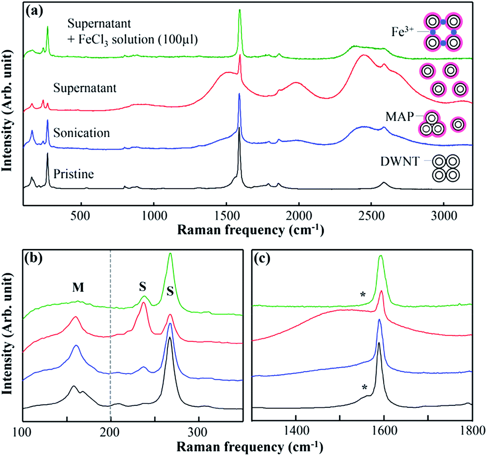 | ||
| Fig. 2 (a) Raman/fluorescence spectra taken with laser excitation of 785 nm for pristine DWNTs and MAP-dispersed DWNT suspensions at different dispersion states (sonicated, supernatant) and the FeCl3 solution added-MAP dispersed DWNT supernatant, and their corresponding (b) radial breathing mode (where S indicates semiconducting and M indicates metallic tubes), and (c) G-band (asterisk indicates the BWF line associated with metallic outer tubes). | ||
 | ||
| Fig. 3 The (a) C1s, (b) O1s, and (c) Fe3+2p XPS spectra of pristine DWNTs, MAP-coated DWNT and FeCl3 solution added-MAP dispersed DWNT complexes (note that the concentration and amount of FeCl3 solution were 100 mM, 30 μl). All the samples were prepared by buckypaper though filtration with an Omnipore membrane filter (JVWP 0.1 μm). | ||
The radial breathing modes (RBMs) of CNTs have been used to evaluate their chirality and diameter as well as their dispersion state.33–38 Strano et al. revealed that the RBM intensity at 267 cm−1 decreased and the RBM intensity at 234 cm−1 increased when SWNTs were isolated completely with sodium dodecyl sulfonate (SDS).33,38 For the MAP-dispersed DWNT supernatant, the RBM peaks from the semiconducting inner tube exhibited higher intensity at 237 cm−1 and relatively lower intensity at 268 cm−1 than those of the sonicated or bundled DWNTs (Fig. 2(b)). However, the addition of FeCl3 solution caused a significant decrease in the RBM intensity at 237 cm−1 and an increase in the RBM intensity at 268 cm−1 (Fig. 2(b)). This result indicated that the electronic structure was largely modified by the formation of strong DOPA–Fe3+ bonds between the individually isolated DWNTs as the 3D-network structure, so that the resonance states of S inner tubes shifted like the bundled DWNTs. In contrast to the UV-Vis-NIR and Raman results, TEM image (Fig. S1(c)†) showed individually isolated DWNTs state, even after the addition of FeCl3 to the MAP-dispersed DWNT suspension. This result indicated that the individually isolated phase of DWNTs that enclosed by MAP–Fe3+ complex was retained, but non-radiative channels were created through the formation of MAP–Fe3+ bonds.
When looking at the RBM peaks of the metallic outer tube, the intensity at 160 cm−1 was maintained in the MAP-dispersed DWNT supernatant (Fig. 2(b)). However, the addition of FeCl3 solution caused the RBM peak at 160 cm−1 to be almost suppressed because the outer tubes were in direct contact with the MAP–Fe3+ complex. This indicated that circumferentially generated and concentrated stress was induced by surface stiffening of the sidewall which suppressed RBM vibrations in the radial direction.39 Moreover, when looking at the G-band (Fig. 2(c)), the broad and asymmetric Breit–Wigner–Fano (BWF) line40 of the pristine DWNT sample was strongly suppressed in the Fe–MAP–DWNT suspension. The metallic outer tube could not resonance with 785 nm excitation and their corresponding metallic shoulder of G′-band was thus reduced, which resulted in the disappearance of the RBM peak (160 cm−1) in the iron–MAP–DWNT suspension. These results supported the protective function of the outer tubes in DWNTs.41,42
X-ray photoelectron spectroscopy measurement was also carried out on pristine, MAP–DWNT, and Fe–MAP–DWNT samples in order to determine the relative quantity of the functional groups attached to the outer walls of the DWNTs (Fig. 3). For the pristine DWNT sample, there was an intense peak at ca. 284.7 eV from the sp2-hybridized carbon atoms and also a broad peak at 285.3 eV due to the sp3-hybridized carbon atoms (Fig. 3(a)).43,44 For MAP–DWNTs, the prominently present sp3-hybridized peaks (e.g., C–N, C![[double bond, length as m-dash]](https://www.rsc.org/images/entities/char_e001.gif) O, CO–N and O–CO carbon atoms) came from the functional groups of MAP,19 indicating that the outer tubes of DWNTs had been well coated by MAP. Some weak peaks (Fig. 3(b)) came from O1s of the hydroxyl- and carboxyl-groups on defects and edges of the pristine DWNT sample; on the other hand, peaks with large intensities, such as from O–CO, O–C and OC at 534.4, 533.3, and 532.2 eV, respectively, were closely associated with MAP.
O, CO–N and O–CO carbon atoms) came from the functional groups of MAP,19 indicating that the outer tubes of DWNTs had been well coated by MAP. Some weak peaks (Fig. 3(b)) came from O1s of the hydroxyl- and carboxyl-groups on defects and edges of the pristine DWNT sample; on the other hand, peaks with large intensities, such as from O–CO, O–C and OC at 534.4, 533.3, and 532.2 eV, respectively, were closely associated with MAP.
When 30 μl of FeCl3 solution was added to the MAP-dispersed DWNT supernatant, the O1s peak shifted to lower energy (ca. 0.4 eV) due to a decrease in the O–C signal, and a new peak appeared at 531.6 eV, which was associated with the O–Fe coordinative bonding.45 From Table 1, it is interesting to note that the ratio of the O–Fe bond increased by ca. 16.40% when the O–C bond was reduced ca. 15.8% even though the atomic ratio of oxygen had not changed. Such result signified a transition from the O–C to O–Fe bond when the FeCl3 solution was added to the supernatant, without any significant change in the N1s (Fig. S3†) and C1s XPS spectra (Fig. 3). Therefore, we are able to say that the hydroxyl groups of the DOPA created coordinative bonds with Fe3+ ion. In addition, the presence of Fe3+ ions was verified by the peaks at 710.8 eV and 724.2 eV associated with 2p3/2 and 2p1/2, and their charge-transfer satellite peaks at 714.4 eV and 727.6 eV, respectively.46 Furthermore, as shown in Table 1, the atomic ratio of Fe3+ ions did not change significantly (ca. 0.5%) on increasing the addition amount of FeCl3 from 30 μl to 50 μl. Thus, this result indicated that the DOPA components within the MAP were saturated in this range because all the hydroxyl groups available to bond with Fe3+ had been fully utilized.
| Sample I.D. | Atomic composition (%) | Bonding composition (%) | ||||||
|---|---|---|---|---|---|---|---|---|
| C | O | N | Fe | O–C | OC |
OCO | O–Fe | |
| Pristine DWNTs | 95.95 | 3.88 | — | — | 46.13 | 19.85 | 34.00 | — |
| MAP–DWNTs | 67.13 | 27.31 | 5.47 | — | 54.66 | 31.04 | 14.20 | — |
| Fe–MAP–DWNTs (30 μl) | 66.67 | 27.86 | 4.67 | 0.79 | 38.86 | 29.77 | 14.60 | 16.40 |
| Fe–MAP–DWNTs (50 μl) | 67.70 | 27.10 | 4.39 | 0.81 | 36.25 | 30.12 | 14.43 | 17.11 |
To confirm the optical sensitivity of the MAP-dispersed DWNT supernatant in regard to the Fe3+ ions, Raman/fluorescent, UV-Vis-NIR absorption spectra and PL maps were measured (Fig. 4) with 3 ml of MAP-dispersed DWNT supernatant to which was added 5 to 100 μl FeCl3 solution sequentially. As shown in Fig. 4(a), with increasing amount of FeCl3 solution, several strong luminescent peaks in the supernatant continuously decreased and simultaneously the RBM peak at 160 cm−1 disappeared, accompanying by a relative change in the RBM intensities at 237 cm−1 and 268 cm−1. When more than 50 μl of FeCl3 solution was added, the luminescent peaks disappeared completely, signifying that the hydroxyl groups within the DOPA were fully saturated with Fe3+ ions. Fig. 4(b) shows that sharp absorption peaks in the UV-Vis-NIR spectra, coming from van Hove singularities of MAP-dispersed DWNT supernatant, were broadened after addition of FeCl3 solution. This depressed absorption peaks can be explained by the formation of MAP–Fe3+ bonds on the surface of DWNTs which largely altered the electronic absorption of the inner tubes. Moreover, the inherent chiral singularities of individual DWNTs were not observed due to the electrical interaction between MAP–Fe3+-linked DWNTs caused by the charge transfer effect. The broadening of the absorption peaks did not progress any more after adding 40 μl of FeCl3 since all DOPA groups were consumed by bonding with Fe3+ ions, similar to the Raman results, as mentioned above. PL maps (Fig. 4(c)) also showed the disappearance of strong emission peaks for the MAP-dispersed DWNT supernatant after FeCl3 solution was added. We observed completely depressed emissions when adding 30 μl and 50 μl of FeCl3 solution. Therefore, from the combined results of Raman, UV-Vis-NIR spectra and PL maps, we observed that 3 ml of MAP-dispersed DWNT supernatant was fully saturated with ca. 50 μl of FeCl3 solution. From this result, the saturation ratio of Fe3+ with regard to MAP was decided to be ca. 9.3 wt%: 3766.7 mol% (Table S3†).
 | ||
| Fig. 4 (a) Raman/fluorescence spectra taken with laser excitation of 785 nm for MAP-dispersed DWNT supernatant with different amounts of FeCl3 solution (100 mM). The inset shows the magnified low-frequency Raman spectra for the corresponding samples. (b) Their corresponding UV-Vis-NIR absorption spectra, and (c) PL maps. | ||
Conclusions
In summary, we prepared individually dispersed DWNTs in aqueous solution using MAP and examined the optical sensitivity of MAP-dispersed DWNT suspensions with regard to the metal ion (Fe3+) using various optical tools. With increasing dispersion state of the MAP-dispersed DWNT suspension, the intensified absorption and luminescence peaks indicated that the DWNTs were individually isolated in an aqueous solution. In comparison with SDBS-dispersed DWNT suspension, the red shift in the absorption and luminescent peaks for the MAP-dispersed DWNT suspension can be explained in terms of both the altered band-gap and the dielectric screening effect. The addition of the FeCl3 solution to MAP-dispersed DWNT supernatant created the formation of coordinative bond between the DOPA of MAP and Fe3+ ion. From XPS spectra, we observed a transition from O–C to O–Fe bond, indicating the formation of DOPA–Fe3+ coordinative bonds, on addition of the FeCl3 solution to MAP dispersed DWNT supernatant. The optical depression in the ferric chloride added MAP dispersed DWNT supernatant was verified by the disappearance of luminescence peaks and relative changes in the RBM intensities due to the formation of DOPA–Fe3+ bonds. The UV-Vis-NIR spectra also showed the loss of van Hove singularities by broadened absorption peaks on the addition of FeCl3 solution. Both the depression of the BWF line and RBM peak at 160 cm−1 associated with metallic outer tubes indicated that the circumferentially coated MAP–Fe3+ complex on the outer tubes largely altered the resonance condition of the outer tubes. We also observed the optical properties to change sensitively from the Raman, UV-Vis-NIR spectra and the PL of MAP-dispersed DWNT supernatant when different amounts of the FeCl3 solution were added. However, when the amount of FeCl3 solution reached 50 μl, the optical activities of DWNTs were completely depressed, indicating that DOPA groups in MAP were fully saturated with ca. 9.3 wt% and ca. 3766.7 mol% of Fe3+ ions. From these results, it is expected that MAP-dispersed DWNT suspensions are promising materials in the development of highly sensitive and responsive luminescence sensors for highly sensitive optoelectronic applications.Experimental
Synthesis of high purity DWNT sample
The synthesis of DWNTs was carried out by the catalytic chemical vapour deposition method in a furnace using Mo/Al2O3 and Fe/MgO as a conditioning catalyst and nanotube catalyst, respectively. Subsequently, a methane/argon (1![[thin space (1/6-em)]](https://www.rsc.org/images/entities/char_2009.gif) :1) mixture was fed into the reactor typically for 10 min at 875 °C. The as-grown products were purified via the following steps. First, an oxidation process (500 °C, 20 min) was carried out to reduce the chemically active SWNTs. Second, a hydrochloric acid (18%, 100 °C, 10 h) treatment was done in order to remove the metal catalysts, followed by air oxidation at 500 °C for 10 min in order to remove all carbonaceous impurities. The detailed experimental procedures were described in our previous papers.7
:1) mixture was fed into the reactor typically for 10 min at 875 °C. The as-grown products were purified via the following steps. First, an oxidation process (500 °C, 20 min) was carried out to reduce the chemically active SWNTs. Second, a hydrochloric acid (18%, 100 °C, 10 h) treatment was done in order to remove the metal catalysts, followed by air oxidation at 500 °C for 10 min in order to remove all carbonaceous impurities. The detailed experimental procedures were described in our previous papers.7
Dispersion of DWNTs using MAP in aqueous solution
The prepared highly pure DWNTs (1 mg) were individually isolated in heavy water (10 ml) with the help of MAP (recombinant mussel adhesive protein, 22.6 kDa, Kollodis & Biosciences) (10 mg) under strong sonication (VCX 750, Sonics & Materials, 750 W) for 1 h at 4 °C, and subsequent ultracentrifugation (Optima Max-XP, Beckman Coulter, 240000g). The supernatant, containing MAP-dispersed DWNT suspension (70%) and rich in isolated nanotubes, was obtained and characterized in our study. To confirm the dispersibility and chemical effect of MAP, SDBS (1 wt%)-dispersed DWNT suspension was also prepared in this study.
The formation of coordinative bonds between DOPA and iron
The iron(III) chloride hexahydrate (FeCl3·6H2O) (270.3 mg) was dissolved in heavy water (10 ml) to produce 100 mM solution. The prepared FeCl3 solution was added to 3 ml of individually isolated MAP-DW solution in different amounts (5–100 μl) and subsequent mild sonication (1510, Branson, 5 min) was applied.Characterizations
The optical spectra for DWNT suspensions were obtained using a UV-Vis-NIR spectrophotometer (Shimadzu soildspec-3700), photoluminescence maps (NIR-PL system, Shimadzu) and a 785 nm excited Raman system (Renishaw, inVia Raman microscope). We also obtained Raman spectra using 532 and 633 nm laser excitations produced by a Kaiser HoloLab5000 system. The chemical bonding and elemental composition of MAP-DW and their metal absorbed composite were characterized by X-ray photoemission spectroscopy (XPS, ESCA-3400, Kratos Analytical) using a MgKα X-ray source at a 10 mA emission current and a 10 kV accelerating voltage. We also obtained the morphology and elemental composition using a field emission scanning electron microscope (FE-SEM) coupled with energy dispersive X-ray spectroscopy (EDS) (JSM-7000f, JEOL). Finally, we have used a high-resolution TEM (JEM-2010FEF, JEOL) to see the dispersion state of the nanotubes as well as the coated mussel protein on the sidewalls of the DWNTs.Acknowledgements
We acknowledge the support from the Shinshu University COI STREAM program (Center of Innovation Science and Technology based Radical Innovation and Entrepreneurship Program) from the Ministry of Education, Culture, Sports, Science and Technology and the Japan Science and Technology Agency. Y. A. K. acknowledges the financial support from the National Research Foundation of Korea (NRF) grant funded by the Korea government (MSIP) (No. NRF-2014R1A2A1A10050585) and from the framework of international cooperation program managed by the National Research Foundation of Korea (NRF-2015K2A2A4000110, FY2015).Notes and references
- K. Besteman, J. O. Lee, F. G. M. Wiertz, H. A. Heering and C. Dekker, Nano Lett., 2003, 3, 727–730 CrossRef CAS.
- P. Asuri, S. S. Karajanagi, E. Sellitto, D. Y. Kim, R. S. Kane and J. S. Dordick, Biotechnol. Bioeng., 2006, 95, 804–811 CrossRef CAS PubMed.
- N. Saito, K. Aoki, Y. Usui, M. Shimizu, K. Hara, N. Narita, N. Ogihara, K. Nakamura, N. Ishigaki, H. Kato, H. Haniu, S. Taruta, Y. A. Kim and M. Endo, Chem. Soc. Rev., 2011, 40, 3824–3834 RSC.
- A. B. Dalton, S. Collins, E. Munoz, J. M. Razal, V. H. Ebron, J. P. Ferraris, J. N. Coleman, B. G. Kim and R. H. Baughman, Nature, 2003, 423, 703 CrossRef CAS PubMed.
- M. Endo, H. Muramatsu, T. Hayashi, Y. A. Kim, M. Terrones and N. S. Dresselhaus, Nature, 2005, 433, 476 CrossRef CAS PubMed.
- W. Shi, Z. Wang, Q. C. Zhang, Y. Zheng, C. Ieong, M. Q. He, R. Lortz, Y. Cai, N. Wang, T. Zhang, H. J. Zhang, Z. K. Tang, P. Sheng, H. Muramatsu, Y. A. Kim, M. Endo, P. T. Araujo and M. S. Dresselhaus, Sci. Rep., 2012, 2, 625 Search PubMed.
- Y. A. Kim, H. Muramatsu, T. Hayashi, M. Endo, M. Terrones and M. S. Dresselhaus, Chem. Vap. Deposition, 2006, 12, 327–330 CrossRef CAS.
- Y. A. Kim, K. S. Yang, H. Muramatsu, T. Hayashi, M. Endo, M. Terrones and M. S. Dresselhaus, Carbon Letters, 2014, 15, 77–88 CrossRef.
- G. M. do Nascimento, T. Hou, Y. A. Kim, H. Muramatsu, T. Hayashi, M. Endo, N. Akuzawa and M. S. Dresselhaus, Carbon, 2011, 49, 3585–3596 CrossRef CAS.
- H. Muramatsu, T. Hayashi, Y. A. Kim, D. Shimamoto, M. Endo, V. Meunier, B. G. Sumpter, M. Terrones and M. S. Dresselhaus, Small, 2009, 5, 2678–2682 CrossRef CAS PubMed.
- N. Kamaraju, S. Kumar, Y. A. Kim, T. Hayashi, H. Muramatsu, M. Endo and A. K. Sood, Appl. Phys. Lett., 2009, 95, 81106 CrossRef.
- T. Hayashi, D. Shimamoto, Y. A. Kim, H. Muramatsu, F. Okino, H. Touhara, T. Shimada, Y. Miyauchi, S. Maruyama, M. Terrones, M. S. Dresselhaus and M. Endo, ACS Nano, 2008, 2, 485–488 CrossRef CAS PubMed.
- Y. Lee and K. E. Geckeler, Adv. Mater., 2010, 22, 4076–4083 CrossRef CAS PubMed.
- S. S. Karajanagi, H. C. Yang, P. Asuri, E. Sellitto, J. S. Dordick and R. S. Kane, Langmuir, 2006, 22, 1392–1395 CrossRef CAS PubMed.
- D. Nepal and K. E. Geckeler, Small, 2006, 2, 406–412 CrossRef CAS PubMed.
- J. H. Waite, Int. J. Adhes. Adhes., 1987, 7, 9–14 CrossRef CAS.
- B. P. Lee, P. B. Messersmith, J. N. Israelachvili and J. H. Waite, Annu. Rev. Mater. Res., 2011, 41, 99–132 CrossRef CAS PubMed.
- S. Ryu, Y. Lee, J. W. Hwang, S. Hong, C. Kim, T. G. Park, H. Lee and S. H. Hong, Adv. Mater., 2011, 23, 1971–1975 CrossRef CAS PubMed.
- Y. C. Jung, H. Muramatsu, K. Fujisawa, J. H. Kim, T. Hayashi, Y. A. Kim, M. Endo, M. Terrones and M. S. Dresselhaus, Small, 2011, 7, 3292–3297 CrossRef CAS PubMed.
- Z. P. Xu, Sci. Rep., 2013, 3, 2914 Search PubMed.
- M. J. Harrington, A. Masic, N. Holten-Andersen, J. H. Waite and P. Fratzl, Science, 2010, 328, 216–220 CrossRef CAS PubMed.
- S. Reich, C. Thomsen and P. Ordejon, Phys. Rev. B: Condens. Matter Mater. Phys., 2002, 65, 153407 CrossRef.
- M. J. O'Connell, S. M. Bachilo, C. B. Huffman, V. C. Moore, M. S. Strano, E. H. Haroz, K. L. Rialon, P. J. Boul, W. H. Noon, C. Kittrell, J. P. Ma, R. H. Hauge, R. B. Weisman and R. E. Smalley, Science, 2002, 297, 593–596 CrossRef PubMed.
- R. B. Weisman and S. M. Bachilo, Nano Lett., 2003, 3, 1235–1238 CrossRef CAS.
- J. Lefebvre, J. M. Fraser, Y. Homma and P. Finnie, Appl. Phys. A: Mater. Sci. Process., 2004, 78, 1107–1110 CrossRef CAS.
- Y. Miyauchi, R. Saito, K. Sato, Y. Ohno, S. Iwasaki, T. Mizutani, J. Jiang and S. Maruyama, Chem. Phys. Lett., 2007, 442, 394–399 CrossRef CAS.
- V. V. Papov, T. V. Diamond, K. Biemann and J. H. Waite, J. Biol. Chem., 1995, 270, 20183–20192 CrossRef CAS PubMed.
- J. Maultzsch, H. Telg, S. Reich and C. Thomsen, Phys. Rev. B: Condens. Matter Mater. Phys., 2005, 72, 205438 CrossRef.
- A. M. Rao, E. Richter, S. Bandow, B. Chase, P. C. Eklund, K. A. Williams, S. Fang, K. R. Subbaswamy, M. Menon, A. Thess, R. E. Smalley, G. Dresselhaus and M. S. Dresselhaus, Science, 1997, 275, 187–191 CrossRef CAS PubMed.
- H. B. Zeng, D. S. Hwang, J. N. Israelachvili and J. H. Waite, Proc. Natl. Acad. Sci. U. S. A., 2010, 107, 12850–12853 CrossRef CAS PubMed.
- S. Y. Ma, L. Liu, V. Bromberg and T. J. Singler, J. Mater. Chem. C, 2014, 2, 3885–3889 RSC.
- T. Akter and W. S. Kim, ACS Appl. Mater. Interfaces, 2012, 4, 1855–1859 CAS.
- D. A. Heller, P. W. Barone, J. P. Swanson, R. M. Mayrhofer and M. S. Strano, J. Phys. Chem. B, 2004, 108, 6905–6909 CrossRef CAS.
- A. Jorio, A. P. Santos, H. B. Ribeiro, C. Fantini, M. Souza, J. P. M. Vieira, C. A. Furtado, J. Jiang, R. Saito, L. Balzano, D. E. Resasco and M. A. Pimenta, Phys. Rev. B: Condens. Matter Mater. Phys., 2005, 72, 075207 CrossRef.
- J. Jiang, R. Saito, G. G. Samsonidze, A. Jorio, S. G. Chou, G. Dresselhaus and M. S. Dresselhaus, Phys. Rev. B: Condens. Matter Mater. Phys., 2007, 75, 035407 CrossRef.
- A. Jorio, M. A. Pimenta, A. G. Souza, R. Saito, G. Dresselhaus and M. S. Dresselhaus, New J. Phys., 2003, 5, 139 CrossRef.
- F. Villalpando-Paez, H. Son, D. Nezich, Y. P. Hsieh, J. Kong, Y. A. Kim, D. Shimamoto, H. Muramatsu, T. Hayashi, M. Endo, M. Terrones and M. S. Dresselhaus, Nano Lett., 2008, 8, 3879–3886 CrossRef CAS PubMed.
- M. S. Strano, V. C. Moore, M. K. Miller, M. J. Allen, E. H. Haroz, C. Kittrell, R. H. Hauge and R. E. Smalley, J. Nanosci. Nanotechnol., 2003, 3, 81–86 CrossRef CAS PubMed.
- J. H. Kim, M. Kataoka, D. Shimamoto, H. Muramatsu, Y. C. Jung, T. Hayashi, Y. A. Kim, M. Endo, J. S. Park, R. Saito, M. Terrones and M. S. Dresselhaus, ACS Nano, 2010, 4, 1060–1066 CrossRef CAS PubMed.
- A. M. Rao, P. C. Eklund, S. Bandow, A. Thess and R. E. Smalley, Nature, 1997, 388, 257–259 CrossRef CAS.
- D. Shimamoto, H. Muramatsu, T. Hayashi, Y. A. Kim, M. Endo, J. S. Park, R. Saito, M. Terrones and M. S. Dresselhaus, Appl. Phys. Lett., 2009, 94, 083106 CrossRef.
- K. Iakoubovskii, N. Minami, T. Ueno, S. Kazaoui and H. Kataura, J. Phys. Chem. C, 2008, 112, 11194–11198 CAS.
- H. Ago, T. Kugler, F. Cacialli, W. R. Salaneck, M. S. P. Shaffer, A. H. Windle and R. H. Friend, J. Phys. Chem. B, 1999, 103, 8116–8121 CrossRef CAS.
- H. Murphy, P. Papakonstantinou and T. I. T. Okpalugo, J. Vac. Sci. Technol., B: Microelectron. Nanometer Struct.--Process., Meas., Phenom., 2006, 24, 715–720 CrossRef CAS.
- L. M. Bronstein, A. Ivanovskaya, T. Mates, N. Holten-Andersen and G. D. Stucky, J. Phys. Chem. B, 2009, 113, 647–655 CrossRef CAS PubMed.
- T. Yamashita and P. Hayes, Appl. Surf. Sci., 2008, 254, 2441–2449 CrossRef CAS.
Footnote |
| † Electronic supplementary information (ESI) available: SEM image, EDX, Raman and XPS result. See DOI: 10.1039/c5ra27842b |
| This journal is © The Royal Society of Chemistry 2016 |