Evaluation of in-tube solid-phase microextraction method for co-extraction of acidic, basic, and neutral drugs

Hamid Asiabia, Yadollah Yamini*a, Shahram Seidib, Meysam Safaria and Maryam Shamsayeia
aDepartment of Chemistry, Tarbiat Modares University, P.O. Box 14115-175, Tehran, Iran. E-mail: yyamini@modares.ac.ir; Fax: +98 21 8288006544; Tel: +98 21 82883417
bDepartment of Analytical Chemistry, Faculty of Chemistry, K.N. Toosi University of Technology, Tehran, Iran

Received 27th December 2015 , Accepted 25th January 2016

First published on 27th January 2016


Abstract

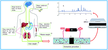
Simultaneous extraction of acidic, basic, and neutral drugs from different biological samples is a considerable and disputable concept in sample preparation strategies. In the present work, a new in-tube solid phase microextraction approach named electrochemically controlled double in-tube solid phase microextraction (EC-DIT-SPME) is introduced for simultaneous extraction of acidic, basic, and neutral drugs from different biological matrices. For this purpose, novel nanostructured coatings based on copolymer of polypyrrole and indole-2-carboxylic acid (PPy-co-PIca) as non-overoxidized and overoxidized forms were electrochemically synthesized on the inner surface of stain-less steel tubes. During the extraction procedure, the PPy-co-PIca and overoxidized PPy-co-PIca coated stain-less steel tubes were used as anode and cathode electrodes, respectively. Extraction of basic and acidic drugs were performed on the surface of overoxidized PPy-co-PIca and PPy-co-PIca, which are connected to the negative and positive potentials, respectively. With regard to the fact that neutral drugs can be absorbed in both electrodes, extraction of neutral drugs will be done in both of them. Satisfactory analytical figures of merit including linear dynamic range and limits of detection in the range of 0.15–500 ng mL−1 and 0.05–1.9 ng mL−1 and good extraction recoveries (38.5–78.6%) were obtained. Finally, the proposed method was successfully applied for simultaneous analysis of acidic, basic, and neutral drugs in some biological samples.


Introduction

The growing worldwide consumption of pharmaceuticals and their entrance in the environment has become an important issue in recent years. Therefore, determination of pharmaceuticals such as analgesics, anti-inflammatories, anti-epileptics, and β-blockers in biological samples has found noticeable importance in medical sciences.1,2 Medicines have a variety of acidic, basic, and neutral forms. Neutral drug analysis constitutes one of the most important fields of the science at present. It is important to provide a simple, sensitive, rapid, and reliable method that can be employed for simultaneous extraction and measurement of the wide range of acidic, basic, and neutral drugs.

So far, many approaches such as low-voltage electrically-enhanced microextraction,3 magnetic nanoparticles based dispersive micro-solid-phase extraction,4 electrochemically controlled solid-phase extraction,5 and electro-membrane extraction6 have been used for extraction of the different types of acidic and basic species at the same time while a wide range of consumed drugs are neutral species. Hence, measuring neutral drugs together with acidic and basic ones is important and no method has been provided for extraction of neutral and ionic species simultaneously until now.

In-tube solid phase microextraction (IT-SPME) is one of the most popular sample preparation methods due to its significant advantages such as improved automation, reproducibility, and high-throughput capability; initially developed by Eisert and Pawliszyn for determination of non-volatile organic pollutants in water samples.7 In this extraction technique, desorption and injection can be performed automatically, allowing a shorter analysis time and better precision and accuracy. Configuration of the IT-SPME technique plays a crucial role in the rate and efficiency of extraction as well as its applications.8–12 To achieve proper operation during simultaneous extraction of cationic, anionic, and neutral species by IT-SPME system, it firstly seems necessary to select an appropriate extraction setup as well as a suitable adsorbent. For simultaneous extraction of neutral species, selecting a suitable adsorbent with a high adsorption capacity for extraction of neutral species that is also conductive is essential to perform uptake and release of ionic species by applying potential. Conductive polymers have many applications as adsorbents in SPME methods.13–16 They have high capacity to adsorb neutral species via establishing different intermolecular interactions such as acid–base, π–π, dipole–dipole, hydrophobic, and hydrogen bonding.17,18 They can also be used for electrochemically controlled SPME (EC-SPME) to extract ionic species by applying an electrical potential because of being conductive.19–22

Copolymers have attracted considerable attentions as adsorbents in solid based extraction techniques due to providing the advantages such as good stability, high surface area, high interaction capability, and consequently high adsorption capacity.23–27 Accordingly, in this work a new method based on electrochemically controlled double in-tube solid phase microextraction (EC-DIT-SPME) was introduced for simultaneous extraction of neutral and ionized species, in which a new nanostructured conductive copolymer based on polypyrrole-co-indole-2-carboxylic acid (PPy-co-PIca) was used as the adsorbent. This new SPME coating was electrochemically deposited on the inner surface of a stain-less steel tube as the unbreakable substrate by providing a simple setup.

To demonstrate the efficiency of the proposed method, thebaine (TEB) as a basic drug, indomethacin (IND) and naproxen (NAP) as acidic drugs, and oxazepam (OXA) and lorazepam (LOR) as neutral drugs were extracted from 30 mL of aqueous sample solutions into 40 μL of the acceptor solution. Extraction of basic drugs was performed on the surface of overoxidized PPy-co-PIca that was connected to a negative potential and extraction of acidic drugs were performed on the surface of PPy-co-PIca connected to a positive potential. With regard to the fact that neutral drugs can be absorbed in both electrodes, extraction of neutral drugs was carried out using both electrodes. During the electrochemical extraction procedure, a low voltage battery (1.0 V) was used, which provides simplicity of automation and an economical way to extract species from biological samples.

Experimental section

Chemicals and reagents

All chemicals used were of analytical-reagent grade. Standards of indomethacin, thebaine, naproxen, oxazepam, and lorazepam were purchased from Sigma-Aldrich (Milwaukee, WI, USA). HPLC-grade methanol and acetonitrile were purchased from Caledon (Georgetown, ON, Canada). Indole-2-carboxylic acid, synthetic pyrrole (98% pure), and oxalic acid (98% pure) were purchased from Sigma-Aldrich.

Apparatus

A potentiostat and galvanostat Autolab system with PGSTAT30 and GPES 4.9 software from Eco Chemie (Utrecht, The Netherlands), was applied for electrochemical preparation of coatings. Particle size and morphology of the synthesized nanostructured composite coating were determined by a scanning electron microscope (SEM) model EM3200 from KYKY Zhongguancun (Beijing, China). The HPLC instrument consisted of an Agilent (Wilmington, DE, USA) 1200 series binary pump, micro vacuum degasser, an injector equipped with a 20 μL sample loop, a 1200 series diode array detector, and B.04.03 Chem Station software. An ODS-3 column (250 mm × 4.6 mm, with 5 μm particle size) from Hector Company (Daejeon, Korea) was applied to separate the drugs. Mixture of acetonitrile (solvent A) and 10 mM sodium dihydrogen phosphate (solvent B) with pH 5.0 under gradient elution condition was used as the mobile phase at a flow rate of 1 mL min−1. The gradient program was 80[thin space (1/6-em)]:[thin space (1/6-em)]20 B in A, from 0 to 6 min; 70[thin space (1/6-em)]:[thin space (1/6-em)]30 B in A, from 6 to 8 min; 60[thin space (1/6-em)]:[thin space (1/6-em)]40 B in A, from 9 to 11 min; 50[thin space (1/6-em)]:[thin space (1/6-em)]50 B in A, from 11 to 14 min; 40[thin space (1/6-em)]:[thin space (1/6-em)]60 B in A, from 14 to 16 min; stay in this condition for 5 min and then, it returned to its initial composition. The analytical signal was recorded between 190 and 400 nm. In the present work, absorption measurements were performed at 230 nm (for oxazepam, lorazepam, naproxen, and indomethacin) and 280 nm (for thebaine).

Preparation of polymer-coated capillary tubes

The copolymer coating of PPy and PIca was synthesized electrochemically via in situ polymerization from acetonitrile solution containing pyrrole (0.2 mol L−1), indole-2-carboxylic acid (0.2 mol L−1), and oxalic acid (0.5 mol L−1). A nanostructured PPy-co-PIca was coated inside a stain-less steel capillary tube (10 cm length and 0.75 mm diameter) using cyclic voltammetry in the potential range between −1.0 and +1.5 V during 15 cycles (scan rate: 50 mV s−1). A stain-less steel tube was employed as the working electrode; Pt and Ag/AgCl electrodes were used as counter and reference electrodes, respectively. A peristaltic pump was used to pass the monomer solution through the inner surface of the stain-less steel tube (Fig. S-1, ESI). According to the literature, overoxidation of the coating was carried out in 0.1 mol L−1 KNO3 solution by application of a constant potential of +1.5 V for 15 min.28

Fig. 1 shows the cyclic voltammograms of the polymerization of pyrrole, indole-2-carboxylic acid, and PPy-co-PIca as a function of the number of cycles. Owing to the differences in the cyclic voltammetric growth profiles of PPy (EAp = 0.3 V, ECp = −0.4 V) and polyindole-2-carboxylic acid (EAp = 1.4 V, ECp = 1.2 V), the multisweep cyclic voltammograms during the growth of the copolymer were recorded. As can be seen in Fig. 1C, in the presence of 0.2 mol L−1 pyrrole and 0.2 mol L−1 indole-2-carboxylic acid, the current value started to increase at +0.3 V which can be attributed to the pyrrole oxidation in the first scan. Increasing of the oxidation potential from +0.4 V to +1.4 V with increasing the number of cycles indicates the growth of copolymer film on the surface of electrode. The comparison of the oxidation potential of polypyrrole and indole-2-carboxylic acid suggests that polypyrrole starts to form earlier than indole-2-carboxylic acid.29 To the best of our knowledge, there is no report in the literature related to the application of electrochemically synthesized PPy-co-PIca as a sorbent in SPME methods. This copolymer provides different intermolecular interactions with various analytes and can be examined as a suitable alternative sorbent in SPME.30


image file: c5ra27825b-f1.tif
Fig. 1 Multi-sweep cyclic voltammograms taken during (A) polymerization of pyrrole (0.2 mol L−1) (B) polymerization of indole-2-carboxylic acid (0.2 mol L−1) (C) copolymerization of pyrrole (0.2 mol L−1) and indole-2-carboxylic acid (0.2 mol L−1) in solution containing oxalic acid (0.5 mol L−1).

On-line in-tube SPME-HPLC procedure

A schematic diagram of the complete assembly and operation mode of the instrument is shown in Fig. 2. During the extraction step, the PPy-co-PIca and overoxidized PPy-co-PIca coated stain-less steel tubes were mounted on valve 1 (V1) in the position where the loop was originally positioned and used as anode and cathode electrodes, respectively. A 1.0 V battery was used as a potential source. Capillary connections were facilitated by 2.5 cm sleeve of 1/16-in polyether ether ketone (PEEK) tubing at each end of the capillary. Both V1 and V2 valves were initially set at the load position (red arrows). Pump A was “On” to flow the sample solution through the tubes at the flow rate of 3.6 mL min−1 and pump B was “Off”. By passing sample solution from the inner surface of the coated capillary electrodes, extraction of basic drugs was performed on the surface of overoxidized PPy-co-PIca, which was connected to the negative potential and extraction of acidic drugs was performed on the surface of PPy-co-PIca, which was connected to the positive potential under flow conditions. With regard to the fact that neutral drugs can be adsorbed in both electrodes, extraction of neutral drugs was carried out in both tubes. The effluent of V1 was poured again into the sample compartment after passing through the coated tube. In other word, this procedure was carried out in a circulating path. Also, the HPLC mobile phase was directly driven by pump C through the analytical column at the flow rate of 1.0 mL min−1 to obtain a stable baseline for chromatographic separation.
image file: c5ra27825b-f2.tif
Fig. 2 Schematic representation of the DT-EC-IT-SPME followed by on-line HPLC/DAD analysis.

After performing the extraction for a given time interval, direction of applied potential was changed. V1 and V2 was directed to the injection position, pump A was turned off while pump B was turned on to flow the desorption solvent (0.1 mol L−1 NaCl in methanol) through the tube at a flow rate of 3.0 mL min−1. By passing desorption solvent from inner surface of the coated capillary electrodes, desorption of drugs occurred by applying potential. The effluent of desorption solvent was circulated as same as the extraction procedure to reach the maximum desorption efficiency (blue arrows). Finally after a given desorption time interval, V2 was directed to load position and desorption solvent is collected in HPLC loop. Then pump B was turned off, V2 was returned to the injection position and the extracted analytes collecting into the loop of V2 were eluted by the mobile phase into the HPLC column for analysis.

Sampling and sample preparation

Blank human plasma samples were collected from healthy volunteers and stored at −20 °C before use. Given amounts of the analytes were spiked into 5.0 mL of each plasma sample, homogenized by a stirrer and then allowed to remain at room temperature for 10 min. The samples were diluted with 0.5 mL of ACN and centrifuged at 5000 rpm for 20 min. The supernatant of samples were diluted with 5.0 mmol L−1 of phosphate buffer (pH = 7.0) to 30.0 mL, and then filtered through a 0.45 μm pore filter prior to EC-DIT-SPME.

Drug-free urine samples were collected from healthy volunteers. Any precipitated material was removed by centrifuging the samples at 5000 rpm for 10 min, then the supernatant of the urine samples was diluted four times with 5.0 mmol L−1 of phosphate buffer solution (pH = 7.0) and spiked with the target analytes for the next extraction procedures.

Results and discussion

In the present study, the applicability of EC-DIT-SPME followed by HPLC/DAD analysis was investigated for quantitative simultaneous analysis of cationic, anionic, and neutral drugs. There are several factors that can affect the extraction efficiency of the analytes, including the sample volume, extraction time, desorption time, flow rates of sample and desorption solutions, and pH of sample solution. An interesting point is that, no substantial change is made in the extraction efficiency of an ionizable species by variation of pH when it has its both ionic and neutral forms attributing to the simultaneous extraction possibility of ionic and neutral analytes using EC-DIT-SPME. Therefore, there is no need to optimize pH in different samples. Initial experimental conditions were sample volume of 20 mL; extraction time of 10 min; desorption time of 5 min; extraction flow rate of 3.0 mL min−1, and desorption flow rate of 3.0 mL min−1.

Characterization of PPy-co-PIca films

Fig. 3(1) shows the SEM micrographs of both PPy-co-PIca and overoxidized PPy-co-PIca coatings. The nanostructures of PPy-co-PIca and overoxidized PPy-co-PIca coatings with diameters in the range of 23–54 nm and 34–93 nm can be observed in Fig. 3(1). SEM was used to estimate the average thickness of each coating, which was found to be about 50 μm for nano-structured films. Fig. 3(1A) shows the SEM micrograph of the PPy-co-PIca coating, which are strikingly different from that of the overoxidized PPy-co-PIca coating (Fig. 3(1B)). It can be seen that the PPy-co-PIca has a regular and granular structure. The homogeneous structure is very helpful for fast adsorption and desorption of the template molecules. The surface of overoxidized PPy-co-PIca is quite interesting; it can be seen that the overoxidized PPy-co-PIca film has more porous structure than PPy-co-PIca film. This is while the regularity of the coating decreased by overoxidation. In addition, decreasing the conductivity and electroactivity of the film by overoxidation process leads to an important difference in anion and cation uptake selectivity.5 Also, nanosized structure of both coatings is a favorable feature to provide large sample loading and consequently high extraction capacity for different ionic and neutral species.
image file: c5ra27825b-f3.tif
Fig. 3 (1) SEM micrograph of PPy-co-PIca coated on the inner surface of the stain-less steel tube. (2) FT-IR spectra: (a) PPy; (b) PPy-co-PIca.

Fig. 3(2) shows the FT-IR spectra of PPy and PPy-co-PIca. As can be seen, their absorption peaks have some differences. PPy shows characteristic peaks at 3050, 1543, 1425, 1259 and 911 cm−1. The peaks at 1543 and 1425 cm−1 are due to the stretching vibration of the C[double bond, length as m-dash]C double bond of PPy and the C–N stretching vibration, respectively (Fig. 3(2a)). The peaks at 1259 and 911 cm−1 are due to ring deformation of PPy and the peak at 3050 cm−1 is assigned to the N–H bond. The typical transmittance FT-IR spectrum of PPy-co-PIca is shown in Fig. 3(2b). The band at 1387 cm−1 is related to the symmetrical stretching of the COO group and the intense band at 1633 cm−1 is the characteristic absorption of –C[double bond, length as m-dash]O stretch mode. The spectrum associated with the PPy-co-PIca is characterized by a very large absorption band located in the spectral domain between 3050 to 3450 cm−1, which is a characteristic of –OH and –NH groups. Other peaks of interest appear at 500–1000 cm−1. The peaks shape change to some extent which can be attributed to the spectral overlapping and the interaction between PIca and PPy so that the bands at 568, 605, 717 and 911 cm−1 become stronger and shift to 599, 695, 794 and 920 cm−1, respectively. The presence of the peaks related to the both PPy and PIca units indicate the formation of PPy-co-PIca.

Effect of sample volume

In EC-DIT-SPME, sample solution is passed through the stain-less steel capillary and the amount of the analyte, retained by the stationary phase at equilibrium, is directly related to the number of moles of analytes in the sample solution (concentration), which can be described by the following equation:31
 
image file: c5ra27825b-t1.tif(1)
where ms is the mass of analyte adsorbed by the stationary phase, Vs and Vp are the volumes of stationary phase and sample solution passing through the capillary column, respectively, Kc is the distribution constant of analyte between stationary phase and sample matrix and Cp is the initial concentration of analyte in the sample. Effect of sample volume (Vs) on the extraction efficiency of the analytes was optimized in the range of 5 to 60 mL. Results showed that the peak areas of most analytes were increased by increasing the sample volume from 5 to 30 mL. Based on eqn (1) by increasing the sample volume (Vp > Vs), its effect in ms can be ignored. According to the results, extraction efficiencies were remained relatively constant by more increasing of sample volume from 30 to 60 mL. Therefore, sample volume of 30 mL was selected as the optimal value for the subsequent experiments.

Extraction and desorption times

Extraction time is one of the key parameters affecting the distribution of analytes between the sorbents and the sample solution. SPME is an equilibrium based method, in which the extraction efficiency is expected to be increased by increasing the time until equilibrium is reached. To reveal the effect of extraction time on the extraction efficiency of the drugs, the extraction times were varied in the range of 5–30 min (Fig. 4A). It was found that extending the extraction time more than 15 min had no effect on the peak area; so, 15 min was selected as the optimal extraction time. After extraction of the analytes, on-line desorption was followed. In order to ensure complete desorption of the drugs from the inner surface of the stain-less steel tubes, determination of suitable desorption time is critical. This parameter was varied within a range of 1–10 min. As shown in Fig. 4B, the amounts of extracted drugs (presented as the peak areas) were rapidly increased by increasing of desorption time from 1 to 7 min. However, with more increase in desorption time from 7 to 10 min, extraction efficiencies remained relatively constant. Therefore, 7 min was selected as the optimal desorption time for the subsequent experiments.
image file: c5ra27825b-f4.tif
Fig. 4 Effect of (A) extraction time (min) (B) desorption time (min) (C) extraction flow rate (mL min−1), and (D) desorption flow rate (mL min−1) on the extraction efficiency of drugs. Initial experimental conditions: extraction time, 10 min; desorption time, 5 min; extraction flow rate, 3.0 mL min−1, and desorption flow rate, 3.0 mL min−1.

Extraction and desorption flow rate

This is a fact that in in-tube SPME methods, the time required to reach extraction equilibrium is proportional to the loop length, the analyte distribution constant, and the volume of the sample solution that passes inside the tubes. The sample volume can be easily and accurately controlled by controlling the flow rate of the sample solution, which guarantees the precision of the extraction. The flow rate of the sample solution was optimized in the range of 0.6–4.8 mL min−1. As shown in Fig. 4C, the peak areas of the most analytes were increased by increasing the extraction flow rate from 0.6 to 3.6 mL min−1. However, increasing of this parameter is limited by practical factors. Air bubbles are formed at high flow rates at the edge of the capillary, which are aspirated into the tube. Also, difficult sample handling because of uncontrolled back flush in the capillaries leads to prolonged equilibrium time. Moreover, high extraction flow rate may lead to decreasing of mass transfer from sample solution to the surface of coatings.32,33 For this reasons, increasing extraction flow rate beyond 3.6 mL min−1 declined the extraction efficiencies of all drugs. Therefore, 3.6 mL min−1 was selected as the optimal extraction flow rate for the subsequent experiments.

Desorption of the extracted analytes was performed by passing the constant volume of desorption solvent (0.1 mol L−1 NaCl in methanol) through the capillary in a similar manner. In this study, the effect of desorption flow rate on the extraction efficiency of the drugs was studied in the range of 0.6–4.2 mL min−1. As seen in Fig. 4D, the extraction efficiencies of total analytes were increased by increasing flow rate of desorption solvent from 0.6 to 3.0 mL min−1. Higher flow rates were not favorable since an increase in the flow resistance occurred. Therefore, 3.0 mL min−1 was selected as the optimal desorption flow rate for the subsequent experiments.

Method evaluation

Quantitative parameters of the proposed method such as linearity, limits of detection (LODs) and quantification (LOQs), extraction recovery (ER%), intra- and inter-assay precision (RSD%), accuracy based on relative recovery (RR%), and matrix effect (ME%) were calculated under the optimized conditions according to the recommendations of Food and Drug Administration (FDA). The results are presented in Tables 1 and 2. Calibration curves were plotted using ten spiking levels of the drugs at the concentrations ranged from 0.15–500, 1.55–500, and 1.75–500 ng mL−1 in water, urine, and plasma samples, respectively. The ME% values obtained were in the ranges of 96.1–99.3%, 71.5–84.0%, and 50.7–67.4% in water, urine, and plasma samples, respectively (Table 1). Thus, the results indicate that the matrix effect exists in urine and plasma samples and the quantitative determinations should be carried out by working curve procedure such as match matrix method. The applicability of this method was investigated by comparing the peak area ratios of each drug from the spiked urine and plasma samples to those obtained in working curve solutions (drug-free urine and plasma samples) at the concentrations of 10, 50, and 100 ng mL−1. The relative recoveries (RRs%) obtained in Table 2 indicate good matching among peak areas and applicability of match matrix method. Moreover, the RR% values demonstrated good accuracies for the determination of the target analytes from complex matrices by the proposed method. Moreover, EC-DIT-SPME displayed good precision to determine the drugs in the water, urine, and plasma samples with intra-day RSD% values in the ranges of 1.5–4.0, 2.5–4.5, and 2.9–5.1 and inter-day RSD% values in the ranges of 2.6–4.9, 3.2–5.3, and 3.4–6.0, respectively (for 5 consecutive days). The tube-to-tube reproducibility was assessed by calculating the relative standard deviation (RSD%) for extraction of drugs. The RSDs% obtained among different tubes in a day (intra-day RSD%) and in three days (inter-day RSD%) were in the range of 5.4–6.1% and 6.5–7.3%, respectively.
Table 1 Figures of merit of the proposed method for simultaneous extraction of some ionized and neutral analytes in water, urine, and plasma samples
Matrix Analyte LOD (ng mL−1) Linearity (ng mL−1) r2 LOQ (ng mL−1) ER% Matrix effecta (ME%)
10 50 100
a All concentrations are in ng mL−1.
Water TEB 0.10 0.40–500 0.9988 0.40 77.2 97.3 96.3 99.3
OXA 0.07 0.15–500 0.9992 0.15 74.0 98.6 97.1 96.1
LOR 0.05 0.15–500 0.9979 0.15 78.6 96.2 97.5 96.4
NAP 0.20 0.60–500 0.9966 0.60 65.1 95.6 98.3 97.2
IND 0.10 0.40–500 0.9983 0.40 63.8 97.7 97.4 98.8
Urine TEB 0.90 1.50–500 0.9977 1.50 58.0 71.5 75.4 83.2
OXA 0.80 1.40–500 0.9973 1.40 50.2 71.9 74.9 82.5
LOR 0.70 1.35–500 0.9981 1.35 56.7 79.2 73.5 83.9
NAP 1.20 1.80–500 0.9990 1.80 45.8 77.0 78.1 84.0
IND 0.95 1.60–500 0.9978 1.60 41.7 78.5 77.3 80.1
Plasma TEB 1.50 2.00–500 0.9964 2.00 53.4 50.7 55.8 62.5
OXA 1.20 1.80–500 0.9973 1.80 45.7 54.3 56.0 64.8
LOR 1.10 1.75–500 0.9982 1.75 51.2 59.2 58.9 66.7
NAP 1.90 2.80–500 0.9985 2.80 40.4 57.7 60.4 65.0
IND 1.40 1.90–500 0.9974 1.90 38.5 54.3 62.3 67.4


Table 2 Accuracy and precision of the proposed method for determination of some ionized and neutral drugs in drug-free water, urine, and plasma samples
Analyte Conc.a (ng mL−1) Accuracy (RR%) Precision (RSD%)
Intra-assay (n = 3) Inter-assay (n = 3) Inter-assay (n = 3) Intra-assay (n = 3)
Wb Uc Pd W U P W U P W U P
a The added concentrations for urine and plasma samples are based on diluted samples.b W: water.c U: urine.d P: plasma.
TEB 10.0 103.5 94.8 95.6 103.7 95.1 95.2 3.8 4.3 4.7 3.3 3.8 4.2
50.0 103.9 94.0 95.0 103.4 94.0 94.7 3.5 4.0 4.3 3.1 3.5 3.8
100.0 102.5 94.0 94.5 102.3 94.2 94.1 3.0 3.7 3.9 2.8 3.0 3.4
OXA 10.0 104.1 95.9 96.3 104.1 96.3 96.8 3.6 4.5 4.9 3.0 3.6 3.9
50.0 103.6 96.3 96.0 103.7 96.0 96.7 3.2 3.9 4.5 2.8 3.5 3.6
100.0 103.1 95.5 95.6 102.7 95.4 96.1 2.6 3.7 4.1 2.0 3.0 3.3
LOR 10.0 104.3 95.7 96.4 104.2 95.4 96.7 3.6 4.0 4.7 2.8 3.3 3.5
50.0 104.8 95.3 96.0 104.6 95.0 96.3 3.2 3.6 3.9 2.5 3.0 3.1
100.0 103.7 94.9 95.4 103.7 94.4 95.4 2.6 3.2 3.4 1.5 2.5 2.9
NAP 10.0 105.5 97.3 99.2 105.3 97.2 99.9 4.9 5.3 6.0 4.0 4.5 5.1
50.0 105.0 97.5 98.8 105.4 97.0 98.4 4.2 5.0 5.8 3.7 4.0 4.6
100.0 105.2 96.8 98.1 105.5 97.0 97.9 3.9 4.0 5.1 3.2 3.6 4.2
IND 10.0 94.6 96.8 99.8 104.5 97.0 99.4 4.2 4.9 5.4 3.7 4.2 4.8
50.0 94.5 96.3 99.1 104.7 96.3 99.7 3.9 4.2 5.2 3.3 3.6 4.4
100.0 93.9 96.0 98.9 104.1 96.5 98.6 3.3 3.8 4.6 2.8 3.2 3.5


Comparison of the proposed method with other existing techniques

A comparison of the proposed method with some of the published techniques for extraction and determination of target analytes is summarized in Table S-1 (ESI). As can be deduced, the method is quite comparable to those mentioned in Table S-1. The proposed method has some advantages in comparison with other extraction methods (LLE, SPME, and SPE) including:

(1) Common sample preparation techniques such as SPE and LLE involve various drawbacks such as time consuming operations, requiring large amounts of sample and organic solvent, and some difficulties in automation. For example, long sample preparation time limits the numbers of samples, and multi-step procedures are prone to loss of analytes. Ideally, sample preparation techniques should be fast, easy to use, inexpensive, and compatible with a range of analytical instruments. The proposed EC-DIT-SPME method, in this work, is suited to developing green extractions by combining miniaturization, automation and reduction of solvent consumption.

(2) This method can be easily used for simultaneous extraction of acidic, basic, and neutral drugs in different biological matrices. This is a considerable supremacy which is rarely provided by other extraction techniques.

(3) Shorter sample analysis time, as well as more accurate quantification, good recovery, suitable sample clean-up and satisfactory reproducibility were achieved by this method, which are favorable for development of a routine method for simultaneous determination of the ionic and neutral drugs in various biological matrices.

Analysis of real samples

In order to investigate practical utility of the proposed extraction method for analysis of the drugs in real samples with complex matrices, performance of the proposed procedure was tested by extraction and determination of the drugs in some plasma and urine samples. Accuracy was calculated as the relative recovery for the analysis of known amounts of target analyte added to real samples using the proposed method (Table 3). Precision, defined as the relative standard deviation (RSD%), was determined by using three determinations in each of the real samples (by spiking 20 ng mL−1 of analytes). Also the method was applied for analysis of four real samples, including two urine and two plasma samples. No target analytes were found in plasma samples, but analysis of urine samples showed presence of low concentration of some drugs (Table 3). As can be seen, satisfactory RR% ranging from 91.0 to 109.5% and RSDs% in the range of 3.6–8.8% were obtained for the real samples. Fig. 5 illustrates the HPLC-DAD chromatogram of urine I before (A) and after (B) spiking target drugs at the concentration level of 10 ng mL−1, which shows the presence of OXA and LOR in the urine sample. The results demonstrated a good performance and accuracy of the presented method for determination of the drugs in complex biological matrices.
Table 3 Analytical results for simultaneous extraction and determination of some ionized and neutral drugs in urine and plasma samples by the proposed methoda
Matrix   TEB OXA LOR NAP IND
a All concentrations in this table are in ng mL−1.
Urine I Cinitial Not detected 2.1 1.6 Not detected Not detected
Cadded 20.0 20.0 20.0 20.0 20.0
Cfound 18.6 23.3 22.7 21.9 21.0
RSD% (n = 3) 4.3 3.6 3.9 5.7 4.7
RR% 93.0 106.0 105.5 109.5 105.0
Urine II Cinitial Not detected Not detected Not detected 5.0 Not detected
Cadded 20.0 20.0 20.0 20.0 20.0
Cfound 18.8 19.2 19.1 26.4 21.1
RSD% (n = 3) 5.0 4.4 4.8 5.9 5.1
RR% 94.0 96.0 95.5 107.0 105.5
Plasma I Cinitial Not detected Not detected Not detected Not detected Not detected
Cadded 20.0 20.0 20.0 20.0 20.0
Cfound 19.1 19.3 18.8 18.2 18.6
RSD% (n = 3) 5.9 4.9 5.1 6.8 6.3
RR% 95.5 96.5 94.0 91.0 93.0
Plasma II Cinitial Not detected Not detected Not detected Not detected Not detected
Cadded 20.0 20.0 20.0 20.0 20.0
Cfound 18.5 18.7 18.4 18.5 19.0
RSD% (n = 3) 6.3 5.2 6.9 8.8 7.0
RR% 92.5 93.5 92.0 92.5 95.0


image file: c5ra27825b-f5.tif
Fig. 5 HPLC/DAD chromatograms of (A) non-spiked and (B) spiked (10 ng mL−1) of analytes from urine sample I.

Conclusions

In the present work, for the first time, a new EC-DIT-SPME was introduced for simultaneous extraction and determination of trace amounts of acidic, basic, and neutral drugs from biological samples. Shorter sample analysis time, as well as more accurate quantification, good recovery, sample clean-up and satisfactory reproducibility were achieved by this method, which are favorable for routine simultaneous analysis of the ionic and neutral drugs in various biological matrices. Also, for the first time, new nanostructured PPy-co-PIca and overoxidized PPy-co-PIca coatings were electrochemically synthesized. The electrodeposited coatings presented porous structure with high specific surface area and adsorption capacity leading to high extraction efficiency for neutral component. Moreover, with conductive polymer as a coating of a capillary column, the established in-tube SPME method will be very attractive because it provides higher extraction efficiency and more convenient manipulation based on change in electrochemical synthesis condition of the polymer. Moreover, the electroactivity of conducting polymers has attracted great interest in development of electrochemically controlled extraction and desorption stages for cationic and anionic species by in-tube SPME method. Under the optimized conditions, all target anionic, cationic, and neutral drugs were detected in sub-ppb amounts. The main limitation of this work is difficulty in separation of the peaks of all neutral, cationic, and anionic analytes when HPLC/DAD was used. If other chromatographic systems and detectors can be used, this method may have the ability to extract a wide range of analytes simultaneously.

Conflict of interest

The authors have declared no conflict of interest.

Acknowledgements

Financial support from Tarbiat Modares University is gratefully acknowledged.

References

  1. C. G. Daughton and T. A. Ternes, Environ. Health Perspect., 1999, 107, 907–938 CrossRef CAS PubMed.
  2. T. Heberer, Toxicol. Lett., 2002, 131, 5–17 CrossRef CAS PubMed.
  3. S. Seidi, Y. Yamini, M. Rezazadeh and A. Esrafili, J. Chromatogr. A, 2012, 1243, 6–13 CrossRef CAS PubMed.
  4. A. A. Asgharinezhad, N. Mollazadeh, H. Ebrahimzadeh, F. Mirbabaei and N. Shekari, J. Chromatogr. A, 2014, 1338, 1–8 CrossRef CAS PubMed.
  5. Y. Şahin, B. Ercan and M. Şahin, Talanta, 2008, 75, 369–375 CrossRef PubMed.
  6. C. Basheer, J. Lee, S. Pedersen-Bjergaard, K. E. Rasmussec and H. K. Lee, J. Chromatogr. A, 2010, 1217, 6661–6667 CrossRef CAS PubMed.
  7. R. Eisert and J. Pawliszyn, Anal. Chem., 1997, 69, 3140–3147 CrossRef CAS.
  8. Y. Moliner-Martínez, H. P. Garcia, A. Ribera, E. Coronado and P. Campíns-Falcó, Anal. Chem., 2012, 84, 7233–7240 CrossRef PubMed.
  9. M. Mullett, W. Martin and P. Pawliszyn, J. Anal. Chem., 2001, 73, 2383–2389 Search PubMed.
  10. X. Wang, X. Li, Z. Li, Y. Zhang, Y. Bai and H. Liu, Anal. Chem., 2014, 86, 4739–4747 CrossRef CAS PubMed.
  11. H. Asiabi, Y. Yamini, F. Rezaei and S. Seidi, Microchim. Acta, 2015, 182, 1941–1948 CrossRef CAS.
  12. H. Asiabi, Y. Yamini, S. Seidi, A. Esrafili and F. Rezaei, J. Chromatogr. A, 2015, 1397, 19–26 CrossRef CAS PubMed.
  13. B. Deore, H. Yakabe, H. Shiigi and T. Nagaoka, Analyst, 2002, 127, 935–939 RSC.
  14. G. Liljegren and L. Nyholm, Analyst, 2003, 128, 232–236 RSC.
  15. C. Zhang, Z. Zhang and G. Li, J. Chromatogr. A, 2005, 1063, 1–8 CrossRef.
  16. J. Wu and J. Pawliszyn, Anal. Chem., 2001, 73, 55–63 CrossRef CAS PubMed.
  17. J. Wu and J. Pawliszyn, J. Chromatogr. A, 2001, 909, 37–52 CrossRef CAS PubMed.
  18. H. Bagheri and A. Mohammadi, J. Chromatogr. A, 2003, 1015, 23–30 CrossRef CAS PubMed.
  19. U. Tamer, B. Yates, A. Galal, T. Gbatu, R. LaRue, C. Schmiesing, K. Temsamani, O. Ceylan and H. B. Mark Jr, Microchim. Acta, 2003, 143, 205–215 CrossRef CAS.
  20. H. Kalhor, A. Ameli and N. Alizadeh, Microchim. Acta, 2013, 180, 783–789 CrossRef CAS.
  21. K. Haupt and K. Mosbach, Chem. Rev., 2000, 100, 2495–2504 CrossRef CAS PubMed.
  22. U. Tamer, N. Ertaş, A. Y. Udum, Y. Şahin, K. Pekmez and A. Yıldız, Talanta, 2005, 67, 245–251 CrossRef CAS PubMed.
  23. B. J. Yates, K. R. Temsamani, Ö. Ceylan, S. Öztemiz, T. P. Gbatu, R. A. LaRue, U. Tamer and H. B. Mark, Talanta, 2002, 58, 739–745 CrossRef CAS PubMed.
  24. A. Babakhanian, S. Kaki, M. Ahmadi, H. Ehzari and A. Pashabadi, Biosens. Bioelectron., 2014, 60, 185–190 CrossRef CAS PubMed.
  25. Y. Kong, X. Shan, J. Ma, M. Chen and Z. Chen, Anal. Chim. Acta, 2014, 809, 54–60 CrossRef CAS PubMed.
  26. A. Kumar, A. K. Malik, D. K. Tewary and B. Singh, Anal. Chim. Acta, 2008, 610, 1–14 CrossRef CAS PubMed.
  27. M. Liu, Z. Zeng and H. Fang, J. Chromatogr. A, 2005, 1076, 16–26 CrossRef CAS PubMed.
  28. M. Ghitaa and D. W. M. Arrigan, Electrochim. Acta, 2004, 49, 4743–4751 CrossRef.
  29. K. Dhanalakshmi and R. Saraswathi, J. Mater. Sci., 2001, 36, 4107–4115 CrossRef CAS.
  30. F. Köleli, Y. Arslan and M. Düdükcu, Synth. Met., 2002, 129, 47–52 CrossRef.
  31. J. Olejniczak and J. Staniewski, Anal. Chim. Acta, 2007, 588, 64–72 CrossRef CAS PubMed.
  32. Y. Fan, Q. Y. Feng, Z. G. Shi and J. B. Wang, Anal. Chim. Acta, 2005, 543, 1–8 CrossRef CAS.
  33. Y. Fan, Y. Q. Feng, S. L. Da and Z. G. Shi, Anal. Chim. Acta, 2004, 523, 251–258 CrossRef CAS.

Footnote

Electronic supplementary information (ESI) available. See DOI: 10.1039/c5ra27825b

This journal is © The Royal Society of Chemistry 2016
Click here to see how this site uses Cookies. View our privacy policy here.