DOI:
10.1039/C5RA27386B
(Paper)
RSC Adv., 2016,
6, 16720-16729
Nanospheres based on PLGA/amphiphilic cyclodextrin assemblies as potential enhancers of Methylene Blue neuroprotective effect†
Received
21st December 2015
, Accepted 22nd January 2016
First published on 26th January 2016
Abstract
Methylene Blue (MB) has recently showed beneficial effects towards neurological disorders such as Alzheimer’s and Parkinson’s diseases. Intravenous administration of MB could be difficult because of its poor cooperation with patients, thus entrapment in a carrier system could improve compliance. PLGA nanospheres have been proposed as a delivery system for MB, but they suffer from low encapsulation efficiency and rapid release of their cargo. Here, we design nanospheres with high affinity for hydrophilic MB based on PLGA and a non-ionic amphiphilic cyclodextrin (SC6OH) as an additional component. Interaction between MB and SC6OH was firstly investigated by UV-vis spectroscopy and steady-state emission fluorescence in aqueous solution. PLGA/SC6OH nanospheres loaded with MB were prepared by a nanoprecipitation/solvent displacement method and characterized by STEM and FTIR-ATR analysis. They display sizes of about 200 nm, and a higher encapsulation efficiency with respect to PLGA nanospheres prepared without SC6OH. This latter modulates the release profiles of MB from the nanospheres, producing a release sustained for five days. In vitro biological studies on human neuroblastoma SH-SY5Y cells demonstrated that PLGA/SC6OH nanospheres did not affect cell viability. In addition, MB loaded-PLGA/SC6OH nanospheres produced significant neuroprotection against the metabolic effects of iodoacetic acid, especially in the presence of NADH electron donor.
1 Introduction
Since the 1930s Methylene Blue (MB), a hydrophilic cationic drug with low MW, has been used to treat urinary tract infections.1 Recently, the effect of MB as an efficient photosensitiser2,3 and inhibitor of reactive oxide species overproduction (ROS),4,5 as well as its potential to attenuate the formation of amyloid plaques and neurofibrillary tangles in vitro have been investigated widely.4–8 These findings suggested MB as a promising candidate for the treatment of many neurological disorders, such as Parkinson’s and Alzheimer’s diseases.9 In this direction, in 2008, Wischik et al. reported the beneficial effects of an MB oral formulation (a gelatin capsule), called Rember™,10 in clinical trials on mild or moderate Alzheimer’s disease patients, being well orally adsorbed with a high bioavailability (≅70%).11 However, i.v. administration to patients that require chronic dosage such as those related to dementia could be difficult, and a non-invasive therapy is desirable. An intranasal route would represent a uniquely direct route of access to the brain,12 even if the low amount of drug able to be transported directly from nose-to-brain is taken into account.13 The strategy of applying drug-loaded polymeric nanoparticles to the olfactory epithelium could potentially improve the direct delivery of drugs to the central nervous system (CNS). PLGA is the most widely investigated polymer for highly biocompatible, biodegradable and non-toxic drug delivery systems based on nanoparticles.14,15 By promoting endocytosis and targeting the blood–brain barrier (BBB) receptors, PLGA nanospheres (NPs) can provide an efficient drug delivery system to the CNS.16 Different methods can be employed to prepare NPs that encapsulate efficiently lipophilic drugs.17 However, some drawbacks are generally present when hydrophilic drugs are incorporated into PLGA nanoparticles,17,18 and a few recent reports have dealt with the preparation of MB/PLGA NPs for MB sustained release.19
A double W/O/W emulsion/solvent evaporation method could be used,20 even if the rapid partition of drug from the inner to the external aqueous phase during evaporation of the organic solvent produces very low encapsulation efficiency.21 To avoid this drawback, combined techniques have been attempted.22 Natural or modified cyclodextrin (CyD) and their complexes with various drugs can be enclosed23 or surface-adsorbed in PLGA NPs,24 generating novel nanotools for imaging25 or combined chemotherapy.23 The data in the literature shows that the presence of CyD within the polymeric matrix can modify some of the properties of PLGA particles, such as the encapsulation efficiency and release rate of highly hydrophobic drugs.26,27 This could allow the encapsulation of hydrophilic drugs to be extended by adding suitably modified CyDs to PLGA NPs, thus improving the entrapment efficiency and release rate of the drug and simplifying the preparation process. Amphiphilic cyclodextrins (ACyDs) are a class of CyDs modified with hydrophobic and hydrophilic side chains at the CyD upper and lower rims, respectively. Actually, the hydrophobic and hydrophilic substituent groups on the CD rims affects the structural properties of these molecules in aqueous media, thus making different nanoaggregate typologies.28–30 Recently, some of us demonstrated that ACyDs grafted with a thioalkyl chain at the primary rim and oligoethylene glycol moieties at the secondary rim are capable of forming nanoassemblies (micelles, micellar aggregates, or vesicles) with potentially low immunogenicity,31,32 dependening on the amphiphilic balance. The interaction of these ACyDs with PLGA NPs could influence the affinity of the polymeric matrix towards hydrophilic drugs. Moreover, due to their low critical aggregation concentration,33 ACyDs could replace the surfactants usually employed to stabilize the emulsion during the preparation process of the NPs. In this perspective, thanks to the tunable amphiphilic balance in ACyDs, our approach should permit to decorate PLGA NPs with differently modified ACyDs. This strategy should create distinct compartments in the nanoassembly, (i.e. NPs in the core and ACyD in surface) which can accommodate drugs selectively, dependent on their polarity.
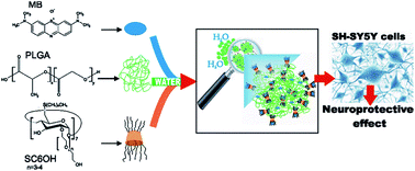
On this basis, in this study, to the best of our knowledge, we introduce for the first time a nanoassembly based on amphiphilic CD [(2-oligo-(ethyleneoxide)-6-hexylthio)-β-CyD] (SC6OH) and PLGA. The new PLGA/ACyD NPs are intended to deliver a hydrophilic drug such as MB in cell models for neuroprotection studies (Scheme 1). MB loaded-PLGA/SC6OH NPs were prepared by the nanoprecipitation/solvent displacement method. The influence of ACyD on the encapsulation parameters was evaluated in comparison with PLGA NPs prepared without SC6OH but using both the same method and the W/O/W emulsion/solvent evaporation method, respectively. Interaction of MB with SC6OH was investigated by UV-vis and fluorescence spectroscopy. MB loaded PLGA/SC6OH NPs were characterized by Scanning Transmission Electron Microscopy (STEM) and Quasi-Elastic Light Scattering (QELS) to elucidate their morphology and size, respectively. FTIR-ATR analysis highlighted the entrapment of MB in the PLGA/SC6OH system. The influence of SC6OH on the in vitro release of the drug from NPs was evaluated and in vitro biological assays were performed on human neuroblastoma cell lines (SH-SY5Y) in order to assess the biocompatibility of PLGA/SC6OH NPs and the neuroprotective effect of MB loaded-PLGA/SC6OH NPs, against cellular metabolic changes.
 |
| Scheme 1 Sketch of nanosphere formation in aqueous dispersion prepared by MB, PLGA and SC6OH by the nanoprecipitation/solvent displacement method. | |
2 Experimental
2.1 Materials
Methylene Blue (tetramethylthionine chloride, MB, MW = 319.86 a.m.u.) was received as gift sample from A.C.E.F. S.p.A. (Fiorenzuola d’Arda, Italy). Polylactic-co-glycolic acid (PLGA, RG502H, 50
:
50; MW range 7000–17
000), polyoxyethylene sorbitan monoleate (Tween® 80), Pluronic® F68, Span® 80 and all other chemicals and solvents were of analytical grade and obtained from Sigma-Aldrich (Milano, Italy). Heptakis(2-O-oligo(ethylene oxide)-6-hexylthio)-β-CyD (SC6OH, MW (nEO = 32, EO = ethylene oxide) = 3250 a.m.u) was synthesized according to general procedures.34 All dispersions used for spectroscopic characterizations were prepared in pure microfiltered water (Galenica Senese, Siena, Italy). Deionized, double distilled water was used throughout the study. All solvents were filtered through 0.22 μm Millipore® GSWP filters (Bedford, USA).
2.2 MB/SC6OH interaction studies
MB and SC6OH were solubilized separately in acetonitrile. After mixing, organic solvent was evaporated under reduced pressure at 40 °C, forming a film of MB/SC6OH. Micro-filtered water (3 ml) was added to the film and vortexed for 1 min to obtain MB/SC6OH nanoaggregates at a 1
:
10 molar ratio ([MB] = 3 μM, [SC6OH] = 30 μM), corresponding to ≅ 1/10 w/w.35 The dispersions were then analyzed by UV-vis spectroscopy, steady-state fluorescence emission, and STEM. UV-vis spectra were performed using a FullTech Instruments (Roma, Italy) double beam spectrophotometer mod. PG T80 (resolution 0.001 × 10⁻³ absorbance units; signal-to noise ratio, 1 × 10⁻⁴). One centimeter (d) rectangular quartz cells (Hellma, Milano, Italy) were employed in the 400–800 nm spectral range. All measurements were carried out at 25.0 ± 0.1 °C and running at least three times. The pH of the MB/SC6OH dispersions was ≅6.8. Steady-state fluorescence measurements were performed on a Jasco model FP-750 spectrofluorimeter. Emission spectra were collected using λexc = 590 nm in a 1 cm path length quartz cell. The spectra have not been corrected for the adsorption at λexc. The depolarized fluorescence spectra were measured using: |
 | (1) |
where ρ is the static anisotropy and IVV, IHH, IVH and IHV are the fluorescence intensities registered with different polarizer orientations (V = vertical, H = horizontal).36 STEM images were carried out at a 200 kV accelerating voltage, using a Jeol JEM 2010F (JEOL Ltd. Japan) microscope. STEM imaging allows a decrease of the electron dose with respect to conventional TEM images, reducing the damage to carbon-based samples even at 200 kV.
2.3 Preparation of NPs
MB loaded-PLGA/SC6OH NPs were prepared by the nanoprecipitation/solvent displacement method. Briefly, MB (0.5 mg), PLGA (20 mg) and different amounts of SC6OH (10, 20, 30 and 40 mg) were dissolved in acetone (6 ml). The organic phase was poured into 20 ml of aqueous solution under magnetic stirring, thus forming a milky colloidal suspension. The suspensions were left to stir overnight to eliminate the organic solvent.37 NPs were then purified by centrifugation at 5000 rpm for 15 min, collecting and centrifuging again the supernatants at 18
000 rpm for 30 min with a Beckman Optima™ XL-100K centrifuge. The supernatants were eliminated. The pellets were re-suspended in 1 ml of water, and then freeze-dried (VirtTis Benchtop K Instrument, SP 127 Scientific, USA) after addition of trehalose (5% w/v) as a lyoprotectant agent. MB loaded-PLGA NPs were prepared dissolving MB (0.5 mg), PLGA (20 mg) and Tween 80® (0.5% w/v) in acetone (6 ml) and adopting the previous procedure.
Additional MB loaded-PLGA NPs were obtained by the W/O/W emulsion/solvent evaporation method by dissolution of MB (0.5 mg) in 1 ml of water. This solution was added dropwise to 5 ml of an acetone/dichloromethane mixture (1
:
1, v/v) containing PLGA (20 mg) and Span® 80 (0.5%, w/v), under sonication over an ice bath for 2 min. This first emulsion was added dropwise to 50 ml of an aqueous solution containing Pluronic® F68 (3%, w/v) under sonication for 1 min.38 The colloidal suspension was stirred overnight to eliminate the organic solvent, and then was subjected to the same procedure described above (purification by centrifugation and lyophilization in presence of trehalose).
2.4 Characterization of NPs
The NPs were weighed and the yield percentage was calculated using the following formula:
% Yield = (actual weight/theoretical weight) × 100 |
Morphology of the NPs was investigated by STEM before and after freeze-drying using the same apparatus previously described. The mean sizes of NPs were determined via QELS experiments by using an He–Ne laser source (λ = 632.8 nm) at a power of 10 mW, linearly polarized orthogonal to the scattering plane. The scattered light, collected in a self-beating mode, is analyzed using a MALVERN 4700 correlator which builds up the normalized intensity autocorrelation function.39 FTIR-ATR measurements were performed at room temperature using a Bomem DA8 Fourier transform spectrometer, operating with a Globar source, in combination with a KBr beamsplitter and a DTGS/KBr detector. Spectra were collected in the 600–3700 cm−1 wavenumber range. The powders were contained with a Golden Gate diamond ATR system, based on the ATR technique.40 The spectra were recorded in a dry atmosphere, in order to avoid contamination, with a resolution of 2 cm−1, automatically adding 100 repetitive scans in order to obtain a good signal-to-noise ratio and high reproducibility. All the IR spectra were normalized for taking into account the effective number of absorbers. No mathematical correction (e.g., smoothing) was done, and spectroscopic manipulation such as baseline adjustment and normalization were performed using the Spectracalc software package GRAMS (Galactic Industries, Salem, NH, USA). An ATR setup exhibits various advantages with respect to an ordinary absorption setup. It is non-destructive, requires only micrograms of sample, and at the origin of the spectra displays a very good signal-to-noise ratio, it being in particular easy to avoid saturation of the bands. In addition, chemical analysis can be performed directly on the ATR spectra, avoiding the implementation of elaborate calculations of optical constants.41
2.5 MB loading and entrapment efficiency
The drug loading (or content) (D.C.%) and encapsulation efficiency (E.E.%) percentages were determined by dissolving each type of NP (3 mg) of in acetone (V = 3 ml). The resulting solutions were filtered (Nylon 0.2 μm, Millipore filter – Bedford, USA) and analyzed by UV-vis spectrophotometry for the quantitative determination of MB, using the same instrument and conditions described before. The drug loading and encapsulation efficiency percentages were calculated, using the following equations, respectively:
Entrapment efficiency (%) = (amount of MB in NPs/amount of MB initially added to formulation) × 100 |
Drug loading (%) = (amount of MB in NPs/weighted amount of NPs) × 100 |
2.6 In vitro release of MB from NPs
Weighted amounts of MB-loaded PLGA/SC6OH40 NPs and MB loaded-PLGA NPs, prepared by nanoprecipitation/solvent displacement methods (1 mg and 10 mg, respectively), were suspended in 10 ml of phosphate buffer solution (PBS, pH 7.4) and poured into a dialysis bag (Spectra/Por dialysis bags, 3500 MWCO – Spectrum Laboratoris, Inc.). The bags were placed under magnetic stirring (100 rpm) into a beaker containing 90 ml of PBS (pH 7.4) maintained at 37.0 ± 0.5 °C. At fixed time intervals (1, 5, 24, 48, 72 and 120 h) the dialysis medium was collected and substituted with 90 ml of fresh PBS. All the collected volume was evaporated under vacuum at 25.0 ± 0.1 °C and the residue was solubilized in acetone (2 ml) and analyzed by UV-vis spectroscopy. The experiments were done in triplicate and are expressed as mean values ± standard deviations (S.D.). The resulting release data were treated according to zero-order (cumulative percentage of released drug vs. time), first-order (log cumulative percentage of remaining drug vs. time), and Higuchi (cumulative percentage of released drug vs. square root of time) equation models.42
2.7 Biological studies
Human neuroblastoma SH-SY5Y cells were cultured in 1
:
1 Ham’s F12 Dulbecco’s modified Eagle’s medium-low glucose, supplemented with 10% fetal calf serum, in a 95% air–5% CO2 humidified incubator at 37.0 ± 0.5 °C. In some experiments, cells were differentiated with 10 μM retinoic acid (RA) for six days as described by Guarneri et al.43 For drug treatment, SH-SY5Y cells (3 × 10⁴) were seeded on 96-well plates. After 48 h, the medium was replaced with fresh medium and cells were treated with free MB, MB-loaded PLGA/SC6OH40 NPs, or empty nanospheres (PLGA or PLGA/SC6OH40), at the indicated concentrations, for 24 hours. To verify the effects on longer incubation times, some treatments lasted 48 or 72 hours. In neuroprotective experiments, cells were treated with free MB or MB loaded-PLGA/SC6OH40 NPs at the indicated concentrations, and exposed to 20 μM iodoacetic acid (IAA). After 2 h, media were removed and fresh media containing free MB or MB loaded-PLGA/SC6OH40 NPs, at the same concentrations previously employed, were again added for further 22 h. To increase the metabolic changes induced by IAA and to stabilize MB effects, cells treated or untreated with IAA and/or MB and MB loaded-NPs were also exposed to NADH (165 μM) at the same conditions as above. Afterwards, cell viability was assessed by using a Cell Counting Kit-8 (CCK-8, Sigma-Aldrich) and an LDH assay kit (Cytotoxicity Detection Kit, Roche) according to the manufacturer’s instructions. All systems were suspended in sterile PBS (10 mM, pH 7.4) and diluted in the medium at the final concentration, just before use. Each experiment was carried out in triplicate and was repeated at least three times. Statistical analyses were performed using GraphPad Prism Version 5 for Windows, with a one-way ANOVA followed by a post hoc Bonferroni’s t test.
3. Results and discussion
3.1 MB/PLGA/SC6OH: interaction studies
Recently, both in vitro and in vivo studies demonstrated that MB is a promising candidate for the treatment of many neurological disorders (e.g., Parkinson’s and Alzheimer’s diseases).4–9 The drug was administered orally, but alternative administration routes with a suitable delivery systems could improve its in vivo efficacy. With the aim to improve the encapsulation efficiency and release rate of this hydrophilic drug, NPs based on PLGA and ACyD (SC6OH) were here proposed as an intriguing novel drug delivery system. Before proceeding to the characterization of the NPs, interaction studies between SC6OH with MB and PLGA were carried out both in aqueous medium and the solid state. As evidenced by STEM analysis, SC6OH/MB forms nanoaggregates with a hydrophilic shell probably constituted by oligo-ethylene groups of the macrocycle (see Fig. S1†).
3.1.1 UV-vis and fluorescence studies. UV-vis studies demonstrated that SC6OH assemblies interact with MB, influencing the drug’s spectrum (Fig. 1A). Free MB shows an absorption band centered around 662 nm, due to the presence in aqueous solution of a monomeric form, and a shoulder at about 615 nm, which is tentatively ascribable to its dimer form.44 In the presence of SC6OH, we observed a slight blue-shift and an increase of the intensity of either band and shoulder. We hypothesize that MB interacts externally with the oligo-ethylene groups of the macrocycle, probably by electrostatic interaction between the negative surface charge of the amphiphile,45 and the positive charge of the drug. On the other hand, the existence in water of an inclusion complex between MB and the cavity of SC6OH can be plausibly excluded on the basis of different results previously obtained by Zhang et al.44 These authors observed in the presence of CyDs a hyperchromicity of the main band at 662 nm and a hypochromicity of the shoulder at around 615 nm, both 10 and 5 nm red-shifted, respectively. They justified these results as a suppression of dimer formation and the inclusion of an MB monomer within the macrocycle cavities. Conversely in our studies MB/SC6OH exhibits a different spectrum than for other MB/CyDs systems, indicating a diverse interaction of MB with SC6OH. It is probable that the electrostatic interaction between the negative surface of ACyD nanoaggregates and cationic MB drives mainly the external positioning of the drug which is situated, probably in the monomeric form, in a good percentage on the surface of the nanospheres.46
 |
| Fig. 1 UV-vis (A) and fluorescence emission spectra (λexc = 590 nm) (B) of free MB (3 μM) (a) and MB/SC6OH system at 1 : 10 ([MB] = 3 μM, [SC6OH] = 30 μM) (b) in aqueous dispersions (T = 25.0 ± 0.1 °C, pH = 6.8). | |
Whereas the emission fluorescence intensity of MB is less affected in the SC6OH/MB system (Fig. 1B), the anisotropy values at the emission maxima (λ = 679 nm and at λ = 705 nm) are higher for SC6OH/MB complexes (ρ679 ≅ 0.12 and ρ705 ≅ 0.10) than in the free drug (ρ679 ≅ 0.09 and ρ705 ≅ 0.08), thus confirming the interaction between MB and SC6OH (inset of Fig. 1B).
3.1.2 FTIR studies. FTIR-ATR spectroscopy was used to characterize the interaction between PLGA and SC6OH and to confirm the entrapment of MB in NPs, based on the observation of the dramatic alterations in shape and position suffered by the vibrations of those functional groups involved in that interaction. FTIR-ATR spectra of all the pure substances, namely MB (a), PLGA (b), SC6OH (c) are described in the ESI (Fig. S2†) and attributed on the bases of previous FTIR-ATR studies on similar systems.47–49In the case of MB, prominent absorption bands are revealed at ∼1593 cm−1, assigned to vibrations of the aromatic ring (CN (lateral) stretching + CH bending out of plane (CH3) + CH bending in plane (ring)), at ∼1332 cm−1 (CC stretching + CH bending in plane (ring) + CH bending out of plane (CH3)), a double peak at ∼1140 cm−1 (CH bending in plane of the ring) and ∼1173 cm−1 (CH bending out of plane (CH3) + CH bending in plane of the ring). Finally, a composite band is observed in the ∼870 ÷ 800 cm−1 zone, corresponding to CH bending out of plane and skeletal deformations (CC and CS).
The pure PLGA spectrum revealed, as the main contributions, bands at ∼3000 cm−1 and ∼2956 cm−1 (C–H stretching of –CH2, –CH3), followed by a shoulder at ∼2918 cm−1 (C–H stretch of –CH–). Again, the strongest absorbance peak is evident at ∼1753 cm−1 (C
O stretching of the ester group), a composite peak in the ∼1470 ÷ 1340 cm−1 zone (CH-bending). Finally, the absorption at ∼1180 ÷ 1050 cm−1 was assigned to C–O stretching, whereas the band at ∼850 cm−1 was assigned to CH-bending.
The FTIR-ATR spectrum of SC6OH showed the presence of a large band from ∼3700 cm−1 to ∼3030 cm−1, reflecting the contribution to the O–H stretching vibration coming from different OH groups. Based on the results of previous FTIR-ATR studies on similar systems,47–49 these contributions have been ascribed, in particular, to come from primary (at ∼3525 cm−1) and secondary (at ∼3280 cm−1) OH groups of SC6OH, clusters of water molecules inside the hydrophobic SC6OH cavity (at ∼3580 cm−1), H2O molecules in the interstices among different SC6OH molecules linked to them via hydrogen bonds (at ∼3410 cm−1 and at ∼3174 cm−1), and hydroxyl groups of oligo-ethylene chains (at ∼3000 cm−1, expected to be overlapped with the C–H stretching vibrations). Going to lower wavenumbers, a double-peak is well evident in the ∼3030 ÷ 2680 cm−1 (CH stretching), and a prominent composite band at ∼1210 ÷ 1000 cm−1 (C–O stretching).
FTIR-ATR spectra of unloaded PLGA/SC6OH40 NPs and of the ternary system MB/PLGA/SC6OH have been collected, in order to investigate, at the solid state, the interaction between PLGA and SC6OH and to confirm the loading of MB in NPs. We decided to focus our attention on the vibrational bands of free MB at ∼1593 cm−1 and ∼1332 cm−1, since they fall in a wavenumber range free from interfering peaks coming from PLGA and/or SC6OH, and so they can constitute excellent candidates to show some variations attributable to MB interaction with the NP matrix. In the same way, the vibrational band from ∼3700 cm−1 to ∼3030 cm−1 of pure SC6OH can be considered as a marker of changes in the H-bond environment of this system as a consequence of the establishment of an interaction, not having MB characteristic peaks in this zone, and the contribution coming from PLGA being negligible.
In Fig. 2A the FTIR-ATR spectrum of unloaded PLGA/SC6OH40 NPs is compared to the calculated weighed addition of the spectra of the two separate components.
 |
| Fig. 2 FTIR-ATR spectrum of (A) unloaded PLGA/SC6OH40 NPs (a) and calculated weighed addition of the spectra of PLGA and SC6OH (b), and (B) MB loaded-PLGA/SC6OH40 NPs (a), and calculated weighed addition of the MB and PLGA/SC6OH40 experimental profiles (b). | |
Evident spectral changes are revealed, especially in the O–H and C–H stretching bands, that appear upshifted and modified in shape and the relative intensity passes from the calculated profile to the experimental one. These changes suggest a modification of bond strengths and lengths as a consequence of the activation of some molecular interactions between these two systems, involving the aforementioned functional groups. In particular, it is reasonable to assume a redistribution of water molecules among the different hydrogen bond sites, because of the formation of hydrogen bonds between PLGA and SC6OH. Again, the observed high-wavenumber shift of the O–H stretching vibration, with an overall diminishing of the bandwidth, allowed us to hypothesize that the observed change in the hydrogen bonding scheme should imply a reduced cooperativity involving shorter lifetimes. Also the C–O stretching region undergoes a significant upshift in passing from the calculated to the experimental spectrum, together with relevant changes in the relative intensity of the peaks falling in this zone. This reveals that, because of the formation of new intermolecular hydrogen bonds between PLGA and SC6OH, the electrostatic environment (in turn related to the first neighbors of the oscillator) experienced by the C–O groups is altered in such a way to increase the overall dipole moment of the CO functional group, i.e. to become less intense, in agreement with the behavior of the O–H stretching band.
The entrapment of MB in PLGA/SC6OH40 NPs has been verified by comparing the FTIR-ATR spectrum of the MB loaded-PLGA/SC6OH40 NPs with the one obtained by a weighed superposition of MB and PLGA/SC6OH40 experimental profiles, as reported in Fig. 2B.
The disappearance, passing from the calculated to the experimental spectrum, of the bands at ∼1593 cm−1 and ∼1332 cm−1, typical of MB, is revealed as the main result, evidencing hindrance of the corresponding vibrational modes because of the close-fitting of MB within the NPs. It is also worth noting that no significant differences are revealed in the O–H stretching band shape, suggesting that the insertion of MB does not significantly alter the hydrogen bond network of the PLGA/SC6OH40 system.
3.3 Overall properties of MB loaded-PLGA/SC6OH NPs
MB-loaded NPs were easily prepared by simple nanoprecipitation/solvent displacement (generally not suitable for hydrophilic drugs), resulting from interaction between SC6OH and both MB and PLGA. This property influences notably all technological parameters of the NPs as reported in Table 1.
Table 1 Overall properties of NPs (sizes), polydispersity index (P.I.), yield percentage and encapsulation efficiency percentage (E.E.%) of MB loaded-PLGA NPs prepared in the presence and in the absence of SC6OH. SD was calculated from at least three different batches
NPs compositiona |
Sizes (nm) ± S.D. |
P. I. ± S.D. |
Yield% ± S.D. |
D.C.% ± S.D. |
E.E.% ± S.D. |
The amounts of MB and PLGA were always maintained at 0.5 mg and 20 mg, respectively. The amount of SC6OH was changed from 10 to 40 mg. NPs were prepared with the nanoprecipitation/solvent displacement method using acetone as the organic phase. NPs were prepared with the W/O/W emulsion/solvent evaporation method using a mixture of acetone/dichloromethane (1 : 1, v/v) as the organic phase; Tween 80 and Pluronic F68® were used as surfactants. |
MB loaded-PLGAb |
220 ± 4 |
0.19 ± 0.02 |
43.21 ± 2.69 |
0.52 ± 0.19 |
3.12 ± 1.12 |
MB loaded-PLGA (W/O/W)c |
266 ± 5 |
0.40 ± 0.10 |
39.98 ± 6.32 |
1.13 ± 0.26 |
6.75 ± 1.54 |
MB loaded-PLGA/SC6OH10b |
230 ± 6 |
0.13 ± 0.06 |
36.12 ± 3.21 |
5.02 ± 0.43 |
30.09 ± 2.58 |
MB loaded-PLGA/SC6OH20b |
221 ± 2 |
0.23 ± 0.08 |
40.58 ± 1.89 |
4.86 ± 0.38 |
29.14 ± 2.31 |
MB loaded-PLGA/SC6OH30b |
226 ± 5 |
0.15 ± 0.04 |
34.14 ± 5.61 |
5.43 ± 0.61 |
32.56 ± 3.65 |
MB loaded-PLGA/SC6OH40b |
201 ± 2 |
0.16 ± 0.08 |
40.85 ± 4.01 |
9.65 ± 0.31 |
57.89 ± 1.86 |
No influence was exerted on the recovery yield (about 40% for all systems), size (always in the nanometric range) or polydispersity index by the utilization of SC6OH. Conversely, a significant effect was observed in the E.E.% values. In the presence of different amounts of ACyD, a large increase of E.E.% was observed with respect to the NPs prepared both with the nanoprecipitation/solvent displacement method (E.E.% of about 3%, w/w) and with the W/O/W double emulsion method (E.E.% of about 7%, w/w) but without SC6OH. Due to the higher affinity of hydrophilic MB for water, rather than for the organic solvent (log
P value in octanol
:
PBS at pH 7 is −0.9),50 it interacts poorlywith the hydrophobic PLGA, leading plausibly, when the PLGA NPs were prepared in the absence of SC6OH, to drug diffusion from the organic phase containing PLGA to the external aqueous environment.18 On the other hand, the amphiphilic characteristics of SC6OH can help the interaction both with hydrophobic PLGA, by means of SC6OH hydrophobic chains, and with hydrophilic MB which interacts with hydrophilic surface rich of oligo-ethylene groups.
It is probable that SC6OH is able to keep MB into the organic phase, along with PLGA, thus reducing MB diffusion. ACyD plays a key-role in the entrapment of MB, as it is evidenced by MB E.E.% in PLGA/SC6OH NPs which have been obtained by nanoprecipitation. By using this technique, MB E.E.% values are higher than those obtained using W/O/W emulsion/solvent evaporation, although this latter method is generally utilized to entrap hydrophilic drugs within PLGA.16
Because of the similar sizes and yield of all investigated systems and provided that the highest E.E.% was observed for the NPs prepared using the highest SC6OH concentration, PLGA/SC6OH40 NPs was selected for the following morphological characterization by STEM analysis and our successive studies. In Fig. 3 STEM images of MB loaded-PLGA/SC6OH40 NPs dispersion (before freeze-drying) are reported. STEM shows the presence of regular shaped NPs (Fig. 3A), sporadically coated by MB/SC6OH micellar aggregates as featured in Fig. 3B. Freeze-dried NP powder has a less-regular shape and a certain degree of aggregation, although still within the nanometric range (data not shown).
 |
| Fig. 3 Representative STEM images of MB-loaded PLGA/SC6OH40 NPs (A) with featured PLGA NPs coated with SC6OH/MB aggregates (B). | |
3.4 Release studies
The release profiles of MB from PLGA and PLGA/SC6OH40 NPs, prepared by the nanoprecipitation/solvent displacement method, were evaluated in PBS at pH 7.4 (Fig. 4). The novel PLGA/SC6OH40 NPs show an appealing release profile, being able to sustain MB release for about 5 days, after a burst effect of about 40% (w/w) in the first 5 h of the experiment. On the contrary PLGA NPs not associated with SC6OH produced a total release of MB within 24 h. Probably, in this latter system the encapsulated drug was principally located on the surface or close to the surface of the NPs as a results of the migration of MB from the organic to aqueous phase during the preparation process. The presence of SC6OH both in the NPs matrix and surface (as demonstrated by STEM analysis, see Fig. 3) confers more homogeneity to the system and improves hydrophilicity at the surface. Actually, we believe that about 40% of the entrapped MB could be superficially located in the NPs by means of electrostatic interaction with SC6OH. The remaining 60% is presumably incorporated in PLGA/SC6OH40 NPs matrix. Hence more time was needed for its diffusion through the polymer matrix to the release medium.
 |
| Fig. 4 In vitro release profiles of MB loaded-PLGA NPs (●) and MB loaded-PLGA/SC6OH40 NPs (■) in PBS (pH 7.4) and at 37.0 ± 0.5 °C. | |
The release data of MB from PLGA/SC6OH40 NPs were treated according to zero-order, first-order and Higuchi equation models in order to determine the release rate and mechanisms of drug release from the NPs. The best model fitting the release data was evaluated by correlation coefficient (r2). The release parameters (r2 and k) reported in Table 2 showed the best correlation by using the Higuchi model,42 highlighting the release of MB from the matrix as a square root of a time-dependent process based on Fickian diffusion.
Table 2 Regression coefficient (r2) and rate constant (k) of MB release data from PLGA/SC6OH40 NPs according to different kinetic models
Zero order |
First order |
Higuchi |
r2 |
k0 (h−1) |
r2 |
k1 (h−1) |
r2 |
kH (h−1/2) |
0.8794 |
0.5358 |
0.9361 |
0.0346 |
0.9799 |
6.7967 |
3.5 In vitro studies
In order to determine the biological activity of MB loaded-PLGA/SC6OH40 NPs, we first carried out cell biocompatibility tests of the empty (both PLGA and PLGA/SC6OH40 NPs) or MB-loaded PLGA/SC6OH40 NPs. Then, a CCK-8 assay was performed to analyze SH-SY5Y cell viability upon a 24 hour treatment with different amounts of NPs. As shown in Fig. 5A and B, cell viability was unaffected by PLGA and was significantly increased by PLGA/SC6OH40 NPs at the concentrations tested, except for a trend towards a reduced viability at the highest investigated concentration (166 μg ml−1) of the combined nanospheres. Cell death assay based on LDH released into the medium confirmed the lack of toxicity of PLGA or PLGA/SC6OH40 NPs, and only revealed cell death of about 15–30% at the concentration of 166 μg ml−1 of PLGA/SC6OH40 NPs (data not shown). Therefore, PLGA alone or combined with SC6OH appeared to be well tolerated over a wide concentration range (1.6–83.0 μg ml−1 of PLGA/SC6OH40 NPs). By analyzing the effects on cell viability of the range of concentrations of PLGA/SC6OH40 NPs assembled with MB and the relative concentrations of free MB (0.025–10 μM), it was found that both free and encapsulated MB had similar toxic effects for the cells when used at concentrations above 0.5 μM, whilst they did not affect cell survival at lower concentrations (0.025–0.5 μM) (Fig. 5C and D).
 |
| Fig. 5 Effects of a 24 hour treatment with various concentrations of PLGA (A), PLGA/SC6OH40 (B), free MB (C) or MB loaded-PLGA/SC6OH40 (D) on SH-SY5Y cell viability. Cells were treated with empty or encapsulated nanospheres at the indicated concentrations, and cell viability was measured at 24 after treatment using CCK-8 assay. The amounts of empty nanospheres tested in (B) is equivalent to those loaded with different concentrations of MB (0.025–10 μM) in (D). Data are expressed as a percentage of the absorbance measured in untreated control cells (CNT), and represent the mean ± SD of three independent experiments performed in triplicate. (*) Indicates significant differences from CNT; *p < 0.05, **p < 0.01, ***p < 0.001. | |
This cell biocompatibility of a 24 hour treatment of MB loaded-PLGA/SC6OH40 NPs was confirmed after longer incubation periods (48 and 72 h treatments; see Fig. S3†). In addition, at the same low concentrations, no toxicity was detected after treatment with MB loaded-NPs or free MB of SH-SY5Y cells differentiated with retinoic acid in a neuronal-like phenotype (Fig. S4†).
MB is suggested to act as a neuroprotective agent, working with self-oxidant properties and effectively restoring mitochondrial actions and brain metabolism, however, it may also have adverse effects upon the cellular metabolic state and its concentration.21,51–53 To verify whether free MB and MB loaded-PLGA/SC6OH40 NPs have neuroprotective actions at their lower concentrations, we employed an experimental paradigm to change the cellular metabolic state of SH-SY5Y cell by using IAA, which inhibits glycolysis and causes cell death,54 with NADH as an electron donor.
This paradigm has been previously used by Poteet et al.51 which suggested antioxidant properties of MB against IAA-induced cell toxicity through the action of NADH. We found that LDH release occurred by 2 h exposure with IAA and further increased by adding NADH (Fig. 6).
 |
| Fig. 6 Neuroprotective effect of MB loaded-PLGA/SC6OH40 NPs (MB-NPs). SH-SY5Y cells were treated with various concentrations of free MB or MB-NPs and exposed to IAA (20 μM) with or without NADH (165 μM) for 2 h. After 24 h, LDH release was assessed as described in the Experimental section. Data represent the mean ± SD of three independent experiments performed in triplicate. ΔΔΔp < 0.001 versus untreated cells; ΦΦΦp < 0.001 versus IAA treated cells; *p < 0.05, **p < 0.01, ***p < 0.001 versus IAA + NADH and IAA plus free MB + NADH treated cells; #p < 0.05 versus IAA and IAA plus free MB treated cells. δp < 0.05 versus MB-NPs treated cells without NADH. | |
However, treatment of SH-SY5Y cells with free MB (0.01–0.05 μM) did not protect against IAA-induced LDH release, as previously reported in HT-22 cells,54 either in the presence or absence of NADH. We instead found significant protection using MB loaded-PLGA/SC6OH40 NPs (0.01–0.025 μM), which reduced IAA-induced cell death and showed to be more effective in the presence of NADH, even at higher concentrations tested (0.01–0.05 μM). Presently, we cannot explain the inability of free MB to prevent SH-SY5Y cell death. It has been shown that MB functioning as an alternative electron carrier, may accept electrons from the NADH of mitochondrial complex I and transfer them to cytochrome c (complex IV); this mechanism allows complex I/III blockage to be bypassed.51,55 Perhaps neuroblastoma SH-SY5Y cells have a different metabolic rate and, certainly, more investigations are needed. However, it is interesting that MB loaded-PLGA/SC6OH40 NPs efficiently protected cells from IAA-induced metabolic alterations. It may be possible that in altered metabolic conditions, the gradual release of MB by NPs, in combination with NADH, facilitates mitochondrial electron transfer and cell survival. Studies on the cellular uptake of MB-loaded NPs could shed light on the eventual potentiating effect of MB.
4 Conclusions
Here, we demonstrated that non ionic ACyD (SC6OH) interacts with PLGA, influencing remarkably the physico-chemical properties of PLGA NPs, thus improving their technological parameters, especially in terms of MB loading, significant increase of encapsulation efficiency and prolonged release. Although more in vitro and in vivo studies are required, our results suggest that PLGA/SC6OH40 NPs may be a promising delivery system for MB which, thus-formulated, can enhance its neuroprotective effect on neuroblastoma SH-SY5Y cells. This result could be important to propose novel potential therapeutic strategies for the treatment of severe neurological disease at elevated medical need.
Acknowledgements
This work was partially supported by University of Messina (Ricerca di Ateneo). Grant support was also provided by EuroBioSAS-OP-009 (ICS project) to A. M., and PNR-CNR Aging program 2012–2016 to P. G. We are grateful to Dr N. Micali CNR-IPCF Messina for light scattering measurements.
Notes and references
- R. H. Schirmer, H. Adler, M. Pickhardt and E. Mandelkow, Neurobiol. Aging, 2011, 32, 2325e7 CrossRef PubMed.
- L. M. Moreira, J. P. Lyon, A. P. Romani, D. Severino, M. R. Rodrigues and H. P. M. de Oliveira, Phenotiazinium dyes as photosensitizers (PS) in photodynamic therapy (PDT): Spectroscopic properties and photochemical mechanisms, in Advanced Aspects of Spectroscopy, ed. M. A Farrukh, InTech Open, 2012, pp. 393–422 Search PubMed.
- J. P. Tardivo, A. Del Giglio, C. S. de Oliveira, D. S. Gabrielli, H. C. Junqueira, D. B. Tada, D. Severino, R. d. F. Turchiello and M. S. Baptista, Photodiagn. Photodyn. Ther., 2005, 2, 175 CrossRef CAS PubMed.
- H. Atamna, A. Nguyen, C. Schultz, K. Boyle, J. Newberry, H. Kato and B. N. Ames, FASEB J., 2007, 22, 703 CrossRef PubMed.
- N. L. Callaway, P. D. Riha, K. M. Wrubel, D. McCollum and F. Gonzalez-Lima, Neurosci. Lett., 2002, 332, 83 CrossRef CAS PubMed.
- D. X. Medina, A. Caccamo and S. Oddo, Brain Pathol., 2011, 21, 140 CrossRef CAS PubMed.
- M. Necula, L. Breydo, S. Milton, R. Kayed, W. E. van der Veer, P. Tone and C. G. Glabe, Biochemistry, 2007, 46, 8850 CrossRef CAS PubMed.
- C. M. Wischik, M. Novak, H. C. Thøgersen, P. C. Edwards, M. J. Runswick, R. Jakes, J. E. Walker, C. Milstein, M. Roth and A. Klug, Proc. Natl. Acad. Sci. U. S. A., 1988, 85, 4506 CrossRef CAS.
- M. Oz, D. E. Lorke and G. A. Petroianu, Biochem. Pharmacol., 2009, 78, 927 CrossRef CAS PubMed.
- C. M. Wischik, P. Bentham, D. J. Wischik and K. M. Seng, Alzheimer’s Dementia, 2008, 4(4), T167 CrossRef.
- I. Walter-Sack, J. Rengelshausen, H. Oberwittler, J. Burhenne, O. Mueller, P. Meissner and G. Mikus, Eur. J. Clin. Pharmacol., 2009, 65, 179 CrossRef CAS PubMed.
- L. Illum, Eur. J. Pharm. Sci., 2000, 11, 1 CrossRef CAS PubMed.
- A. Mistry, S. Stolnik and L. Illum, Int. J. Pharm., 2009, 379, 146 CrossRef CAS PubMed.
- T. Musumeci, L. Vicari, C. A. Ventura, M. Gulisano, R. Pignatello and G. Puglisi, J. Nanosci. Nanotechnol., 2006, 6, 3118 CrossRef CAS PubMed.
- A. Takami, B. Masanori and A. Mitsuru, Adv. Polym. Sci., 2012, 247, 31 Search PubMed.
- L. Costantino, F. Gandolfi, L. Bossy-Nobs, G. Tosi, R. Gurny, F. Rivasi, M. A. Vandelli and F. Forni, Biomaterials, 2006, 27, 4635 CrossRef CAS PubMed.
- J. M. Barichello, M. Morishita, K. Takayama and T. Nagai, Drug Dev. Ind. Pharm., 1999, 25, 471 CrossRef CAS PubMed.
- I. Bala, S. Hariharan and M. N. Kumar, Crit. Rev. Ther. Drug Carrier Syst., 2004, 21, 387 CrossRef CAS PubMed.
- U. K. Jinwal, A. Groshev, J. Zhang, A. Grover and V. B. Sutariya, Curr. Drug Delivery, 2013, 11, 541 CrossRef.
- F. Danhier, E. Ansorena, J. M. Silva, R. Coco, A. Le Breton and V. Préat, J. Controlled Release, 2012, 161, 505 CrossRef CAS PubMed.
- F. Tewes, E. Munnier, B. Antoon, L. Ngaboni Okassa, S. Cohen-Jonathan, H. Marchais, L. Douziech-Eyrolles, M. Souce, P. Dubois and I. Chourpa, Eur. J. Pharm. Biopharm., 2007, 66, 488 CrossRef CAS PubMed.
- E. Cohen-Sela, M. Chorny, N. Koroukhov, H. D. Danenberg and G. Golomb, J. Controlled Release, 2009, 133, 90 CrossRef CAS PubMed.
- G. U. Ruiz-Esparza, S. Wu, V. Segura-Ibarra, F. E. Cara, K. W. Evans, M. Milosevic, A. Ziemys, M. Kojic, F. Meric-Bernstam, M. Ferrari and E. Blanco, Adv. Funct. Mater., 2014, 24, 4753 CrossRef CAS.
- L. García-González, L. Yépez-Mulía and A. Ganem, Eur. J. Pharm. Sci., 2016, 81, 113 CrossRef PubMed.
- G. Rigaux, V. G. Roullin, C. Cadiou, C. Portefaix, L. Van Gulick, G. B. œuf, M. C. Andry, C. Hoeffel, L. Vander Elst, S. Laurent, R. Muller, M. Molinari and F. Chuburu, Nanotechnology, 2014, 25, 445103 CrossRef CAS PubMed.
- A. Monza da Silveira, G. Ponchel, F. Puisieux and D. Duchêne, Pharm. Res., 1998, 15, 1051 CrossRef CAS.
- C. Cannavà, S. Tommasini, R. Stancanelli, V. Cardile, F. Cilurzo, I. Giannone, G. Puglisi and C. A. Ventura, Colloids Surf., B, 2013, 111, 289 CrossRef PubMed.
- E. Bilensoy and A. A. Hincal, Expert Opin. Drug Delivery, 2009, 6, 1161 CrossRef CAS PubMed.
- M. Roux, B. Perly and F. Djedaini-Pilard, Eur. Biophys. J., 2007, 36, 861 CrossRef CAS PubMed.
- J. B. G. Yaméogo, A. Gèze, L. Choisnard, J.-C. Putaux, R. Semdé and D. Wouessidjewe, Curr. Top. Med. Chem., 2014, 14, 526 CrossRef.
- P. Falvey, C. W. Lim, R. Darcy, T. Revermann, U. Karst, M. Giesbers, A. T. M. Marcelis, A. Lazar, A. W. Coleman, D. N. Reinhoud and B. J. Ravoo, Chem.–Eur. J., 2005, 11, 1171 CrossRef CAS PubMed.
- D. Lombardo, A. Longo, R. Darcy and A. Mazzaglia, Langmuir, 2004, 20, 1057 CrossRef CAS PubMed.
- A. Mazzaglia, B. J. Ravoo, R. Darcy, P. Gambadauro and F. Mallamace, Langmuir, 2002, 18, 1945 CrossRef CAS.
- A. Mazzaglia, R. Donohue, B. J. Ravoo and R. Darcy, Eur. J. Org. Chem., 2001, 1715 CrossRef CAS.
- R. Stancanelli, M. Guardo, C. Cannavá, G. Guglielmo, P. Ficarra, V. Villari, N. Micali and A. Mazzaglia, J. Pharm. Sci., 2010, 99, 3141 CrossRef CAS PubMed.
- J. R. Lackowicz, Principles of Fluorescence Spectroscopy, KluwerAcademic, Plenum Publishers, New York, 1999 Search PubMed.
- J. P. Rao and K. E. Geckeler, Prog. Polym. Sci., 2011, 36, 887 CrossRef CAS and references therein.
- V. Lassalle and M. L. Ferreira, Macromol. Biosci., 2007, 7, 767 CrossRef CAS PubMed.
- V. Villari and N. Micali, J. Pharm. Sci., 2008, 97, 1703 CrossRef CAS PubMed.
- Y. Maréchal, J. Mol. Struct., 2003, 648, 27 CrossRef.
- L. Lewiner, J. P. Klein, F. Puel and G. Fevotte, Chem. Eng. Sci., 2001, 56, 2069 CrossRef.
- P. Costa and J. M. S. Lobo, Eur. J. Pharm. Sci., 2001, 13, 123 CrossRef CAS PubMed.
- P. Guarneri, C. Cascio, T. Piccoli, F. Piccoli and R. Guarneri, J. Neurosci. Res., 2000, 60, 656 CrossRef CAS PubMed.
- G. Zhang, S. Shuang, C. Dong and J. Pan, Spectrochim. Acta, Part A, 2003, 59, 2935 CrossRef.
- A. Mazzaglia, M. L. Bondì, A. Scala, F. Zito, G. Barbieri, F. Crea, G. Vianelli, P. Mineo, T. Fiore, C. Pellerito, L. Pellerito and M. A. Costa, Biomacromolecules, 2013, 14, 3820 CrossRef CAS PubMed.
- H. C. Junqueira, S. Divinomar, L. G. Dias, S. M. Gugliotti and M. S. Baptista, Phys. Chem. Chem. Phys., 2002, 4, 2320 RSC.
- C. Cannavà, V. Crupi, P. Ficarra, M. Guardo, D. Majolino, A. Mazzaglia, R. Stancanelli and V. Venuti, J. Pharm. Biomed. Anal., 2010, 51, 1064 CrossRef PubMed.
- V. Crupi, R. Ficarra, M. Guardo, D. Majolino, R. Stancanelli and V. Venuti, J. Pharm. Biomed. Anal., 2007, 44, 110 CrossRef CAS PubMed.
- N. Motlekar, M. A. Khan and B. B. C. Youan, J. Appl. Polym. Sci., 2006, 101, 2070 CrossRef CAS.
- I. Walker, S. A. Gorman, R. D. Cox, D. I. Vernon, J. Griffiths and S. B. Brown, Photochem. Photobiol. Sci., 2004, 3, 653 CAS.
- A.-L. Lin, E. Poteet, F. Du, R. C. Gourav, R. Liu, Y. Wen, A. Bresnen, S. Huang, P. T. Fox, S. H. Yang and T. Q. Duong, PLoS One, 2012, 7, e46585, DOI:10.1371/journal.pone.0046585.
- J. C. Rojas, A. K. Bruchey and F. Gonzalez-Lima, Prog. Neurobiol., 2012, 96, 32 CrossRef CAS PubMed.
- M. Yamashita, T. Nonaka, T. Arai, F. Kametani, V. L. Buchman, N. Ninkina, S. O. Bachurin, H. Akiyama, M. Goedert and M. Hasegawa, FEBS Lett., 2009, 583, 2419 CrossRef CAS PubMed.
- E. Poteet, A. Winters, L. J. Yan, K. Shufelt, K. N. Green, J. W. Simpkins, Y. Wen and S. H. Yang, PLoS One, 2012, 7, e48279, DOI:10.1371/journal.pone.0048279.
- F. Gonzalez- Lima and A. Auchter, Front. Cell. Neurosci., 2015, 9, 179, DOI:10.3389/fncel.2015.00179.
Footnote |
† Electronic supplementary information (ESI) available. See DOI: 10.1039/c5ra27386b |
|
This journal is © The Royal Society of Chemistry 2016 |
Click here to see how this site uses Cookies. View our privacy policy here.