A multifunctional resealable perfusion chip for cell culture and tissue engineering

Qi Langa, Yukun Ren*a, Yanshuang Wub, Yongbo Guoa, Xin Zhaoc, Ye Taoa, Jiangwei Liua, Hong Zhaod, Lei Lei*b and Hongyuan Jiang*a
aSchool of Mechatronics Engineering, Harbin Institute of Technology, Harbin 150001, China. E-mail: rykhit@hit.edu.cn; jhy_hit@sina.com
bDepartment of Histology and Embryology, Harbin Medical University, Harbin 150081, China. E-mail: leiys2002@yahoo.com
cThe Key Laboratory of Biomedical Information Engineering of Ministry of Education, School of Life Science and Technology, Xi'an Jiaotong University, Xi'an 710049, China
dThe State Key Lab of Chemical Engineering, Tsinghua University, Beijing 100084, China

Received 18th December 2015 , Accepted 7th March 2016

First published on 11th March 2016


Abstract

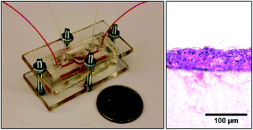
We describe a multifunctional resealable perfusion chip to mimic the human environment for cell or tissue culture in vitro and to increase the efficiency of culture in this study. To meet the culture requirement of a two-dimensional (2D) submerged cultures and an air–liquid interface perfusion cultures, a multifunctional resealable perfusion chip was designed and fabricated. Human embryonic kidney cells (HEK293T) and human colon carcinoma cells (SW620) were submerged cultured in the chip for 72 h. Cell viability and cell proliferation tests were used to evaluate the performance of the chip. Moreover, an artificial epidermis was developed, and it lasted for 7 days for the submerged culture immortal human keratinocyte (HaCaT) monolayer and 35 days for the air–liquid interface differentiated culture. The artificial epidermis cultured in the chip and in a conventional transwell were evaluated by the Live/Dead Viability/Cytotoxicity kit, histology, and hematoxylin and eosin staining. The stability and the benefits of our resealable chip were demonstrated by culture cells and epidermis tissue. Unlike a conventional static culture, no obvious advantage was observed in the 2D submerged culture of HEK293T and SW620 cells in the chip, but an outstanding advantage was observed in the 2D HaCaT cell culture with high density and a reconstructed epidermis. The cells grew well, and the epidermis was successfully reconstructed. This result implies that our multifunctional chip has great potential in cell culture and tissue engineering applications.

Introduction

With the deterioration of the environment, the avian-origin influenza A (H7N9) virus,1 the Ebola virus,2 and the Middle East Respiratory Syndrome (MARS) coronavirus3 have emerged and caused thousands of human infections and even deaths. Thus, these infections seriously threaten the health of human beings. However, an average of 15 years and US $800 million are required to bring a single drug to the market.4 Therefore, developing new drugs is much slower than the occurrence of new disease outbreaks. Presently, new drugs are tested by animal experiments and clinical tests.5 Animal experiments with ethical issues present poorly predictive evaluation generated by the phylogenetic distance between animals and humans,6,7 and clinical tests may endanger trial participants.8,9 Therefore, building a standard in vitro testing platform that enables drug testing on human cells or tissue samples is necessary.

Cell culture and tissue engineering are essential techniques in biological science and biotechnology, and they can be used in drug test in vitro.10,11 However, conventional cell culture and tissue engineering in well-plates or Petri dishes require large cell numbers, large tissue samples, bulky incubators, and other equipment. Moreover, static culture in well-plates cannot supply a fluid dynamic environment to mimic the human body.12,13 Therefore, culturing cells or tissues in a miniature dynamic device, i.e., a perfusion chip is sensible. Currently, the organ chips of bone,14 lung,15,16 liver,17–19 and heart20,21 tissues have been designed to supply a dynamic environment to cells or tissues. These chips are miniature devices with complex structures. However, most of the above chips are closed, and the cells or tissues cannot be further tested or studied out of the chip. Developing a multifunctional resealable perfusion chip with a lid that can be removed during the cell seeding process, reattached during tissue culture to keep the cell microenvironment sealed and sterile, and opened again to extract the cells or tissue for further off-chip studies is necessary.

Most of the cells or tissues submerged cultured in a medium are the same as human tissues or organs. However, some tissues, i.e., epidermis tissue,22–24 bronchial epithelial tissue,25–27 etc., should be cultured at an air–liquid interface (ALI).28 In the ALI system, the apical surface of the epidermis cells or bronchial epithelial cells is exposed to air, which mimics the conditions found in the human epidermis or human airway.29 A multifunctional chip integrated with the function of both submerged culture and ALI should be developed.

In this paper, a multifunctional perfusion chip was designed and fabricated to mimic the dynamic human environment for cell and tissue culture in vitro. The chip is resealable using the polydimethylsiloxane (PDMS) lid sealed by polymethyl methacrylate (PMMA) clamps and bolts. We ran several types of cells to verify the wide application of our chip and examined the effect of the dynamic environment on the cells and tissues. Submerged low-density human embryonic kidney cells (HEK293T) and human colon carcinoma cells (SW620), i.e., normal and cancer cells, respectively, grew well in the chip for 72 h. This condition presented the same culture result as when static culture was conducted. High-density immortal human keratinocytes (HaCaT) cultured in the chip had more advantages in cell viability and cell proliferation than when they were cultured in well plates. The multifunctional perfusion chip integrated the function of ALI, which is used in culture epidermis. An artificial epidermis was developed in our chip for long-term culturing (42 d), and it demonstrated the stability and benefits of the multifunctional perfusion resealable chip.

Materials and methods

Design of the resealable perfusion chip

One of the design requirements for our chip is resealability. Fig. 1a and b show the style and the structure of the multifunctional resealable perfusion chip. The culture chamber (6.5 mm in diameter and 8 mm in height) and the two channels (1 mm in width and 1.5 mm in height) are composed of two PDMS layers clamped by two pieces of PMMA sheets with the aid of four pairs of bolts. The two PDMS layers are not bonded, and the chamber can be open by loosening the bolts. Once the bolts are retightened, the chip is sealed again.
image file: c5ra27102a-f1.tif
Fig. 1 Design and fabrication of the resealable chip. (a) Photograph of the perfusion chip. (b) Schematic of the perfusion chip. (c) Photograph of the sponge scaffold. (d) Schematic of the air–liquid interface perfusion culture system. (e) Fabrication, sterilization, and assembly of the resealable perfusion chip. (I) Pouring PDMS, (II) curing and punch holes, and (III) assembling.

For the 2D submerged cell culture, the cells or tissues are cultured at the bottom of the chamber, with one channel open and the other closed. In the ALI culture, the tissue or the hydrogel is supported using a sponge scaffold (6 mm in diameter and 3 mm in height) (Fig. 1c). The upper surface of the sponge scaffold and the upper surface of the lower channel are at the same level. The lower channel pumps the medium, while the upper channel is connected with a syringe filled with air (5% CO2). Pressure in the culture chamber is controlled by the syringe to keep the liquid level from being higher than the tissue or hydrogel. Fig. 1d illustrates the ALI perfusion culture system.

Fabrication of the resealable perfusion chip

Fig. 1e shows the fabrication, sterilization, and assembly of the resealable perfusion chip. A PMMA sheet (1.5 mm thickness) was cut using a laser cutter (K3020, Huitian) to form the channels and the chamber mold, which was pasted onto a glass slide. Next, an elastomer base and a PDMS curing agent (Dow Corning) were mixed at a ratio of 10[thin space (1/6-em)]:[thin space (1/6-em)]1 (v/v), poured onto the mold, and degassed in a vacuum drying chamber for 30 min (I in Fig. 1e). Afterwards, the PDMS mixture was cured at 80 °C in an oven for 120 min. The chambers and holes of the inlet or outlet were mechanically punched by a biopsy punch (PSAM14, Acuderm) (II in Fig. 1e). The lower channel layer and the chamber II layer of the PDMS were aligned and bonded together by treatment with oxygen plasma (ZEPTO, Diener). Another PMMA sheet with 3 mm thickness was cut by a laser cutter to fabricate the PMMA clamps. Before assembling the chip, all the units of the chip were sterilized by soaking in 75% ethanol for 1 h and subsequently applying ultraviolet (UV) irradiation for 1 h. III in Fig. 1e illustrates the assembly process.

Cell culture

HEK293T and SW620. HEK239T and SW620 cells were purchased from the American Type Culture Collection (ATCC). Two types of freezing first or second passage cells were maintained in a freeze medium (12648-010, Invitrogen) and stored in liquid nitrogen. The quickly thawed cells were cultured in Dulbecco's Modified Eagle's Medium (11965-118, Invitrogen) supplemented with 10% fetal bovine serum (FBS) (10099-141, Invitrogen) and 1% penicillin/streptomycin (P/S) (10378-016, ThermoFisher).
HaCaT. HaCaTs were obtained from ADDexBio Technologies. The thawing method and the culture medium of HaCaTs were the same as those of HEK293T and SW620. However, the cells should be passaged once the cells reached 70% confluency.

Hydrogel scaffolds

Collagen I (A10644-01, ThermoFisher) is a type of natural hydrogel obtained from bovine. The hydrogels were fabricated according to the manufacturer's instruction. Collagen, sterile 1 mol L−1 NaOH, sterile 10× PBS, and sterile distilled water were mixed at a ratio of 8[thin space (1/6-em)]:[thin space (1/6-em)]0.2[thin space (1/6-em)]:[thin space (1/6-em)]1[thin space (1/6-em)]:[thin space (1/6-em)]0.8. The solution was then pipetted into a PDMS mold (round hole with a diameter of 6 mm and depth of 1 mm) and covered with a glass slide that ensured that the hydrogel scaffolds would have a flat surface. The hydrogels were incubated at 37 °C for 40 min. Finally, the solidified hydrogels were gently taken out of the mold with tweezers and soaked in the culture medium at 37 °C for 1 h to remove the unreacted NaOH in hydrogels.

Perfusion culture

HEK293T and SW620 submerged culture. HEK293T cell suspension was seeded on the glass slide (ly1301-6, Liangyi) in 96-well plates at a seeding density of 50[thin space (1/6-em)]000 cells per cm2. The cells were incubated for 4 h to adhere to the glass. Half of the samples were transferred to the bottom of the chip chamber by sterile forceps and were assemble rapidly. For the HEK293T submerged culture, the lower channel was used while the upper channel was closed. The chamber was filled with the culture medium using a syringe in the lower channel, and the perfusion flow rate was maintained at 41.67 μL h−1 (1 mL d−1) for culture of 72 h by a syringe pump (PHD ULTRA, Harvard Apparatus). At each time point (24 h, 48 h and 72 h), the chips were disassembled and the cells were off-chip studied. Some of the samples were stained while the others were put back to the chip for continue culturing. The other half of the samples was cultured in 96-well plates as a control experiment. The medium was changed every 12 h (500 μL each time). Images were taken every 24 h by a microscope (TE2000-U Nikon) to record the cell growth status.

SW620 cells were cultured under the same conditions as in HEK293T. Experiments were conducted in triplicate.

HaCaT monolayer submerged culture. The collagen scaffold was placed on the bottom of the 96-well plates. HaCaT cell suspension was seeded on the collagen scaffold at a density of 100[thin space (1/6-em)]000 cells per cm2. The cells were incubated for 24 h, and most of the cells adhered to the hydrogel scaffold. Half of the samples were transferred to the sponge scaffold. The chamber was filled with the medium by a pipette and assembled in the chip. For the HaCaT submerged culture, upper channel was used while the lower channel was closed. The medium was pumped at a flow rate of 41.67 μL h−1 (1 mL d−1) for 7 d. At each time point (1 d, 4 d, and 7 d), the chips were disassembled and the cells were off-chip studied. The other half of the samples was transferred from the 96-well plates to the cell inserts with a diameter of 6.5 mm and 0.4 μm pore polycarbonate membrane (Corning Transwell 24-well, Corning) as the control experiment. The medium was changed every 24 h (1 mL each time). Experiments were conducted in triplicate.
Reconstruction of epidermis. After submerged culture for 7 d, the HaCaT monolayer was developed, and the cell monolayer was lifted to ALI to induce differentiation. The differentiation medium was EpiGRO Human Epidermal Keratinocyte Complete Media Kit (SCMK001, Millipore) which contains 6 mmol L−1 L-glutamine, 0.40% EpiFactor P, 1.0 μM epinephrine, 0.5 ng mL−1 transforming growth factor α (TGF-α), 100 ng mL−1 hydrocortisone hemisuccinate, 5 μg mL−1 apo-transferrin, and 5 μg mL−1 rh-insulin, supplemented with 1.8 mmol L−1, 1.5 ng mL−1 TGF-α, 10% FBS, and 1% P/S. The medium was emptied into the culture chamber, and the inlet and outlet of the upper channel were closed. Then, the differentiation medium was pumped from the lower channel at a flow rate of 30 μL h−1 (720 μL d−1). The syringe connected with the upper channel was used to control the pressure in the culture chamber and to adjust the height of the liquid level (the same height as in the lower channel). In the control experiment, 360 μL of differentiation medium was added to the 24-well plates to maintain the ALI, and the medium was changed every 12 h. The reconstruction of the epidermis took 35 d. Experiments were conducted in triplicate.

Cell viability

HEK293T and SW620. A cell viability imaging kit (R37610, ThermoFisher) was used according to the manufacturer's instructions to examine the viability of HEK293T and SW620 cultured in the chip and 96-well plates at 24, 48, and 72 h. To stain the cells, the cells were soaked in 500 μL of viability dye solution (NucBlue Live reagent, propidium iodide, and DPBS were mixed at a ratio of 1[thin space (1/6-em)]:[thin space (1/6-em)]1[thin space (1/6-em)]:[thin space (1/6-em)]40) for 30 min at room temperature. Then, the cells were washed three times by DPBS. A dark condition was necessary. The stained cells were imaged using a fluorescence microscope (TE2000-U Nikon). Total number of live and dead cells was quantified using NIH Image J software and the cell viability was determined as the ratio of live cells relative to the total cell number.
HaCaT. LIVE/DEAD® Viability/Cytotoxicity Kit (L-3224, ThermoFisher) was used for examining the viability of HaCaT. The samples cultured in the chip and transwell were stained at 1, 4 and 7 d. The staining process according to the manufacturer's instructions is as follows: calcein-AM, ethidium homodimer, and DPBS were mixed at a ratio of 0.5[thin space (1/6-em)]:[thin space (1/6-em)]2[thin space (1/6-em)]:[thin space (1/6-em)]1000; culture medium was replaced with 200 μL of the dye solution for 20 min in the incubator under a dark condition; and after washing three times by DPBS, the cells were imaged by a Nikon fluorescence microscope. The cell viability was measured as the same method as the viability of HEK293T and SW620.

Cell proliferation

HEK293T and SW620. Both HEK293T and SW620 cells were cultured in a low density on glass slide that could be easily to count. The cell number and cell density were measured using NIH Image J software.
HaCaT. Picogreen® dsDNA quantification kits (P7589, ThermoFisher) was used for examining the proliferation of HaCaT. The samples cultured in the chip and transwell at 1, 4 and 7 d were collected and respectively lysed with 500 μL of 50 μL mL−1 proteinase k at 37 °C for 2 h, and pelleted by centrifugation at 14[thin space (1/6-em)]000 rpm at 4 °C for 10 min. 100 μL of each sample was then mixed with 100 μL PicoGreen working solution (1[thin space (1/6-em)]:[thin space (1/6-em)]200) and incubated in the dark at room temperature for 5 min. Fluorescence of the sample mixtures were measured in 96-well plates at excitation and emission wavelengths of 485 and 520 nm respectively using a microplate reader (Fluostar). The standard curve was created by mixing 100 μL of each DNA standard concentration (2 μg mL−1, 200 ng mL−1, 20 ng mL−1, 2 ng mL−1 and 0 ng mL−1) with 100 μL PicoGreen working solution (1[thin space (1/6-em)]:[thin space (1/6-em)]200) in a 96-well plate and incubated at room temperature for 5 min. Fluorescence of the standard curve mixtures were measured as described above.

Histological analysis of the artificial epidermis

The samples cultured 14, 21, 28, and 35 d after ALI cultured in the chip and the transwell were fixed in 4% paraformaldehyde for 20 min and then washed three times by DPBS. The samples were soaked in a 15% sucrose solution for 4 h and then in a 30% sucrose solution for another 4 h at room temperature. The samples were placed in the Biopsy Cryomold (25608-922, VWR), filled with the Optimal Cutting Temperature (OCT) (14-373-65, Fisher Scientific), and soaked for 10 min. The samples were quickly frozen with OCT in a mixture of dry ice and 100% ethanol. After the OCT was completely frozen, the samples were cut to 5 μm-thick sections by a research cryostat (CM3050s, Leica) and mounted on superfrost plus glass slides (22-037-246, Fisher Scientific).

Hematoxylin–eosin (H & E) staining was used to examine the structure of the artificial epidermis. The glass slide with the samples of 5 μm-thick sections was dipped into distilled water two times and then immersed into a hematoxylin solution (3801575, Leica Biosystems) for 5 min. The glass slide was washed with tap water for 1 min, dipped in 1% acid alcohol two times, and then immersed in bluing solution (28621, ScyTek Laboratories) for 2 min. The samples were stained in eosin solution (HT110116, Sigma Aldrich) for 20 s and washed with tap water for 1 min. The samples were sequentially immersed in 70%, 95%, and 100% two times and in xylene (X3p, Fisher Scientific) two times for 2 min each. The samples were sealed with a cover glass by a mounting medium (Sp15, Fisher Scientific). Images were taken by an Olympus microscope. Experiments were conducted in triplicate.

Results and discussions

Perfusion chip characterization

The 2D submerged cell culture takes no more than 7 d, and the three-dimensional (3D) or tissue engineering usually takes 30 d or more. Stability of the chip is one of the necessary conditions. PDMS, a type of nontoxic material, is casted to the chamber and channels. The chip is resealable with the help of four pairs of bolts and two pieces of PMMA sheets. Four inlets/outlets in the upper and lower channels can be chosen as a requirement for the experiment. In the 2D submerged cell culture, the perfusion method depends on which two inlets/outlets are working. One perfusion method, for instance, is close to the two upper inlets/outlets and uses the lower channel. The cells attach to the laminar flow. Another perfusion method uses the left lower channel as the inlet and the right upper channel as the outlet to prevent air bubbles from damaging the cells. Moreover, the left and right lower channels can set both inlets, while one of the upper channels serves as the outlet. One channel pumps the culture medium, and the other pumps the drug. Thus, the cells at the bottom attach to different drug concentrations.

In the ALI culture, the liquid level is a necessary condition. If the liquid level becomes higher than the cells or tissue, the ALI condition is destroyed. If the liquid level is too low, the cells or tissue will lose the medium. In our chip, pressure in the chamber affects the liquid level, and it can be controlled using a syringe connected with the upper channel. The medium is perfused through the lower channel.

Before the cells were cultured in our perfusion chip, four sets of chips were tested for leakage. The results showed that no leakage occurred. However, small air bubbles formed on top of the culture chamber because air could pass through the PDMS. Nevertheless, the bubbles were disregarded because they did not come in contact with the cells.

Viability and proliferation of HEK293T and SW620

Compared with static culture, cell or tissue culture in a dynamic chip is an outstanding method to mimic the human physiological environment. In the static culture, the transport method of nutrition and cell metabolites is diffusion, and fresh medium for the cells is supplied only after changing the medium. Fresh medium is pumped into the culture chamber in the perfusion chip in real time. Moreover, harmful cell metabolites and dead cells are washed away, while nutrition is supplied instantly.30

To examine the stability of the 2D submerged perfusion culture of the chip, HEK293T and SW620 cells were cultured in the resealable chip, respectively. Fig. 2a and b show the cell density and the cell morphology of HEK293T and SW620 cells cultured in the chip and 96-well plates after culture for 72 h. The status, form, and concentration of the cell culture in the two conditions are almost the same. The cells have the capacity to adapt to the environment because the dynamic culture in the chip with a low flow rate provides an environment similar to that in the 96-well plates. However, if the cells are seeded in high density or in a drug test, the concentration of nutrition and the drug dynamic culture are kept at a level. Fig. 2c and d shows the cell viability images of HEK293T and SW620 cells after culture of 72 h. NucBlue Live reagent (blue) stains the nuclei of all cells, and propidium iodide (red) stains the nuclei of cells with compromised plasma membrane integrity. Therefore, in the merged image, nuclei with the blue fluorescent color indicate live cells, whereas nuclei with purple fluorescent color (red and blue combined) indicate dead cells. Only a few of the cultured cells were dead in the chip and in the 96-well plates.


image file: c5ra27102a-f2.tif
Fig. 2 Cell morphology, cell viability, and cell proliferation of HEK293T and SW620 cells cultured in the chip and 96-well plates. (a) The micrograph of HEK293T in the chip (ai) and the 96-well plates (aii). (b) Micrograph of SW620 in the chip (bi) and 96-well plates (bii). (c) Representative cell viability fluorescent images of HEK293T cells in the chip (ci) and the 96-well plates (cii) after 72 h of culture. Blue fluorescent cells are alive, and purple fluorescent cells are dead. (d) Cell viability fluorescent images of SW620 cells in the chip (di) and the 96-well plates (dii). (e) Quantification of the staining of the live and dead cells of the 2D submerged cultures of HEK293T and SW620 cells at different conditions. (f) Cell proliferation of HEK293T and SW620 in the chip and the 96-well plates at each time point.

Fig. 2e and f show the statistical results of the viability and the proliferation of HEK293T and SW620 cultured in 96-well plates and in the chip. The data showed that two types grew well in both 96-well plates and the chip for 72 h. The quantified viabilities of HEK293T and SW620 were observed at over 95% at each time point in all conditions. Compared with the cells cultured in the 96-well plates, those cultured in the 2D submerged culture in the chip for 72 h had no obvious advantage.

Development of the HaCaT monolayer

Reconstruction of the epidermis takes a long time at 5–8 weeks. A total of 7 d are required to develop a monolayer from HaCaT cells. During this period, HaCaTs in the submerged culture condition grow fast throughout the whole 2D space. The second period induces the HaCaT differentiation and stratification for 4–7 weeks.

To accelerate the culture progress of developing a monolayer, high-density cells were seeded on the hydrogel. In this way, more cells could adhere to the hydrogel. However, the density of cells was so high that some of the cells missed the space and weakened in activity. Fig. 3a and b show the live/dead images of HaCaT cells after culture of 7 d. Green fluorescent cells indicate live cells, and red fluorescent cells are dead cells. An obvious advantage of the chip is that the HaCaTs survival rate in the chip is much higher than that in the 24-well plates. During the period of perfusion culture, shear stress of fluid washed away the dead cells, and the live cells had more space to proliferate. In static culture, some of the dead cells remained around the live cells, thus adversely affecting the function of cell growth and proliferation. Furthermore, the fluidic medium supplied fresh medium to the cells and washed the cell metabolites.


image file: c5ra27102a-f3.tif
Fig. 3 Cell viability and cell proliferation of HaCaTs cultured in the transwell and the chip. Representative live/dead fluorescence images of HaCaT cells cultured in the transwell (a) and the chip (b) after culture of 7 d. The live cells are green, and the dead cells are red. (c) Quantification of the staining of the live and dead cells of the 2D submerged cultures of HaCaTs in the transwell and the chip at 1, 4, and 7 d. (d) Statistical results of HaCaT proliferation in the transwell and the chip at 1, 4, and 7 d. * indicates p < 0.05.

Fig. 3c and d show the statistical results of the viability and the proliferation of HaCaT cells after culture of 7 d. Cell viability (Fig. 3c) of the HaCaT cells cultured in the chip was approximately 80%, 93%, and 92% at 1, 4, and 7 d, much higher than the 78%, 76%, and 81% at 1, 4, and 7 d in the static culture, respectively. Fig. 3d shows that the cell proliferation of HaCaT cells cultured in the transwell is lower than that in the chip between day 1 and day 4, because some of the dead cells were washed away by fluid in the chip. However, the cell proliferations of HaCaT cells by two culture methods have no significant difference. The HaCaTs cultured in the chip filled almost the whole 2D space of hydrogel scaffold by 7 days, so the cell proliferation in the chip could not be much higher because of the limited culture space. At the same time, the density of HaCaTs in transwell is lower than that in the chip, so there is more space for the cells to proliferate in the transwell in last three days (day 4 to day 7). Although no obvious advantage of the chip was found in the 2D cell culture with low density for 72 h, an obvious advantage of the chip was demonstrated in the 2D cell culture with high density for a long time (7 d).

Epidermal development in the perfusion chip

Artificial epidermis is a 3D tissue with multilayers. The epidermis serves as a barrier to protect the body against microbial pathogens, oxidant stress (UV light), and chemical compounds.31 The thickness of the epidermis is an important parameter for the function of the barrier. The artificial epidermis samples at 14, 21, 28, and 35 d cultured in the chip and the transwell were cut into 5 μm-thick sections using cryostat. H & E-stained images revealed that the artificial epidermis was reconstructed in both the chip (Fig. 4a) and in the transwell (Fig. 4b). Flattening and stratification of the epidermis on the hydrogel scaffolds could be clearly seen. The cell nuclei were stained a purple-black color, the epidermis was stained purple, and the hydrogel scaffolds were stained pink. The stratum corneum layer, which is the main function of the barrier, was generated at 35 d in two culture condition. However, the thickness and the number of cell layers cultured in the chip at each time point were greater than those of the cell layers cultured in the transwell.
image file: c5ra27102a-f4.tif
Fig. 4 Reconstructed epidermis in the chip and the transwell. Images of H & E-stained sections of the artificial epidermis cultured in the chip (a) and the transwell (b) for 14 d (i) and 35 d (ii). (c) Quantification of the thickness of the artificial epidermis in the chip and the transwell at 14, 21, 28, and 35 d after ALI. * indicates p < 0.05. (d) Statistical results of the number of epidermis layers at 14, 21, 28, and 35 d after ALI.

Fig. 4c shows the statistical results of the thickness of the epidermis. With a significant increase in thickness, the artificial epidermis was approximately 23, 42, 50, and 56 μm in the chip after culture of 14, 21, 28, and 35 d, but it was only 21, 34, 45, 50 μm in the transwell at each time point, respectively. Fig. 4d shows the quantification of the number of epidermis layers. A difference of 0.5–0.8 layers was found between culture in the transwell and culture in the chip. In the period of developing the epidermis, culture in the chip also presented its obvious advantages. Compared with the previous works of static culture epidermis22 and epidermis would healing,32 our dynamic culture results present the advantages in the thickness and epidermis layers.

Conclusions

In this study, we designed and fabricated a multifunctional resealable perfusion chip for cell and tissue culture in vitro. Both submerged perfusion culture and ALI culture performed well in our chip, which presents high stability in culture for more than 42 d. Compared with cells cultured in the 96-well plates, HEK293T and SW620 cells cultures 2D submerged in the chip for 72 h had no obvious advantage. However, the submerged perfusion culture of 2D HaCaT cells with high density in the chip had more advantages in cell viability and cell proliferation. Moreover, the artificial epidermis was successfully reconstructed in the perfusion chip culture and the conventional static culture. The reconstruction of the epidermis in the chip was faster than that in the transwell. The authors foresee that the chip could be used for more kinds of cells or tissues and believe that it has great potential for cell culture and tissue engineering applications.

Acknowledgements

Yukun Ren and Hongyuan Jiang gratefully acknowledge the National Natural Science Foundation of China (No. 51305106 and No. 11372093), the Fundamental Research Funds for the Central Universities (Grant No. HIT. NSRIF. 2014058 and Grant No. HIT. IBRSEM. 201319), the Self-Planned Task (No. 201510B) of State Key Laboratory of Robotics and System (HIT), and the Programme of Introducing Talents of Discipline to Universities (Grant No. B07018) for funding this research.

References

  1. R. Gao, B. Cao, Y. Hu, Z. Feng, D. Wang, W. Hu, J. Chen, Z. Jie, H. Qiu and K. Xu, N. Engl. J. Med., 2013, 368, 1888–1897 CrossRef CAS PubMed.
  2. W. E. R. Team, N. Engl. J. Med., 2014, 371, 1481–1495 CrossRef PubMed.
  3. A. Zumla, D. S. Hui and S. Perlman, Lancet, 2015, 386, 995–1007 CrossRef.
  4. C. R. Chong and D. J. Sullivan, Nature, 2007, 448, 645–646 CrossRef CAS PubMed.
  5. P. Perel, I. Roberts, E. Sena, P. Wheble, C. Briscoe, P. Sandercock, M. Macleod, L. E. Mignini, P. Jayaram and K. S. Khan, Br. Med. J., 2007, 334, 197 CrossRef CAS PubMed.
  6. I. Wagner, E.-M. Materne, S. Brincker, U. Süßbier, C. Frädrich, M. Busek, F. Sonntag, D. A. Sakharov, E. V. Trushkin and A. G. Tonevitsky, Lab Chip, 2013, 13, 3538–3547 RSC.
  7. M. Zimmermann, Pain, 1983, 16, 109–110 CrossRef CAS PubMed.
  8. J. Moss and E. S. Berner, Int. J. Med. Eng. Inf., 2015, 84, 308–318 CrossRef PubMed.
  9. D. Huh, G. A. Hamilton and D. E. Ingber, Trends Cell Biol., 2011, 21, 745–754 CrossRef CAS PubMed.
  10. S. Vieira, S. Vial, F. R. Maia, M. Carvalho, R. L. Reis, P. L. Granja and J. M. Oliveira, RSC Adv., 2015, 5, 77996–78005 RSC.
  11. R. Ng, R. Zang, K. K. Yang, N. Liu and S.-T. Yang, RSC Adv., 2012, 2, 10110–10124 RSC.
  12. Š. Selimović, M. R. Dokmeci and A. Khademhosseini, Curr. Opin. Pharmacol., 2013, 13, 829–833 CrossRef PubMed.
  13. U. Fernekorn, J. Hampl, C. Augspurger, C. Hildmann, F. Weise, M. Klett, A. Läffert, M. Gebinoga, A. Williamson and A. Schober, RSC Adv., 2013, 3, 16558–16568 RSC.
  14. Y.-s. Torisawa, C. S. Spina, T. Mammoto, A. Mammoto, J. C. Weaver, T. Tat, J. J. Collins and D. E. Ingber, Nat. Methods, 2014, 11, 663–669 CrossRef CAS PubMed.
  15. D. Huh, D. C. Leslie, B. D. Matthews, J. P. Fraser, S. Jurek, G. A. Hamilton, K. S. Thorneloe, M. A. McAlexander and D. E. Ingber, Sci. Transl. Med., 2012, 4, 159ra147 Search PubMed.
  16. M. Baker, Nature, 2011, 471, 661–665 CrossRef CAS PubMed.
  17. D. YoonáNo, Lab Chip, 2013, 13, 3529–3537 RSC.
  18. U. Sarkar, D. Rivera-Burgos, E. M. Large, D. J. Hughes, K. C. Ravindra, R. L. Dyer, M. R. Ebrahimkhani, J. S. Wishnok, L. G. Griffith and S. R. Tannenbaum, Drug Metab. Dispos., 2015, 43, 1091–1099 CrossRef CAS PubMed.
  19. K.-H. Lee, J. Lee and S.-H. Lee, Lab Chip, 2015, 15, 3822–3837 RSC.
  20. A. Grosberg, P. W. Alford, M. L. McCain and K. K. Parker, Lab Chip, 2011, 11, 4165–4173 RSC.
  21. A. Agarwal, J. A. Goss, A. Cho, M. L. McCain and K. K. Parker, Lab Chip, 2013, 13, 3599–3608 RSC.
  22. X. Zhao, Q. Lang, L. Yildirimer, Z. Y. Lin, W. Cui, N. Annabi, K. W. Ng, M. R. Dokmeci, A. M. Ghaemmaghami and A. Khademhosseini, Adv. Healthcare Mater., 2016, 5, 108–118 CrossRef CAS PubMed.
  23. É. De Vuyst, S. Giltaire, C. L. de Rouvroit, A. Chrétien, M. Salmon and Y. Poumay, Arch. Dermatol. Res., 2015, 307, 309–318 CrossRef PubMed.
  24. M. Rimann, E. Bono, H. Annaheim, M. Bleisch and U. Graf-Hausner, J. Lab. Autom., 2015, 2211068214567146 Search PubMed.
  25. A. Vaughan, S. Stevanovic, L. E. Morrison, A. M. Pourkhesalian, M. M. Rahman, A. Zare, B. Miljevic, F. Goh, V. Relan and R. Bowman, J. Environ. Anal. Toxicol., 2015, 5, 1000316 Search PubMed.
  26. A. C. Schamberger, C. A. Staab-Weijnitz, N. Mise-Racek and O. Eickelberg, Sci. Rep., 2015, 5, 8163 CrossRef CAS PubMed.
  27. S. Ashraf, R. Brockman-Schneider and J. E. Gern, in Rhinoviruses, Springer, 2015, pp. 63–70 Search PubMed.
  28. M. Pruniéras, M. Régnier and D. Woodley, J. Invest. Dermatol., 1983, 81, 28–33 CrossRef.
  29. S. M. Casalino-Matsuda, M. E. Monzón and R. M. Forteza, Am. J. Respir. Cell Mol. Biol., 2006, 34, 581–591 CrossRef CAS PubMed.
  30. Q. Lang, Y. Wu, Y. Ren, Y. Tao, L. Lei and H. Jiang, ACS Appl. Mater. Interfaces, 2015, 7, 26792–26801 CAS.
  31. P. M. Elias, Semin. Immunopathol., 2007, 29, 3–14 CrossRef PubMed.
  32. H. Duan, B. Feng, X. Guo, J. Wang, L. Zhao, G. Zhou, W. Liu, Y. Cao and W. Zhang, Int. J. Nanomed., 2013, 8, 2077–2084 Search PubMed.

This journal is © The Royal Society of Chemistry 2016
Click here to see how this site uses Cookies. View our privacy policy here.