
High mechanical strength bioactive wollastonite bioceramics sintered from nanofibers
Kaili Lin*a,
Chucheng Linb and
Yi Zengb
aSchool & Hospital of Stomatology, Tongji University, Shanghai Engineering Research Center of Tooth Restoration and Regeneration, 399 Middle Yanchang Road, Shanghai, 200072, China. E-mail: lklecnu@aliyun.com; Fax: +86-21-66524025; Tel: +86-21-56722215
bAnalysis and Testing Center for Inorganic Materials, Shanghai Institute of Ceramics, Chinese Academy of Sciences, 1295 Dingxi Road, Shanghai 200050, China
First published on 27th January 2016
Abstract
The biodegradable wollastonite (CaSiO3, CS) bioceramics can stimulate the growth and osteogenic differentiation of osteoblasts and bone marrow mesenchymal stem cells (BMSCs), and possess excellent bone regeneration ability in vivo. However, their mechanical properties are apparently lower than that of the cortical bone which has hindered their wider clinic applications. In the present study, CS bioceramics with high bending strength were fabricated by pressureless sintering using wollastonite nanofibers as raw materials. The bending strength of the samples sintered at 1100 °C for 3 h reached 145.70 ± 2.74 MPa, i.e., the upper limit value of human cortical bone. This results from the good sinterability of nanofibers and the fiber self-reinforcement in the sintered matrices. The in vitro simulated body fluid soaking result showed that the fabricated CS bioceramics could induce a fast formation of bone-like apatite layers. Our results suggest that CS bioceramics sintered from nanofibers might be used as bioactive materials under load-bearing applications in the clinical field.
1 Introduction
The wollastonite (CaSiO3, CS) bioceramic is considered as a new type of material for bone regeneration applications due to its biocompatibility, excellent bioactivity and biodegradability.1–8 CS bioceramics and the composites with a CS component have been shown to induce a rapid formation of bone-like apatite layers on their surfaces in simulated body fluid,2,9 cell culture medium,10 artificial saliva,11 and in vivo environments.12 It has been suggested that the formation of bone-like apatite layers plays a critical role in tight bonding between implants and host bone tissues.13 Previous studies have shown that the chemical components released from CS could stimulate the proliferation and osteogenic differentiation of osteoblasts and bone marrow stem cells (BMSCs).3,14,15 Our recent studies suggested that the chemical components released from CS could also stimulate the BMSCs and vascular endothelial cells (VECs) to secrete greater amounts of angiogenic factors such as vascular endothelial growth factor (VEGF).15–17 As one of the most important growth factors, VEGF is widely applied to stimulate angiogenesis during bone regeneration.18–20 It is well known that angiogenesis plays an extraordinarily important role during the bone regeneration process.18 Rapid vascularization is especially beneficial for repairing large bone defects and constructing tissue-engineered bones because it promotes the diffusion of oxygen and nutrients from the surrounding tissues and enhances cell viability and function.18,21 More importantly, significant stimulation of osteogenic and angiogenic differentiation was observed even in the absence of extra osteogenic and angiogenic reagents.6 In addition, in vivo experimental results have further demonstrated that CS bioceramics possessed faster bone regeneration capacity and induced improved angiogenesis when compared with the traditional calcium phosphate bioceramics.3,6,7,12To date, most bioceramics and their composites have typically been implanted in the form of granules, scaffolds or bulk materials. However, the mechanical properties of pure CS bioceramics are lower than those of cortical bone, which has hindered the wide use of CS bioceramics for clinical applications.1
Therefore, there is a need to increase the mechanical properties of CS bioceramics. Several methods have been used to overcome this obstacle, such as using two-dimensional graphene nanosheets and Si3N4 particles as reinforcements.22,23 However, graphene and Si3N4 are bioinert and non-biodegradable, which might hinder the bioactivity and biodegradability of final products. Sintering method is another effective approach for improving the mechanical properties of ceramic-based materials.24–26 The spark plasma sintering (SPS) technique has been successfully applied to improve the mechanical properties of CS bioceramics.25,26 During the SPS process, an ultrahigh, pulsed electric current in a high-pressure environment yields high ceramic powder sintering activity, which accelerates the densification process and ultimately produces samples with a smaller grain size and higher density.27 Decreasing grain size and increasing density lead to improved mechanical properties. However, the SPS technique requires special and extremely expensive equipment.
Herein, pure CS bioceramics with a high bending strength of 145.70 ± 2.74 MPa, i.e., the upper limit value of human cortical bone, were fabricated via a simply pressureless sintering using wollastonite nanofibers as raw materials. In addition, the in vitro bioactivity of the fabricated samples was evaluated using the SBF-soaking method.
2 Materials and methods
2.1 Synthesis of the CS nanofibers
In the present study, wollastonite (CS) nanofibers were hydrothermally synthesized on a large scale using cheap and simple inorganic salts as reagents, as previously described.28 In brief, the analytical-grade Ca(NO3)2·4H2O and Na2SiO3·9H2O reagents were dissolved in distilled water without further purification to obtain 0.5 M solutions, respectively, and the reactant molar ratio of Ca/Si was set at 1.0. Then, the Ca(NO3)2 solution was added drop wise into the Na2SiO3 solution at room temperature under vigorous stirring to precipitate a white suspension. After complete addition, the suspension was transferred into Teflon-lined stainless-steel autoclaves and heated at 200 °C for 24 h, followed by cooling down to room temperature naturally.28 After the hydrothermal treatment, the suspension was filtered and washed with distilled water eight times and washed once with anhydrous ethanol. The resulting white powders were then dried at 120 °C for 24 h and calcined at 800 °C for 2 h in a furnace to obtain CS nanofibers. The obtained CS nanofibers were used as raw materials to fabricate the CS bioceramics after passing through the sifter with 200 meshes.2.2 Fabrication of the CS bioceramics
The obtained CS nanofibers were uniformly mixed with 6 wt% polyvinyl alcohol binder. The mixtures were uniaxially compacted at 14 MPa and subsequently isostatically cold-pressed into rectangular-prism-shaped specimens (44 × 8 × 5 mm) under a pressure of 200 MPa for 15 min.1 Subsequently, the specimens were sintered in air at selected temperatures in the range between 1050 and 1200 °C for 3 h at a heating rate of 2 °C min−1. To avoid product distortion during sintering, specimens were embedded in ZrO2 powders. After sintering, the samples were cooled to room temperature in the furnace.2.3 Characterization of the CS nanofibers and bioceramics
The synthetic CS nanofibers were characterized using X-ray diffraction (XRD: D/max 2550 V, Rigaku, Japan) with monochromatic CuKα radiation. The morphology and size of the nanofibers were characterized by field emission transmission electron microscopy (FETEM: JEM-2100F, JEOL, Japan). The residual amount of sodium element in the synthetic CS nanofibers were determined by X-ray fluorescence spectrometer (XRF; PW-2404, Philips, Eindhoven, The Netherlands). The fracture surfaces of the sintered ceramic samples were observed by field emission scanning electron microscopy (FESEM; JSE-6700F, JEOL, Japan). The porosity of the sintered ceramic samples was determined by the Archimedes method.1 The three-point bending strength of the sintered ceramic samples was measured using a mechanical testing machine at a loading rate of 0.5 mm min−1 according to the JIS R1601 standard (Shimadzu AG-5kN, Japan); rectangular-prism-shaped specimens (36 × 6 × 3 mm) with surfaces polished by a 0.5 μm diamond powder were tested with a span length of 30 mm. In this study, five samples were used to test the average porosity, linear shrinkage and bending strength of each group.2.4 Evaluation of the bioactivity of CS bioceramics in simulated body fluid (SBF)
The bioactivity of the sintered CS bioceramics was evaluated by examining bone-like apatite formation ability via SBF soaking method,2 as recommended by Kokubo et al.; the SBF contained ion types and concentrations similar to those found in human plasma.13 To prepare the SBF, analytical reagent grade NaCl, NaHCO3, KCl, K2HPO4, MgCl2, CaCl2, and Na2SO4 were dissolved in distilled water, and the solution was buffered to pH 7.4 at 37 °C with tris(hydroxymethyl)aminomethane and hydrochloric acid (Tris–HCl).2,13The sintered CS bioceramics were soaked in SBF for 3 days at 37 °C with a surface area (cm2) to solution volume (cm3) ratio of 0.1. The SBF solution was refreshed every day. After soaking, the CS bioceramics were removed from the SBF solution, gently rinsed with distilled water, and then dried at room temperature prior to further characterization.2 The formation of bone-like apatite layers on the surfaces of the CS bioceramics was demonstrated by FESEM, XRD and Fourier transform infrared spectroscopy (FTIR; Nicolet Co., USA).
3 Results
Fig. 1 shows an image of the synthetic CS powders produced via hydrothermal method. The image illustrates that the synthetic CS powders were composed of smooth-surfaced, fiber-shaped particles in diameter of 10–30 nm and length up to tens of micrometers. The XRD characterization result shown in Fig. 2 indicates that the synthetic powders were pure β-wollastonite (β-CaSiO3, β-CS) phase (JCPDS card: no. 43-1460). The sharp XRD peaks suggested that the obtained β-CS nanofibers were highly crystallized. In addition, the XRF determination result showed that the obtained CS nanofibers contained 0.68 wt% sodium (Na). The residual Na element came from the Na2SiO3 using as raw material in the synthetic process of wollastonite nanofibers.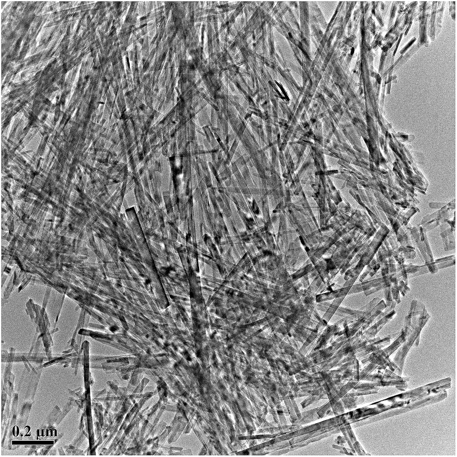 | ||
| Fig. 1 FETEM image of the obtained wollastonite powders synthesized by hydrothermal method. | ||
 | ||
| Fig. 2 XRD pattern of the obtained nanofibers synthesized by hydrothermal method. | ||
The synthetic CS nanofibers were used as raw materials to fabricate the CS bioceramics. Table 1 presents the effect of sintering temperature on the linear shrinkage, porosity and bending strength of the ceramic samples. The linear shrinkage of the samples increased from 10.65% to 14.94% with an increase in sintering temperature from 1050 to 1150 °C; linear shrinkage slightly decreased to 13.07% with a further increase of sintering temperature to 1200 °C. As expected, the porosity of the samples was observed to decrease with the increase of sintering temperature from 1050 to 1100 °C, and porosity decreased slightly with further increase of sintering temperature. While the bending strength of the fabricated CS bioceramics increased with the sintering temperature increase from 1050 to 1100 °C, reaching a maximal value of 145.70 ± 2.74 MPa, it then decreased with a further increase the sintering temperature up to 1200 °C. However, it is hard to understand that the linear shrinkage decreased with the increase of the sintering temperature from 1150 to 1200 °C, which needs to be further investigated in details.
| Sintering temperature (°C) | 1050 | 1100 | 1150 | 1200 |
| Linear shrinkage (%) | 10.65 ± 0.62 | 11.41 ± 0.80 | 14.94 ± 0.97 | 13.07 ± 1.20 |
| Porosity (%) | 22.79 ± 0.33 | 11.39 ± 0.49 | 9.67 ± 0.28 | 9.49 ± 0.53 |
| Bending strength (MPa) | 51.12 ± 3.36 | 145.70 ± 2.74 | 93.90 ± 5.15 | 52.93 ± 8.42 |
Fig. 3 shows the fracture surfaces of the ceramic samples sintered at various temperatures ranging from 1050 to 1200 °C for 3 h. The samples sintered at 1050 °C were highly porous with evenly distributed micropores in range of 0.1–0.4 μm in diameter, and the samples were completely constructed of fiber-like crystals with diameters of 30–100 nm and lengths up to several micrometers (Fig. 3A). The number of the micropores decreased remarkably with the increase of sintering temperature to 1100 °C. Moreover, a few fiber-like grains with diameters of 0.4–0.8 μm were still distributed among the sintered matrices. In addition, fiber debonding and pulling-out phenomena could be clearly observed from the fracture surface (Fig. 3B). Increasing the sintering temperature to 1150 °C, the fracture surfaces presented continuous phases accompanied by the formation of spherical pores with diameters of 0.3–1 μm, which demonstrated that a liquid phase had been formed. Meanwhile the amount of the fiber-like crystals decreased remarkably (Fig. 3C). With a further increase of sintering temperature to 1200 °C, the fracture structure with liquid phase phenomenon was maintained. While the sizes of the sphere-like micropores decreased apparently to 0.2–0.4 μm, and the micropores were uniformly distributed within the sintered matrices. Moreover, the fiber-like crystals disappeared completely (Fig. 3D).
 | ||
| Fig. 3 The fracture images of samples sintered at 1050 °C (A), 1100 °C (B), 1150 °C (C) and 1200 °C (D) for 3 h. The black arrows were the fiber-like grains distributed among the sintered matrices. | ||
Fig. 4 shows the surface morphologies of the ceramic samples sintered at 1100 °C for 3 h before and after soaking in SBF. It can be clearly observed that large numbers of fiber-like crystals with diameters of 0.2–1 μm and lengths up to 5 μm were uniformly distributed within the sintered samples before soaking (Fig. 4A). After soaking in SBF for 3 days, the samples were completely covered by a dense layer (Fig. 4B). The high magnification image inserted in Fig. 4B illustrates that the newly formed layer consisted of finely structured worm-like particles with a diameter of 50 nm and lengths up to 200 nm.
 | ||
| Fig. 4 FESEM images for the surface of the samples after soaking CS bioceramics sintered at 1100 °C for 3 h in SBF for 3 days. The white arrows were the fiber-like grains distributed among the sintered matrices. The high magnification image inserted on top right corner of (B) revealed a large amount of worm-like particles deposited on the surface after soaking CS bioceramics in SBF. | ||
Fig. 5 presents the XRD spectra of the sintered ceramic samples before and after soaking in SBF for 3 days. The results indicated that the samples sintered at 1100 °C for 3 h were β-wollastonite (β-CaSiO3, β-CS) phase (JCPDS card: no. 84-0655) (Fig. 5A). After soaking the samples in SBF for 3 days, the intensity of the β-CS peaks decreased, while the typical diffraction peaks of hydroxyapatite [Ca10(PO4)6(OH)2, HAp] (JCPDS card: no. 09-0432) appeared, suggesting that the newly formed layers were bone-like apatite.
 | ||
| Fig. 5 XRD for the samples after soaking CS bioceramics sintered at 1100 °C for 3 h in SBF for 0 (A) and 3 days (B). The wide diffraction peaks appeared around 30–33 deg. indicated that the deposited worm-like particles on CS bioceramics after soaking in SBF was hydroxyapatite phase. | ||
FTIR was used to further characterize the surface properties of the fabricated β-CS bioceramics before and after soaking in SBF (Fig. 6). Before soaking, the intense peaks between 908 and 1085 cm−1 could be attributed to silicate (Si–O–Si) absorption peaks, and the peaks at 686, 645, 565, and 454 cm−1 were attributable to the bending mode of Si–O–Si.2,9 After soaking in SBF for 3 days, the intensity of the silicate (Si–O–Si) absorption peaks decreased remarkably. The newly appeared peaks at 1113 and 1035 cm−1 could be attributed to the phosphate groups (PO43−) of the deposited apatite layers, the peaks at 1505 and 1420 cm−1 corresponded with the absorption peaks for carbonate group (CO32−), and those at 3403 and 1644 cm−1 resulted from adsorbed water molecules.2,9 The FTIR spectra further demonstrated that the layers deposited on the sintered CS bioceramics after soaking in SBF were bone-like hydroxycarbonate apatite (HCA). The formation of a bone-like HCA layer on the surface of a bioactive material is considered to play an essential role in tight bone bonding between bioactive materials and neighboring tissues.2,9,13 In the present study, the results suggested that the CS bioceramics sintered from nanofibers could induce the rapid formation of bone-like HCA layers, indicating the excellent bioactivity of the fabricated samples.
 | ||
| Fig. 6 FITR spectra for the samples after soaking CS bioceramics sintered at 1100 °C for 3 h in SBF for 0 (A) and 3 days (B). | ||
4 Discussion
The wollastonite (CS) ceramic is considered as a promising new biomaterial for bone regeneration due to its excellent biocompatibility, bioactivity and biodegradability. However, its mechanical properties are apparently lower than that of cortical bone, which has hindered its wider use for clinical applications, especially in load-bearing situations. Our previous study showed that the porosity and bending strength of the CS bioceramics pressurelessly sintered from micro-sized particles at 1100 °C for 3 h were 18.60% and 66 MPa, respectively.1 The poor mechanical strength of the fabricated wollastonite (CS) bioceramics was due to the high porosity remained in the sintered matrices, which come from the low sintering ability of the micro-sized powders. It is well known that the ceramic powders in nano-size possess excellent sintering ability. In our previous study, the CS nanofibers have been successfully synthesized in large-scale by the combination of the chemical precipitation and hydrothermal treatment.28 However, their sintering ability and the mechanical strength of the sintered bodies have not yet been investigated. Our hypothesis is that the CS nanofibers might possess excellent sintering ability. In addition, the fiber-like crystals might be reserved in the final products which can further perform as the self-reinforcement in the sintered matrices.The present study confirmed that the bending strength of pure CS bioceramics fabricated from CS nanofibers reached 145.70 ± 2.74 MPa, i.e., the upper limit value of human cortical bone, which was approximately 2.2 times as high as that of the samples sintered from micro-sized particles by a similar sintering process, and was similar to that of the samples reinforced by the bioinert Si3N4 particles. Recently, Zeng et al.29 reported the improved mechanical properties of CS bioceramics using Si3N4 as reinforcement. In their study, the optimal bending strength reached 157.2 MPa with the addition of 3 wt% Si3N4. However, the inert reinforcement might hinder the bioactivity and biodegradability of final products.
The excellent bending strength observed in the present study resulted from the high sinterability of the nanofibers and from the fiber self-reinforcement in the sintered matrices. It is well known that nano-sized powders possess a much higher driving force for densification due to their enormous surface area compared with micro-sized powders, and this driving force accelerates rates of densification and grain boundary motion.30 Differences in raw powder sintering ability lead to significant differences in the relative densities and microstructures of the resulting sintered matrices; these differences ultimately influence the mechanical properties of the sintered matrices.30
In general, the porosity and grain morphology are the key factors affecting the strength of the ceramics.2 The bending strength of the ceramic materials appeared to increase with decreasing the porosity of the sintered matrices. In the present study, the porosity of the CS bioceramics pressurelessly sintered from nanofibers at 1100 °C for 3 h was 11.39%, which was apparently lower than that of the samples sintered from micro-sized particles due to the better sintering ability of the nanofibers. However, it is still difficult to fabricate the fully dense CS bioceramics, even at higher sintering temperatures, using nanofibers as raw materials due to low sintering ability for CS material.1 The higher sintering temperatures might decrease the porosity of the samples. In contrast, the over-high sintering temperatures resulted in liquid phase formation, which ultimately decreased the bending strength.
The XRF determination result showed that the obtained CS nanofibers contained 0.68 wt% sodium (Na). The residual Na element came from the Na2SiO3 using as raw material in the synthetic process of wollastonite nanofibers. The residual sodium component might play as the driving force for the formation of liquid phase during sintering, which apparently enhanced the densification process and also modified the microstructures of the sintered matrices.31 The sodium element always presents in the final calcium silicate products when the raw materials containing sodium component were used.31–33. However, the previous studies have shown that it is still difficult to obtain fully dense CS bioceramics using this kinds of CS powders containing sodium element or even using Na2O as sintering additive.1,34 In the present study, the CS bioceramics with higher density could be facilely fabricated using nanofibers as raw materials comparing with the micro-sized powders. Therefore, it can be concluded that using the nanofibers as raw materials can accelerate the densification process.
Moreover, the fiber-like grains that remained in the sintered matrices further increased the bending strength of the obtained samples by acting as self-reinforcements. The fiber-like grains serve as reinforcements in the fabricated CS bioceramics by not only hindering crack propagation but also resisting the interfacial shear stress that otherwise results in fiber debonding and pulling-out during bending strength tests.1,30,35–37 It is believed that the fiber-like grains played a dominative role in the mechanical properties among the factors of porosity, grain size, and grain morphologies.9 The present study confirmed that pure CS bioceramics with high mechanical strength could be facilely fabricated by pressureless sintering using CS nanofibers as raw materials. It can be predicted that the mechanical strength of the nanofiber derived CS bioceramics might be further improved via increasing their density. Our present study showed that the CS bioceramics with a relative density of about 95% fabricated using spark plasma sintering (SPS) technique expresses excellent bending strength of about 294 MPa.25
The SBF soaking results demonstrated that the CS bioceramics sintered from nanofibers could induce the rapid formation of bone-like HCA layers, indicating the excellent bioactivity of the fabricated CS bioceramics. The formation of a bone-like HCA layer is considered to play an essential role in tight bone bonding between bioactive materials and neighboring tissues.13,38–40 Our results suggest that CS bioceramics sintered from nanofibers might be used as a bioactive material for clinical load-bearing applications. In addition, it is considered that the major drawback of CS bioceramics is their high dissolution rate. More importantly, the dissolution rate of calcium silicate bioceramics can be modified by incorporation of different elements such as Zn, Mg, Sr, Ti and Zr etc.41,42 Therefore, we can expected that the high mechanical strength calcium silicate bioceramics with regulated degradation rate might be fabricated using the calcium silicate nanofibers containing these kinds of elements. However, the degradation rate of the CS bioceramics fabricated from nanofibers and the effect of the element incorporation on their degradation should be further investigated in details.
5 Conclusions
In this study, the sinterability of the wollastonite (CS) nanofibers, and the microstructure, bending strength and in vitro bioactivity of the fabricated CS bioceramics were investigated. The results showed that CS bioceramics with a high bending strength of 145.70 ± 2.74 MPa, i.e., the upper limit value of human cortical bone, could be fabricated via the low-cost pressureless sintering using CS nanofiber as raw materials. The SBF soaking evaluation revealed a rapid formation of bone-like apatite on the surfaces of the CS bioceramics, which indicated that the fabricated materials possessed excellent bioactivity. Our results suggest that CS bioceramics sintered from nanofibers might be potential candidates for load-bearing bone implant materials.Acknowledgements
The authors gratefully acknowledge the Fund of the Science and Technology Commission of Shanghai Municipality (No. 15441905300), the Fund of Shanghai Municipal Commission of Health and Family Planning (201540369), and the Fundamental Research Funds for the Central Universities (1504219035).Notes and references
- K. Lin, W. Zhai, S. Ni, J. Chang, Y. Zeng and W. Qian, Ceram. Int., 2005, 31, 323–326 CrossRef CAS.
- K. Lin, D. Zhai, N. Zhang, N. Kawazoe, G. Chen and J. Chang, Ceram. Int., 2014, 40, 3287–3293 CrossRef CAS.
- K. Lin, L. Xia, H. Li, X. Jiang, H. Pan, Y. Xu, W. W. Lu, Z. Zhang and J. Chang, Biomaterials, 2013, 34, 10028–10042 CrossRef CAS PubMed.
- F. Baino and C. Vitale-Brovarone, Ceram. Int., 2015, 41, 11464–11470 CrossRef CAS.
- H. C. Li, D. G. Wang and C. Z. Chen, Ceram. Int., 2015, 41, 10160–10169 CrossRef CAS.
- C. Wang, K. Lin, J. Chang and J. Sun, Biomaterials, 2013, 34, 64–77 CrossRef CAS PubMed.
- S. Liu, F. Jin, K. Lin, J. Lu, J. Sun, J. Chang, K. Dai and C. Fan, Biomed. Mater., 2013, 8, 025008 CrossRef PubMed.
- K. Lin, J. Chang and R. Shen, Biomed. Mater., 2009, 4, 065009 CrossRef PubMed.
- K. Lin, M. Zhang, W. Zhai, H. Qu and J. Chang, J. Am. Ceram. Soc., 2011, 94, 206–212 Search PubMed.
- C. Sarmento, Z. B. Luklinska, L. Brown, M. Anseau, P. N. De Aza and S. De Aza, J. Biomed. Mater. Res., Part A, 2004, 69, 351–358 CrossRef PubMed.
- P. N. De Aza, Z. B. Luklinska, M. R. Anseau, F. Guitian and S. De Aza, J. Dent., 1999, 27, 107–113 CrossRef CAS PubMed.
- S. Xu, K. Lin, Y. Hu, Z. Wang, J. Chang, L. Wang, J. Lu and C. Ning, Biomaterials, 2008, 29, 2588–2596 CrossRef CAS PubMed.
- T. Kokubo and H. Takadama, Biomaterials, 2006, 27, 2907–2915 CrossRef CAS PubMed.
- K. Xiong, J. Zhang, H. Shi, J. Liu, H. Wu, H. Li and J. Ye, RSC Adv., 2015, 5, 8329–8339 RSC.
- C. Wang, K. Lin, J. Chang and J. Sun, J. Biomed. Mater. Res., Part A, 2014, 102, 2096–2114 CrossRef PubMed.
- L. Fei, C. Wang, Y. Xue, K. Lin, J. Chang and J. Sun, J. Biomed. Mater. Res., Part B, 2012, 100, 1237–1244 CrossRef PubMed.
- C. Wang, Y. Xue, K. Lin, J. Lu, J. Chang and J. Sun, Acta Biomater., 2012, 8, 350–360 CrossRef CAS PubMed.
- K. Lin, P. Liu, W. Zhang, L. Wei, Z. Zou, Y. Qian, Y. Shen and J. Chang, Chem. Eng. J., 2013, 222, 49–59 CrossRef CAS.
- H. J. Chung and T. G. Park, Adv. Drug Delivery Rev., 2007, 59, 249–262 CrossRef CAS PubMed.
- P. Tayalia and D. J. Mooney, Adv. Mater., 2009, 21, 3269–3285 CrossRef CAS PubMed.
- E. C. Novosel, C. Kleinhans and P. J. Kluger, Adv. Drug Delivery Rev., 2011, 63, 300–311 CrossRef CAS PubMed.
- C. Shuai, C. Gao, P. Feng and S. Peng, RSC Adv., 2014, 4, 12782–12788 RSC.
- M. Mehrali, E. Moghaddam, S. F. S. Shirazi, S. Baradaran, M. Mehrali, S. T. Latibari, H. S. C. Metselaar, N. A. Kadri, K. Zandi and N. A. A. Osman, ACS Appl. Mater. Interfaces, 2004, 6, 3947–3962 CrossRef PubMed.
- K. Lin, L. Chen and J. Chang, Int. J. Appl. Ceram. Technol., 2012, 9, 479–485 CrossRef CAS.
- L. H. Long, L. D. Chen, S. Q. Bai, J. Chang and K. L. Lin, J. Eur. Ceram. Soc., 2006, 26, 1701–1706 CrossRef CAS.
- E. K. Papynov, V. Y. Mayorov, A. S. Portnyagin, O. O. Shichalin, S. P. Kobylyakov, T. A. Kaidalova, A. V. Nepomnyashiy, T. A. Sokol'nitskaya, Y. L. Zu and V. A. Avramenko, Ceram. Int., 2015, 41, 1171–1176 CrossRef CAS.
- F. Zhang, K. Lin, J. Chang, J. Lu and C. Ning, J. Eur. Ceram. Soc., 2008, 28, 539–545 CrossRef CAS.
- K. Lin, J. Chang, G. Cheng and M. Ruan, J. Cryst. Growth, 2007, 300, 267–271 CrossRef CAS.
- Y. Pan, K. Zuo, D. Yao, J. Yin, Y. Xin, Y. Xia, H. Liang and Y. Zeng, J. Mech. Behav. Biomed. Mater., 2015, 55, 120–126 CrossRef PubMed.
- K. Lin, J. Chang, J. Lu, W. Wu and Y. Zeng, Ceram. Int., 2007, 33, 979–985 CrossRef CAS.
- X. H. Huang and J. Chang, J. Nanopart. Res., 2007, 9, 1195–1200 CrossRef CAS.
- X. Wang, J. Zhuang, Q. Peng and Y. Li, J. Solid State Chem., 2005, 178, 2332–2338 CrossRef CAS.
- A. B. Y. Hazar, Ceram. Int., 2007, 33, 687–692 CrossRef CAS.
- H. C. Li, D. G. Wang, C. Z. Chen, F. Weng and H. Shi, Ceram. Int., 2016, 42, 1439–1445 CrossRef CAS.
- F. P. Knudsen, J. Am. Ceram. Soc., 1959, 42, 376–387 CrossRef CAS.
- Y. Dou, K. Lin and J. Chang, Nanoscale, 2011, 3, 1508–1511 RSC.
- K. Lin, J. Chang, X. Liu and C. Ning, Int. J. Appl. Ceram. Technol., 2010, 7, 178–183 CrossRef CAS.
- C. Turdean-Ionescu, B. Stevensson, J. Grins, I. Izquierdo-Barba, A. García, D. Arcos, M. Vallet-Regí and M. Edén, RSC Adv., 2015, 5, 86061–86071 RSC.
- S. Liu, W. Gong, Y. Dong, Q. Hu, X. Chen and X. Gao, RSC Adv., 2015, 5, 38830–38836 RSC.
- Y. Shen, Z. Hua, L. Zhang and X. Hao, RSC Adv., 2015, 5, 18788–18795 RSC.
- X. Jin, J. Chang, W. Zhai and K. Lin, J. Am. Ceram. Soc., 2011, 94, 173–177 CrossRef.
- M. Zhang, C. Wu, K. Lin, W. Fan, L. Chen, Y. Xiao and J. Chang, J. Biomed. Mater. Res., Part A, 2012, 100, 2979–2990 CrossRef PubMed.
| This journal is © The Royal Society of Chemistry 2016 |