Selective separation and identification of metabolite groups of Polygonum cuspidatum extract in rat plasma using dispersion solid-phase extraction by magnetic molecularly imprinted polymers coupled with LC/Q-TOF-MS

Zaiyue Yanga, Qizhi Caia, Ning Chenab, Xuemin Zhou*a and Junli Hong*a
aSchool of Pharmacy, Nanjing Medical University, Nanjing 210029, PR China. E-mail: xueminzhou001_001@hotmail.com; hongjunli1102@163.com; Fax: +86 25 86868476; Tel: +86 25 86868476
bJiangsu Province Institute of Materia Media, Nanjing University of Technology, Nanjing 210029, PR China

Received 14th December 2015 , Accepted 19th January 2016

First published on 22nd January 2016


Abstract

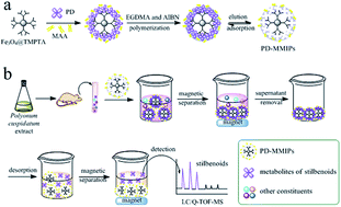
In this work, magnetic molecularly imprinted polymers (MMIPs) were successfully prepared for specific recognition and selective enrichment of metabolite groups of Polygonum cuspidatum extract in rat plasma. Two kinds of MMIPs were obtained using polydatin (PD) and emodin-8-O-β-D-glucoside (EG) as template molecules, respectively, methacrylic acid as functional monomer, ethylene glycol dimethacrylate as cross-linker and 2,2′-azobisisobutyronitrile as initiator. The membrane thickness of both MMIPs was approximately 10 nm. With the dendritic-grafting modification, magnetic nanoparticles (MNPs) possessed the outstretched branched structure, orderly open cavities and abundant functional groups with a good specific surface area (47.99 m2 g−1). And both MMIPs obtained high adsorption capacity (221 μmol g−1 for PD and 190 μmol g−1 for EG) and fast kinetics. Dispersion solid-phase extraction using MMIPs (MMIPs-DSPE) was applied to selectively separate and enrich the target metabolite groups of stilbenoids and anthraquinones, respectively. Furthermore, three classical pretreatment methods including protein precipitation (PP), liquid–liquid extraction (LLE) and solid phase extraction (SPE) were compared with MMIPs-DSPE in terms of matrix effect, efficiency of extraction and capacity using LC/Q-TOF-MS. The result showed that MMIPs obtained the lowest matrix effect from 1.52 to 8.96% and the most constituents of target metabolite groups with 17 stilbenoids and 19 anthraquinones, respectively. The proposed analysis platform has been proven to be selective, sensitive and simple for the enrichment of target metabolite groups, and has great significance for discovering minor bioactive constituents and elucidating integrative mechanism of traditional Chinese medicines.


1. Introduction

The metabolomics of traditional Chinese medicines (TCMs) have gained extensive attention worldwide, for the metabolites are comprehensive and potentially active ingredients or biomarkers for disease diagnosis and therapeutic response.1–3 Multiple metabolites investigation, especially the target metabolite groups research, has great important significance for clarifying the pharmacodynamic material basis and the active constituents of TCMs. However, the knowledge about the chemical compositions of metabolomics of TCMs is far from sufficient, because of the complex and trace constituents, matrix interferences, multi-component systems of TCMs and the lack of holistic approaches and appropriate analytical methodologies. Therefore, the integrated evaluation method and the advanced analytical strategy are essential for target metabolite groups study of TCMs.

Polygonum cuspidatum Sieb. et Zucc., a traditional Chinese medicinal herb, is widely distributed in Asia and North America and officially listed in the Chinese Pharmacopoeia (2015 version).4 The dry roots have been widely used for treatment of various inflammatory diseases, hepatitis, tumors, hypertension, hyperlipemia and menopausal symptoms.5–8 Phytochemical studies indicated that stilbenoids and anthraquinones are high-content constituents that generally regarded as an index for quality control of this herb. As the representative stilbenoids in Polygonum cuspidatum, resveratrol and polydatin (PD) are much higher than in grape and other plants, which have many protecting properties such as tumor inhibition, antioxidant, inhibitor of platelet aggregation and modulator of lipoprotein metabolism.9–12 The predominant anthraquinones are emodin-type anthraquinones which have antileukemic, antiseptic and antitumor activity, etc.13–15

TCMs are mostly administered orally, so the investigation on metabolites in serum is important for active target finding and the deeper pharmacological mechanism study.16,17 For Polygonum cuspidatum, the stilbenoid and anthraquinone metabolite groups were the major forms of metabolites in plasma. However, many metabolite analysis were concerned with only several pure compounds above.18,19 There are lack of research on the integrated effects and multi-component profiling of target metabolite groups. Thus, establishment of a reliable method to enrich and rapid identify multiple bioactive metabolites is essential and helpful for elucidating integrative mechanism of TCMs.

Presently, various analytical techniques, such as gas chromatography coupled with mass spectrometry (GC-MS)20,21 and high performance liquid chromatography with mass spectrometry (HPLC-MS)22,23 have already been widely used for the metabolic investigation. However, the main problems concerning metabolic analysis are aimless, the loss of trace constituents and the matrix interferences. Thus some clean-up pretreatments including protein precipitation (PP),24 liquid–liquid extraction (LLE)25 and solid phase extraction (SPE)26 are widely reported. However, these classical extraction methods suffer from many disadvantages or limitations, such as lack of selectivity, a high volume of organic solvents requirement and the low extraction recoveries. Therefore, an efficient clean-up process and pre-concentration step is crucially important to minimize interferences and improve the sensitivity and selectivity for the target metabolite groups.

Molecular imprinting technology (MIT) has been developed rapidly with the property of predetermination, specific recognition and extensive practicability.27–30 Molecular imprinting polymers (MIPs), the highly stable polymers which possess recognition sites of the three-dimensional (3D) network by synthetic materials, are able to specifically rebind the target analytes, or enrich structurally related compounds.31,32 Currently, with the introduction of magnetic nanoparticles (MNPs)33–35 and highly branched dendrimer polymers,36,37 the MIPs have showed remarkable advantages of selective, cost-effective, time saving, convenient (without additional centrifugation or filtration) and high adsorption capacity, which can highly enrich and separate trace constituents from the complex matrixes.

At the present work, the method of selective enrichment and recognition of target metabolite groups in rat plasma was developed by dispersion solid-phase extraction using magnetic molecularly imprinted polymers (MMIPs-DSPE) with dendritic-grafting combined with the LC/Q-TOF-MS technique. Furthermore, compared with traditional pretreatment methods of PP, LLE and SPE, our work provided superior selectivity, enrichment capacity and simplicity for “fishing” and “capturing” the target metabolite groups in complex biological samples. The molecular targeting research of our work is more meaningful for the metabolomics and active constituents discovery of TCMs.

2. Experimental

2.1 Reagents and equipment

Polydatin (PD), oxyresveratrol, resveratrol, emodin-8-O-β-D-glucoside (EG), emodin, rhein, physcion, chrysophanol, aloe emodin and genistein were purchased from Nanjing Jingzhu biological technology Co., Ltd. (Nanjing, China). Genistein was used as reference compound. Tetraethyl orthosilicate (TEOS), acetic acid, methanol, and absolute alcohol were from Sinopharm Chemical Reagent Co., Ltd. (Shanghai, China). Ethylenediamine (EDA), methyl acrylate (MA), and methacrylic acid (MAA) were obtained from Shanghai Linfeng Chemical Reagent Co., Ltd. (Shanghai, China). 2,2′-Azobisisobutyronitrile (AIBN) was from Shanghai No. 4 Reagent & H.V. Chemical Co., Ltd. (Shanghai, China) and purified through recrystallization in ethanol before use. 3-Amino-propyl-triethoxysilane (APTES) and ethylene glycol dimethacrylate (EGDMA) were obtained from Sigma-Aldrich (St. Louis, USA). Milli-Q water was used throughout the experiments. All other chemicals were analytical grade and obtained commercially.

Analysis was performed on an Agilent 1200 Series (Agilent Technologies, Germany) LC system coupling to a 6538 QTOF MS with ESI source (Agilent Technologies, Santa Clara, CA, USA). SEM images were recorded with S-4800 Field-Emission (Hitachi, Japan). FT-IR spectras were obtained on a TENSOR27 infrared scanner (Bruker, Germany). BET analysis was performed on BELSORP-MINI (BEL, Japan). Characteristics were also measured by X-ray diffraction (XRD) (Dmax22500, Rigaku, Japan) and vibrating sample magnetometry (VSM) (Lake Shore).

2.2 Molecular modeling studies

Computational simulation was employed to calculate binding energy (ΔEbinding) between templates and monomers, and solvation energy (ΔEsolvation) between templates and solvents. The full geometry optimization was carried with the 6-31G basis set at Density Functional Theory (DFT) level by Gaussian 09. ΔEbinding and ΔEsolvation were finally computed by following formulas:
ΔEbinding = |EcomplexEtemplateEmonomer|

ΔEsolvation = |EsolventEtemplate|
where Ecomplex, Etemplate, Emonomer and Esolvent are the potential energies of the complex of monomer and template, template, monomer and solvent, respectively.

2.3 Preparation of PD-MMIPs and EG-MMIPs

2.3.1 Dendritic-grafting modification of Fe3O4@SiO2 MNPs. The synthesis of Fe3O4@SiO2 was published in our previous work.37 Subsequently, Fe3O4@SiO2 were modified as follows (Fig. S1):

(a) 1 g Fe3O4@SiO2 was dispersed in 100 mL toluene by ultrasonic vibration and 20 mL APTES was added under nitrogen gas. The mixture was stirred for 24 h at 120 °C. Then Fe3O4@APTES with amino groups on the surface were obtained by the magnetic separation.

(b) Fe3O4@APTES were dispersed in 100 mL methanol and 15.32 mL MA was added. The solution was stirred at 50 °C with nitrogen gas. After 24 h, Fe3O4@MA were obtained.

(c) Fe3O4@MA were dispersed in methanol and 11.36 mL EDA. The reaction was stirred for 24 h at 50 °C, and then Fe3O4@EDA were obtained by the magnetic separation.

(d) 0.5 g Fe3O4@EDA was dispersed in methanol and then 44 mL TMPTA was added. The mixture was stirred vigorously and purged with nitrogen gas at 30 °C. After 6 h, Fe3O4@TMPTA with the double bond groups were obtained and dried in vacuum.

2.3.2 Preparation of PD-MMIPs and EG-MMIPs. As the representative compounds of stilbenoids and anthraquinones in Polygonum cuspidatum, PD and EG were chosen as templates to synthesize MMIPs, respectively.

Fe3O4@TMPTA MNPs (50 mg), PD (100 mg) and a prescribed amount of MAA were dispersed in 50 mL acetonitrile–methanol (9[thin space (1/6-em)]:[thin space (1/6-em)]1, v/v) solution and stirred at room temperature. Then a certain volume of EGDMA and 30 mg AIBN were added. The mixture was continuously stirred for 6 h at 50 °C and 24 h at 60 °C under the protection of nitrogen. After the reaction completed, polymers were separated by an external magnetic field and then eluted by 10% (v/v) acetic acid–methanol solution to remove templates and rinsed with methanol. Finally, PD-MMIPs were obtained after dried in vacuum. PD magnetic non-molecular imprinted polymers (PD-MNIPs) were prepared by the same manner in the absence of PD. Fig. 1a illustrated the preparation process of PD-MMIPs. The preparation processes of EG-MMIPs and EG-MNIPs were the same as above, respectively, while the template was EG.


image file: c5ra26695e-f1.tif
Fig. 1 Schematic representation of (a) the synthesis of PD-MMIPs and (b) the possible pretreatment process of MMIPs-DSPE represented by PD-MMIPs.

2.4 Preparation of Polygonum cuspidatum extract

Rhizoma and radix of Polygonum cuspidatum crude drugs (5 kg) were cut to pieces and immersed in 75% ethanol (1[thin space (1/6-em)]:[thin space (1/6-em)]10, w/v) for 24 h. Then the pieces were extracted for 1 h under thermal reflux twice. The two ethanol solutions were filtrated and combined, then evaporated to dryness in a rotary evaporator (BUCHI ltd., Labortechinik AG, Switzerland) at 60 °C under reduced pressure. The contents of PD and EG in the extract were determined to be 9.91% and 16.09%, respectively. The extract was kept in 4 °C for further oral administration to rats.

2.5 Experimental animals

Male Sprague-Dawley rats (200–220 g) were supplied by Department of Experimental Animals, Nantong University (Jiangsu, China), and acclimated at 25 °C and 55% of humidity under natural light/dark conditions for 1 week before experiment. All animal experiments were carried out in accordance with guidelines evaluated and approved by the Institutional Animal Care and Use Committee of Nanjing Medical University, Jiangsu, China (IACUC-1403070).

2.6 Drug administration and biological samples collection

Polygonum cuspidatum extract was dissolved in 0.5% sodium carboxymethyl cellulose at 350 mg mL−1 and given orally to six rats (4.0 g kg−1). The control group was administered by the same dose of sodium carboxymethyl cellulose. Blood was collected by retroorbital bleeds at 5, 30, 60, 90, 120 and 180 min. Heparinized blood samples were immediately centrifuged to separate plasma. All plasma samples were took out same amount to make a mixed sample and stored at −80 °C until analysis. Mixed blank sample was obtained from the control group by the procedure described above.

2.7 Sample preparation

2.7.1 Performance of MMIPs-DSPE. Three times volume of acetonitrile was added to the mixed plasma sample and 300 μL supernatant was pipetted after centrifuged. Subsequently, the supernatant was dried by nitrogen and redissolved with 1 mL acetonitrile–methanol (9[thin space (1/6-em)]:[thin space (1/6-em)]1, v/v). 20 mg PD-MMIPs and EG-MMIPs were added to above solution and incubated at room temperature, respectively. After 20 min, both MMIPs were obtained by magnetic separation, and a small amount of methanol was used to wash the surface of MMIPs. Desorption was carried out by using 10% (v/v) acetic acid–methanol as eluant under ultrasonication for 20 min to extract the metabolites in PD-MMIPs and 15 min for EG-MMIPs, respectively. After magnetic separation, the supernatant was dried and redissolved with 100 μL mobile phase and 0.1 μg reference compound genistein for further LC/Q-TOF-MS analysis. The mixed blank sample was processed in the same way. This procedure represented by PD-MMIPs was demonstrated in Fig. 1b. Performance of MNIPs-DSPE was carried out by the same process above.
2.7.2 Classical pretreatment methods.
PP. Three times volume of acetonitrile was added to the mixed plasma sample and 300 μL supernatant was pipetted and dried by nitrogen.38 Then the residue was redissolved with 100 μL mobile phase and 0.1 μg reference compound.
LLE. 300 μL supernatant was dried and redissolved with 500 μL Milli-Q water and 2 mL ethyl acetate was added to extract the analytes,39 then the organic solvent was dried and redissolved with 100 μL mobile phase and 0.1 μg reference compound.
SPE. An Oasis HLB cartridge (1 cm3, 30 mg, Waters, USA) was pre-treated with 2 mL methanol followed by 2 mL water. Then 300 μL supernatant was dried and redissolved with 1 mL water and then loaded on the cartridge. The cartridge was washed with 3 mL water and 2 mL methanol[thin space (1/6-em)]:[thin space (1/6-em)]water (15[thin space (1/6-em)]:[thin space (1/6-em)]85, v/v), and then the analytes were eluted with 2 mL methanol.40 Finally, the eluent was dried and redissolved as above.

2.8 Matrix effect

Matrix effect (ME) of different pretreatment methods was performed for each standard analyte. Standard solutions (2 μg mL−1) of PD, oxyresveratrol, resveratrol, EG, emodin, rhein, physcion, chrysophanol and aloe emodin were prepared in methanol. And matrix-matched solutions were obtained by adding same concentration standard solutions after the blank plasma dealt with different methods. ME was calculated as ME (%) = (1 − (Am/As)) × 100%, where As is the average of the peak area of standard in solvent and Am is the average of the peak area of standard in matrix-matched solution.41 The results close to zero indicate low ME, while values more or less than zero indicate suppression or enhancement of ionization by the matrix components.

2.9 LC/Q-TOF-MS conditions

Analysis was performed on an Agilent 1200 Series (Agilent Technologies, Germany) LC system equipped with a binary pump, an online degasser, an auto sampler, a thermostatically controlled column compartment, coupling to a 6538 QTOF MS with ESI source (Agilent Technologies, Santa Clara, CA, USA). Samples were separated on a Hedera ODS-2 C18 column (4.6 × 250 mm, 5 μm) at a temperature of 25 °C with flow rate was 0.5 mL min−1. The injection volume was 10 μL. Separation was obtained using a gradient mobile phase consisting of water (0.1% aqueous formic acid) (A) and methanol (B). The gradient elution was 15–25% B from 0–5 min, 25–35% B from 5–15 min, 35–60% B from 15–25 min, 60–70% B from 25–45 min, 70–100% B from 45–60 min. A 15 min post run time back to the initial mobile phase composition was used after each analysis.

Conditions of ESI source operated in negative-ion mode were as follows: drying gas (N2) flow rate, 10 L min−1; drying gas temperature, 320 °C; nebulizer gas, 45 psig; capillary voltage, 3500 V; fragmentor voltage, 120 V; sample collision energy, 30 V.

2.10 Data processing

All data were acquired using Mass Hunter Workstation Software Version B.02.00 (Agilent Technologies). Mass range was set at m/z 100–1000. The TOF was calibrated every day before sample analysis, using reference masses at m/z 112.9885 and 1033.9881 to obtain high-accuracy mass measurements.

3. Results and discussion

3.1 Preparation of PD-MMIPs and EG-MMIPs

Selection of suitable functional monomer is a crucial factor in the study of MMIPs.42 In this work, three widely used functional monomers (MAA, AA, TFMAA) were compared theoretically by Gaussian 09. ΔEbinding of complexes between templates and monomers (mole ratio from 1[thin space (1/6-em)]:[thin space (1/6-em)]1 to 1[thin space (1/6-em)]:[thin space (1/6-em)]3) was calculated (Table S1). It obviously showed that ΔEMAA > ΔETFMAA > ΔEAA at these mole ratios. Therefore, MAA was chosen as the optimal functional monomer for both PD and EG. ΔEbinding of PD/EG and MAA was also presented as follows: ΔE(1[thin space (1/6-em)]:[thin space (1/6-em)]1) < ΔE(1[thin space (1/6-em)]:[thin space (1/6-em)]2) < ΔE(1[thin space (1/6-em)]:[thin space (1/6-em)]3) < ΔE(1[thin space (1/6-em)]:[thin space (1/6-em)]4) ≈ ΔE(1[thin space (1/6-em)]:[thin space (1/6-em)]5). Considering the excessive amount of functional monomer would have an adverse impact on the binding and desorption kinetics, complexes 1[thin space (1/6-em)]:[thin space (1/6-em)]3 and 1[thin space (1/6-em)]:[thin space (1/6-em)]4 were chosen in the following experiment.

EGDMA, the widely used cross-linking agent43 was investigated with serial molar ratio of template[thin space (1/6-em)]:[thin space (1/6-em)]monomer[thin space (1/6-em)]:[thin space (1/6-em)]cross linker from 1[thin space (1/6-em)]:[thin space (1/6-em)]3[thin space (1/6-em)]:[thin space (1/6-em)]3 to 1[thin space (1/6-em)]:[thin space (1/6-em)]4[thin space (1/6-em)]:[thin space (1/6-em)]16 (Fig. S2). The result showed both MMIPs possessed a best adsorption capacity when the molar ratio was 1[thin space (1/6-em)]:[thin space (1/6-em)]3[thin space (1/6-em)]:[thin space (1/6-em)]6.

ΔEsolvation in different solvents was compared in our work (Table S2). Information indicated the values of ΔEsolvation in acetonitrile and methanol were higher than others for both PD and EG. After optimizing the solubility conditions, acetonitrile–methanol (9[thin space (1/6-em)]:[thin space (1/6-em)]1, v/v) was chosen as solvent.

3.2 Characterization

3.2.1 SEM characterization. The representative SEM images were provided in Fig. 2. It was obvious that all MNPs were regular spheres with an appropriate size. The diameters of Fe3O4 (in the illustration) and Fe3O4@SiO2 were 90 and 180 nm, respectively. After a series of modification, Fe3O4@TMPTA were obtained with about 200 nm. PD-MMIPs and EG-MMIPs were about 220 nm which revealed the obtained thickness of the MMIPs membrane was approximately 10 nm.
image file: c5ra26695e-f2.tif
Fig. 2 SEM images of (a) Fe3O4@SiO2 (Fe3O4 MNPs were in the illustration), (b) Fe3O4@TMPTA, (c) PD-MMIPs and (d) EG-MMIPs.
3.2.2 Modification of SiO2-coated MNPs and FT-IR spectrum. Fe3O4@SiO2 were firstly modified by APTES to produce amino groups, and then dendritic MNPs–NH2 were successfully divergent synthesized by Michael addition reaction and amidation reaction with MA and EDA. Finally, MNPs with double bonds were obtained by reacted with TMPTA. To affirm the presence of functional groups on the surface of Fe3O4@SiO2, each modification stage was tracked by FT-IR spectra.

As shown in curve 1 (Fig. 3a), the absorption band at 469 cm−1 corresponded to Fe–O stretching mode of the tetrahedral and octahedral sites. The broad high-intensity band at 1092 cm−1 was due to the asymmetric stretching bonds of Si–O–Si.44 In curve 2, the two bands at 2947 and 3416 cm−1 can be ascribed to the stretching of C–H and NH2 band for Fe3O4@APTES, respectively.45 Curve 3 showed MNPs–OCH3 were obtained since the stretching vibration absorption of COO bond (1738 cm−1) and the peak of methyl group (1442 cm−1) were appeared.46 After reacted with EDA, the band at 1442 cm−1 in curve 4 disappeared and the characteristic peak of –NH2 group was observed.47 For curve 5, the re-appearance of COO bond at 1738 cm−1 proved TMPTA was modificated on MNPs.48 All data above confirmed the successful modification of SiO2-coated MNPs.


image file: c5ra26695e-f3.tif
Fig. 3 (a) FT-IR spectrum of Fe3O4@SiO2 (1), Fe3O4@APTES (2), Fe3O4@MA (3), Fe3O4@EDA (4) and Fe3O4@TMPTA (5); (b) VSM analysis of Fe3O4, Fe3O4@TMPTA, PD-MMIPs and EG-MMIPs; (c) XRD patterns of the Fe3O4 and Fe3O4@TMPTA; (d) nitrogen adsorption/desorption isotherms of Fe3O4@TMPTA and Fe3O4@SiO2.
3.2.3 Magnetic properties of nanoparticles. VSM was employed to measure magnetic properties (Fig. 3b). The values of saturation magnetization of Fe3O4, Fe3O4@TMPTA, PD-MMIPs and EG-MMIPs were 82.60, 34.80, 16.14 and 15.88 emu g−1, respectively. Fast separation of PD-MMIPs from the solution in the presence of an external magnetic field was easily visible in the illustration.
3.2.4 XRD analysis. XRD patterns of Fe3O4 MNPs and Fe3O4@TMPTA were shown in Fig. 3c. For Fe3O4, peaks at 2θ values of 30.1°, 35.5°, 43.1°, 53.5°, 57.0° and 62.6° were indexed as the diffractions of (220), (311), (400), (422), (511) and (440), respectively, which were the same as the standard diffraction spectrum of Fe3O4. After dendritic-grafting modification, Fe3O4@TMPTA still showed the same signals suggesting that process of modification did not affect the crystal structure of magnetite core.
3.2.5 Nitrogen adsorption/desorption analysis. Experiment of nitrogen adsorption/desorption was carried out to between Fe3O4@TMPTA and Fe3O4@SiO2. As shown in Fig. 3d, both curves exhibited an IV-type curve according to IUPAC classification. The specific surface area of Fe3O4@TMPTA is 47.99 m2 g−1, which was 6.54 times as much as Fe3O4@SiO2. The high surface area further confirmed that modification on the surface of Fe3O4@SiO2 could provide greater specific surface area and more effective sites.

3.3 Evaluation of adsorption characteristics

3.3.1 Adsorption isotherm. The absorption isotherm experiments were carried out in a series of standard solutions of PD or EG in acetonitrile–methanol (9[thin space (1/6-em)]:[thin space (1/6-em)]1, v/v) for 20 min. The results were shown in Fig. 4a and S3a, respectively. The maximum adsorption capacities of PD-MMIPs and PD-MNIPs were 221 and 60 μmol g−1, and EG-MMIPs and EG-MNIPs were 190 and 52 μmol g−1, respectively. Both MMIPs showed much stronger memory function and higher adsorption capacities for target compounds than MNIPs.
image file: c5ra26695e-f4.tif
Fig. 4 (a) Adsorption isotherm of PD-MMIPs and PD-MNIPs; (b) adsorption and desorption kinetic curve of PD-MMIPs; (c) selective recognition property of each compound with PD-MMIPs and PD-MNIPs at the concentration of 2 mmol L−1.

Freundlich isotherm model (eqn (1)) was applied for the adsorption ability evaluation of both MMIPs (Fig. S4).49 Where Q was the amount of adsorbed analyte and C was the concentration of analyte. The parameter α and m were the two Freundlich isotherm constants while α was related to the binding affinity and m was the heterogeneity index with values from 0 to 1. Furthermore, the number of binding sites (NKminKmax) and the weighted average affinity constant ([K with combining macron]KminKmax) for the polymers were calculated by eqn (2) and (3), respectively.

 
log[thin space (1/6-em)]Q = log[thin space (1/6-em)]α + m[thin space (1/6-em)]log[thin space (1/6-em)]C (1)
 
NKminKmax= α(1 − m2)(KminmKmaxm) (2)
 
image file: c5ra26695e-t1.tif(3)

All the data were listed in Table 1. The parameter m was 0.563 and 0.716 for PD-MMIPs and EG-MMIPs, respectively, which demonstrated the existing heterogeneous-binding sites in both MMIPs. Also, the numbers of binding sites measured by NKminKmax were 144.513 mg g−1 for PD-MMIPs (38.636 mg g−1 for PD-MNIPs) and 101.292 mg g−1 for EG-MMIPs (25.459 mg g−1 for EG-MNIPs), respectively.

Table 1 Freundlich fitting parameters, weighted average affinity, and number of sites for MMIPs and MNIPs
  m α ((mg g−1) (L g−1)m) R2 NKminKmax (mg g−1) [K with combining macron]KminKmax (mL g−1)
PD-MMIPs 0.563 60.982 0.9961 144.513 1.240
PD-MNIPs 0.516 17.159 0.9904 38.636 1.367
EG-MMIPs 0.716 35.777 0.9961 101.292 0.802
EG-MNIPs 0.740 9.108 0.9855 25.459 0.762


3.3.2 Adsorption and desorption kinetics. The adsorption kinetics of PD-MMIPs (Fig. 4b) and EG-MMIPs (Fig. S3b) was investigated with 2 mmol L−1 PD and EG, respectively. Time-dependent evolution bound with both MMIPs was exhibited. The adsorption equilibriums were reached within 20 min, which demonstrated the fast adsorption kinetics ability for MMIPs. Meanwhile, the data of desorption kinetics experiments (in the illustrations) showed that desorption could reach equilibrium in short time (20 min for PD-MMIPs and 15 min for EG-MMIPs). The excellent kinetics was mainly because of the outstretched dendritic nanostructures and open cavities of Fe3O4@TMPTA made the templates easily in or out from the recognition sites, which decreased the diffusional resistance.
3.3.3 Selectivity. Several analogs and other compounds were selected to estimate the selectivity. Taking PD-MMIPs for example (Fig. 4c), the rebinding capacities of PD-MMIPs to PD, resveratrol and oxyresveratrol at 2 mmol L−1 level were 88, 79 and 70 μmol g−1, respectively, which were higher than that of emodin and EG. The data indicated PD-MMIPs had a high selectivity and good extraction capacities for analogs but a poor affinity for the other compounds owning to the large difference in molecular and size. PD-MNIPs exhibited inconspicuous difference among them. Similar results were obtained from EG-MMIPs (Fig. S3c). Therefore, the results showed us the high selectivity of PD-MMIPs and EG-MMIPs could be applied to metabolite groups analysis.

3.4 Reproducibility and stability

Reproducibility was investigated by five batches MMIPs prepared with the same method and immersed in the PD and EG standard solutions (2 mmol L−1), respectively. Both RSDs of the adsorption amounts were less than 8%, which indicated a satisfied reproducibility.

Stability was performed in 2 mmol L−1 standard solutions. A good stability was observed with RSD of 7.34% for PD-MMIPs and 8.02% for EG-MMIPs for 5 times. The long-term stability was examined by monitoring the adsorption amount in 1 month. The response did not show obvious decline (9.57% for PD-MMIPs and 10.23% for EG-MMIPs).

3.5 Identification of different metabolite groups in rat plasma using LC/Q-TOF-MS

A total of 43 compounds were identificated in our work, including 18 stilbenoids, 20 anthraquinones and 5 other compounds. Total ion chromatograms (TICs) with numbered peaks for different methods were illustrated in Fig. 5. Information including retention time (RT), negative MS fragmentation behaviors and types of metabolites were listed in Table 2. Details of identification of different metabolite groups using LC/Q-TOF-MS were given in ESI.
image file: c5ra26695e-f5.tif
Fig. 5 Total ion chromatograms of rat plasma samples after oral administration of Polygonum cuspidatum extract using protein precipitation (PP), liquid–liquid extraction (LLE), solid phase extraction (SPE) and MMIPs-DSPE pretreatment by LC/Q-TOF-MS (* referred to the reference compound genistein).
Table 2 Characterization of the metabolites in rat plasma after oral administration of Polygonum cuspidatum extract using MMIPs-DSPE and the classical pretreatment methods coupled with LC/Q-TOF-MS
Peak no. RT (min) Observed m/z ([M − H]) Calculated m/z ([M − H]) Molecular formula ([M − H]) Error (ppm) Major fragments m/z ([M − H]) Compounds PP LLE SPE PD-MMIPs EG-MMIPs
a Further confirmation in comparison with authentic standards.
Stilbenoids
S1 11.9 565.1545 565.1563 C26H29O14 3.21 389.1539, 227.0759 Polydatin-O-glucuronide     + +  
S2 14.8 469.0818 469.0810 C20H21O11S −1.69 389.1528, 227.0747 trans-Polydatin sulfate + +   +  
S3 16.9 469.0816 469.0810 C20H21O11S −1.27 389.1515, 227.0701 cis-Polydatin sulfate +   + +  
S4 18.8 403.1025 403.1035 C20H19O9 2.26 227.0703, 185.0797 Resveratrol-O-glucuronide I + +   +  
S5 19.5 389.1238 389.1242 C20H21O8 1.01 227.0718, 185.0584, 143.0497 Polydatina + + + + +
S6 21.4 309.0435 309.0438 C14H13O6S 0.90 229.0460, 187.0217, 145.0037 7,8-Dihydro-resveratrol sulfate +     +  
S7 21.8 307.0278 307.0282 C14H11O6S 0.75 227.0714, 185.0605, 143.0511 Resveratrol sulfate I       +  
S8 23.4 307.0274 307.0282 C14H11O6S 2.53 227.0762, 185.0663, 143.0348 Resveratrol sulfate II   +   +  
S9 23.7 467.1220 467.1195 C21H23O12 [M + HCOOH − H] −5.35 421.1006, 259.0958 Pentahydroxystilbene-O-glucoside I + +   +  
S10 24.1 389.1241 389.1242 C20H21O8 0.31 227.0724, 185.0538 Resveratrol-O-glucoside I       +  
S11 24.4 389.1236 389.1242 C20H21O8 1.44 227.0712, 185.0602, 143.0507 Resveratrol-O-glucoside II + +      
S12 25.9 467.1190 467.1195 C21H23O12 [M + HCOOH − H] 1.07 421.1083, 259.0825 Pentahydroxystilbene-O-glucoside II     + +  
S13 26.6 307.0292 307.0282 C14H11O6S −3.26 227.0707, 185.0596, 143.0494 Resveratrol sulfate III + + + + +
S14 28.3 403.1026 403.1035 C20H19O9 2.00 227.0708, 185.0609, 143.0464 Resveratrol-O-glucuronide II +   + + +
S15 29.0 227.0712 227.0714 C14H11O3 0.91 185.0605, 143.051 trans-Resveratrola +   + +  
S16 34.6 541.1356 541.1351 C27H25O12 −0.83 313.1045, 227.0718, 169.0914 Resveratrol-O-(6′-galloyl)-glucoside +     +  
S17 39.4 447.1285 447.1297 C22H23O10 2.62 243.1361, 225.1583, 215.1191, 149.1165 Tetrahydroxystilbene-O-(acetyl)-glucoside       +  
S18 47.1 405.1206 405.1191 C20H21O9 −3.69 243.1290, 225.1563, 215.1189 Tetrahydroxy-stilbene-O-glucoside     + +  
[thin space (1/6-em)]
Anthraquinones
A1 20.3 525.0337 525.0344 C21H17O14S 1.43 349.0444, 269.0449, 241.0523, 225.0554, 197.0583 Emodin-O-glucuronide sulfate +       +
A2 27.8 445.0769 445.0776 C21H17O11 1.65 269.0491, 241.0578 Emodin-O-glucuronide I +       +
A3 31.5 431.0976 431.0984 C21H19O10 1.83 269.0453, 225.0550, 210.0460, 182.0008, 154.0425 Emodin-8-O-β-D-glucosidea + + + + +
A4 32.2 349.0023 349.0024 C15H9O8S 0.18 269.0489 Emodin sulfate I         +
A5 32.8 445.1131 445.1140 C22H21O10 2.07 283.0834, 268.0528 Physcion-O-glucoside + + +   +
A6 33.8 285.0398 285.0405 C15H9O6 2.42 268.0400, 257.0706, 255.0517 ω-Hydroxyemodin +       +
A7 34.9 313.0359 313.0354 C16H9O7 −1.67 269.1332 Carboxyl emodin I         +
A8 35.2 445.0772 445.0776 C21H17O11 0.96 269.049, 241.0773, 225.0585, 197.1275 Emodin-O-glucuronide II     +    
A9 36.3 459.0918 459.0933 C22H19O11 3.11 283.0676, 240.0433 Physcion-O-glucuronide I + + + + +
A10 37.1 349.0035 349.0024 C15H9O8S −3.26 269.0478, 225.0785 Emodin sulfate II   +     +
A11 37.6 285.0403 285.0405 C15H9O6 0.04 267.0566, 241.0485 Hydroxyemodin         +
A12 38.2 445.0774 445.0776 C21H17O11 0.56 269.0487, 225.0807 Emodin-O-glucuronide III + + +   +
A13 39.6 269.0453 269.0455 C15H9O5 0.97 240.0471, 211.0318 Aloe-emodina         +
A14 42.3 445.0771 445.0776 C16H11O6 1.2 283.0638 Rhein-O-glucoside   + +   +
A15 45.8 459.0936 459.0933 C22H19O11 −0.77 283.0892, 240.0266 Physcion-O-glucuronide II +       +
A16 47.7 429.0818 429.0827 C21H17O10 2.14 253.1857, 225.1760, 210.1969, 182.9779 Chrysophanol-O-glucuronide   +     +
A17 50.3 431.0974 431.0984 C21H19O10 2.25 269.1037, 241.1825, 225.1146, 182.9984 Emodin-O-glucoside   + + + +
A18 54.9 349.0031 349.0024 C15H9O8S −2.12 269.0495, 225.0871, 197.0570 Emodin sulfate III         +
A19 55.6 313.0351 313.0354 C16H9O7 0.88 269.2178, 224.9879 Carboxyl emodin II +   +   +
A20 57.7 269.0457 269.0455 C15H9O5 −0.6 241.0555, 225.0557 Emodina + + + + +
[thin space (1/6-em)]
Other compounds
C1 49.2 451.1236 451.1246 C21H23O11 2.18 289.1591, 245.1089, 205.0892 Catechin-O-hex +   +    
C2 52.5 407.1347 407.1348 C20H23O9 0.14 245.1071, 230.1180 Torachryson-O-glucoside +   +   +
C3 53.3 463.0848 463.0882 C21H19O12 7.34 301.0385, 272.0351, 179.0478, 151.0874 Quercetin-O-glucoside +   +    
C4 56.3 449.1443 449.1453 C22H25O10 2.27 245.1117, 230.1048 Torachrysone-O-(6′-acetyl)-glucoside     +    
C5 59.7 301.0349 301.0354 C15H9O7 1.58 272.0248, 179.0647, 151.0155 Quercetin   + +    


3.6 Comparison of different extraction procedures

The MMIPs-DSPE and the classical pretreatment methods were compared in terms of ME, extraction efficiency and capacity.
3.6.1 ME. Fig. 6 summarized the values of ME of the four methods. For PP method, high MEs were observed ranging from 12.43 to 43.95% for standards. And the suppression of LLE was still strong with ME (%) values in the 7.88–25.94% range. Furthermore, SPE could reduce ME effectively with the range from 3.41 to 16.00%. Compared with these classical pretreatment methods, MMIPs-DSPE provided a satisfactory performance with the ME (%) values from 1.52 to 8.96% (PD-MMIPs for standards 1–3 and EG-MMIPs for 4–9). This result indicated that MMIPs-DSPE could decrease ME greatly in the process of the plasma sample pretreatment and had a good advantage of capturing trace bioactive constituents in complex matrix.
image file: c5ra26695e-f6.tif
Fig. 6 Matrix effect (ME%) evaluation of different pretreatment methods according to nine standards at the concentration of 2 μg mL−1. (1) PD; (2) oxyresveratrol; (3) resveratrol; (4) EG; (5) aloe emodin; (6) rhein; (7) emodin; (8) chrysophanol; (9) physcion.
3.6.2 Extraction efficiency. The efficiency of extraction for different pretreatment methods was evaluated by the number of metabolites. 24 metabolites were detected in PP with 11 stilbenoids, 10 anthraquinones and 3 other compounds, which indicated no obvious selectivity for stilbenoid or anthraquinone metabolite groups. LLE obtained 17 metabolites with 7 stilbenoids, 9 anthraquinones and one other compound. Similarly, SPE identified 22 metabolites but without selectivity. Moreover, there were no obvious chromatographic peaks observed in blank plasma by MMIPs.

Comparing MMIPs-DSPE with the classical methods, PD-MMIPs captured 21 metabolites including 17 stilbenoid metabolites. Furthermore, 4 anthraquinone metabolites were also obtained, but with very low peak responses (Fig. 5). Similarly, EG-MMIPs obtained 23 metabolites including 19 anthraquinones. DSPE with MNIPs obtained no prominent peaks of metabolites. The information suggested that both MMIPs acquired the most number of target metabolites for each corresponding group with a satisfactory selectivity. This result might because MMIPs could effectively minimize matrix interferences or ion suppression and possess a remarkable selectivity for structural analogues, which made MMIPs-DSPE much easier to “fish” and “capture” target metabolite groups with good selectivity.

3.6.3 Extraction capacity. The properties of extraction capacity were evaluated by the semi-quantitative analysis.50,51 The relative content of each compound was obtained by the response ratios of the metabolite peak responses to that of reference compound genistein (set as 1) based on the TICs, respectively. The total relative contents of different extraction procedures were calculated by the sum of ratios, respectively.

As shown in Fig. 7, compared with the classical pretreatment methods, PD-MMIPs obtained the most sum of ratios of stilbenoid metabolite groups (7.29), while the least sum of ratios of anthraquinones (0.36). Furthermore, the relative contents of the stilbenoids including S5, S13, S14 and S15 using PD-MMIPs were much higher than other methods. The result suggested the good selectivity of PD-MMIPs. Similarly, the total relative ratio of anthraquinone metabolite groups captured by EG-MMIPs was 6.19 while that of stilbenoids was 0.34. The higher relative contents of anthraquinones such as A3, A9, A14 and A17 were also obtained by EG-MMIPs. For the classical pretreatment methods, there were no good performance on extraction capacity and selectivity. The result suggested that the MMIPs-DSPE method could capture and enrich target metabolite groups with the superior selectivity and the best adsorption capacity, which originated from the dendritic structures and imprinting sites on the surface of MNPs. The molecular targeting technique by MMIPs-DSPE had been proved to be effective, selective, simple and convenient for the research of active metabolite groups in complex matrix of biological samples.


image file: c5ra26695e-f7.tif
Fig. 7 Extraction capacity evaluation of different pretreatment methods for target metabolite groups by the relative contents. (a) Stilbenoid metabolite groups (b) anthraquinone metabolite groups.

4. Conclusion

Analysis of target metabolite groups of TCMs is one of the major challenges because of the non-selectivity, complex matrix interferences and complicated procedures. In the present study, an efficient, reliable and simple approach was developed for selective enrichment of target metabolite groups from complex biological samples. Firstly, PD-MMIPs and EG-MMIPs were synthesized and applied to separate stilbenoid and anthraquinone metabolite groups in rat plasma after oral administration of Polygonum cuspidatum extract for the first time. Secondly, the dendritic-grafting modification was developed to obtain the specific surface area and large adsorption capacity. Thirdly, the magnetic assisted separation made it more efficient, fast and economical without additional centrifugation or filtration. The results showed that MMIPs had the characteristics of biomimetic pattern recognition, high selectivity, high enrichment capacity and widely practicality. Coupled with LC/Q-TOF-MS technology, 17 stilbenoid and 19 anthraquinone metabolites were rapidly identified. Comparing with classical pretreatment methods of PP, LLE and SPE, MMIPs-DSPE method showed good resisting disturbance capacity, superior selectivity and large adsorption capacity according to ME, extraction efficiency and capacity, respectively. This strategy has great significance for selectively enrichment of target metabolite groups, discovery of trace bioactive constituents and profiling of multi-components in TCMs or other complicated matrixes.

Acknowledgements

This work was supported by National Natural Science Foundation of China (No. 81202853 and 21175070) and Natural Science Foundation of Jiangsu Province (BK2012444).

References

  1. Y. M. Li, Q. F. Liao, M. N. Lin, D. M. Zhong, L. Wei, B. Han, H. Miao, M. C. Yao and Z. Y. Xie, RSC Adv., 2015, 5, 79329–79341 RSC.
  2. W. X. Li, Y. P. Tang, J. M. Guo, E. X. Shang, Y. F. Qian, L. Y. Wang, L. Zhang, P. Liu, S. L. Su, D. W. Qian and J. A. Duan, J. Chromatogr. A, 2014, 1346, 49–56 CrossRef CAS PubMed.
  3. N. Dhami and A. D. Mishra, J. Herb. Med., 2015, 5, 118–127 CrossRef.
  4. W. Peng, R. X. Qin, X. L. Li and H. Zhou, J. Ethnopharmacol., 2013, 148, 729–745 CrossRef CAS PubMed.
  5. C. C. Lee, Y. T. Chen, C. C. Chiu, W. T. Liao, Y. C. Liu and H. M. D. Wang, J. Biosci. Bioeng., 2015, 119, 464–469 CrossRef CAS PubMed.
  6. J. A. Shin, J. H. Shim, J. G. Jeon, K. H. Choi, E. S. Choi, N. P. Cho and S. D. Cho, Oral Dis., 2011, 17, 162–170 CrossRef PubMed.
  7. C. J. Lin, H. J. Lin, T. H. Chen, Y. A. Hsu, C. S. Liu, G. Y. Hwang and L. Wan, PLoS One, 2015, 10, e0117602,  DOI:10.1371/journal.pone.0125288.
  8. J. Kim, M. Y. Kim, K. H. Leem, S. Moon, N. Jamakattel-Pandit, H. Choi, H. Kim and Y. Bu, Nat. Prod. Res., 2010, 24, 1214–1226 CrossRef CAS PubMed.
  9. E. J. Park and J. M. Pezzuto, Biochim. Biophys. Acta, 2015, 1852, 1071–1113 CrossRef CAS PubMed.
  10. J. Hao, C. Chen, K. P. Huang, J. Y. Huang, J. Li, P. Q. Liu and H. Q. Huang, Eur. J. Pharmacol., 2014, 745, 152–165 CrossRef CAS PubMed.
  11. J. Sih, S. S. Bansal, S. Filippini, S. Ferrati, K. Raghuwansi, E. Zabre, E. Nicolov, D. Fine, M. Ferrari, G. Palapattu and A. Grattoni, Anal. Bioanal. Chem., 2013, 405, 1547–1557 CrossRef CAS PubMed.
  12. M. H. Lee, J. L. Thomas, H. Y. Wang, C. C. Chang, C. C. Lin and H. Y. Lin, J. Mater. Chem., 2012, 22, 24644–24651 RSC.
  13. C. Tabolacci, M. Cordella, L. Turcano, S. Rossi, A. Lentini, S. Mariotti, R. Nisini, G. Sette, A. Eramo, L. Piredda, R. De Maria, F. Facchiano and S. Beninati, Eur. J. Pharmacol., 2015, 762, 283–292 CrossRef CAS PubMed.
  14. K. T. Kongstad, C. Özdemir, A. Barzak, S. G. Wubshet and D. Staerk, J. Agric. Food Chem., 2015, 63, 2257–2263 CrossRef CAS PubMed.
  15. X. M. Jia, F. Yu, J. F. Wang, S. Iwanowycz, F. Saaoud, Y. Z. Wang, J. Hu, Q. Wang and D. P. Fan, Breast Cancer Res. Treat., 2014, 148, 291–302 CrossRef CAS PubMed.
  16. S. Jönsson, U. S. Simonsson, R. Miller and M. O. Karlsson, J. Clin. Pharmacol., 2015, 55, 1268–1279 CrossRef PubMed.
  17. Y. Wang, S. Y. Liu, Y. J. Hu, P. Lia and J. B. Wan, RSC Adv., 2015, 5, 78728–78737 RSC.
  18. P. H. Fan, A. Marston, A. E. Hay and K. Hostettmann, J. Sep. Sci., 2009, 32, 2979–2984 CrossRef CAS PubMed.
  19. H. He, Y. Zhao, X. J. Chen, Y. T. Zheng, X. L. Wu, R. Wang, T. T. Li, Q. L. Yu, J. Jing, L. Ma, W. C. Ren, D. Han and G. J. Wang, J. Chromatogr. B: Anal. Technol. Biomed. Life Sci., 2007, 855, 145–151 CrossRef CAS PubMed.
  20. J. W. Fang, W. Y. Wang, S. J. Sun, Y. Wang, Q. H. Li, X. Lu, Z. H. Hao and Y. Y. Zhang, RSC Adv., 2015, 5, 75612–75621 RSC.
  21. T. H. Kim, S. D. Yoo, H. S. Lee, K. M. Lee, S. H. Seok, M. G. Kim, B. H. Jung, M. G. Kim and B. S. Shin, Drug Metab. Pharmacokinet., 2015, 30, 168–173 CrossRef PubMed.
  22. L. R. Sun, L. F. Jia, X. Xie, K. Z. Xie, J. F. Wang, J. Y. Liu, L. L. Cui, G. X. Zhang, G. J. Dai and J. Y. Wang, Food Chem., 2016, 192, 313–318 CrossRef CAS PubMed.
  23. J. Johänning, G. Heinkele, J. C. Precht, H. Brauch, M. Eichelbaum, M. Schwab, W. Schroth and T. E. Mürdter, Anal. Bioanal. Chem., 2015, 407, 7497–7502 CrossRef PubMed.
  24. M. Meng, B. Smith, B. Johnston, S. Carter, J. Brisson and S. E. Roth, J. Chromatogr. B: Anal. Technol. Biomed. Life Sci., 2015, 1002, 78–91 CrossRef CAS PubMed.
  25. A. Basit, D. Piomelli and A. Armirotti, Anal. Bioanal. Chem., 2015, 407, 5189–5198 CrossRef CAS PubMed.
  26. F. Michopoulos, H. Gika, D. Palachanis, G. Theodoridis and I. D. Wilson, Electrophoresis, 2015, 36, 2170–2178 CrossRef CAS PubMed.
  27. C. Y. Huang, D. O'Hare, I. J. Chao, H. W. Wei, Y. F. Liang, B. D. Liu, M. H. Lee and H. Y. Lin, Biosens. Bioelectron., 2015, 67, 208–213 CrossRef CAS PubMed.
  28. G. J. Guan, S. S. Wang, H. B. Zhou, K. Zhang, R. Y. Liu, Q. S. Mei, S. H. Wang and Z. P. Zhang, Anal. Chim. Acta, 2011, 702, 239–246 CrossRef CAS PubMed.
  29. C. Xue, Q. Han, Y. Wang, J. H. Wu, T. T. Wen, R. Y. Wang, J. L. Hong, X. M. Zhou and H. J. Jiang, Biosens. Bioelectron., 2013, 49, 199–203 CrossRef CAS PubMed.
  30. R. Jalili and M. Amjadi, RSC Adv., 2015, 5, 74084–74090 RSC.
  31. J. H. Wu, Z. Y. Yang, N. Chen, W. Y. Zhu, J. L. Hong, C. G. Huang and X. M. Zhou, J. Colloid Interface Sci., 2015, 442, 22–29 CrossRef CAS PubMed.
  32. S. Wang, Y. Li, X. L. Wu, M. J. Ding, L. H. Yuan, R. Y. Wang, T. T. Wen, J. Zhang, L. N. Chen, X. M. Zhou and F. Li, J. Hazard. Mater., 2011, 186, 1513–1519 CrossRef CAS PubMed.
  33. H. C. Liu and W. Chen, RSC Adv., 2015, 5, 27034–27042 RSC.
  34. W. Y. Zhu, G. Y. Jiang, L. Xu, B. Z. Li, Q. Z. Cai, H. J. Jiang and X. M. Zhou, Anal. Chim. Acta, 2015, 886, 37–47 CrossRef CAS PubMed.
  35. H. M. Duan, X. J. Wang, Y. H. Wang, J. B. Li and C. N. Luo, RSC Adv., 2015, 5, 88492–88499 RSC.
  36. R. J. Dong, Y. F. Zhou and X. Y. Zhu, Acc. Chem. Res., 2014, 47, 2006–2016 CrossRef CAS PubMed.
  37. R. Y. Wang, Y. Wang, C. Xue, T. T. Wen, J. H. Wu, J. L. Hong and X. M. Zhou, J. Sep. Sci., 2013, 36, 1015–1021 CrossRef CAS PubMed.
  38. S. H. Gao, G. R. Fan, Z. Y. Hong, X. P. Yin, S. L. Yang and Y. T. Wu, J. Pharm. Biomed. Anal., 2006, 41, 240–245 CrossRef CAS PubMed.
  39. S. P. Lin, P. M. Chu, S. Y. Tsai, M. H. Wu and Y. C. Hou, J. Ethnopharmacol., 2012, 144, 671–676 CrossRef CAS PubMed.
  40. D. G. Wang, Y. R. Xu and W. Y. Liu, Phytomedicine, 2008, 15, 859–866 CrossRef CAS PubMed.
  41. A. L. Capriotti, C. Cavaliere, P. Foglia, R. Samperi, S. Stampachiacchiere, S. Ventura and A. Lagana, J. Chromatogr. A, 2014, 1343, 69–78 CrossRef CAS PubMed.
  42. M. E. Corman, C. Armutcu, S. Ozkara, L. Uzun and A. Denizli, RSC Adv., 2015, 5, 45015–45026 RSC.
  43. J. Li, X. L. Hu, P. Guan, X. Y. Zhang, L. W. Qian, R. Y. Song, C. B. Du and C. L. Wang, RSC Adv., 2015, 5, 62697–62705 RSC.
  44. S. Wang, R. Y. Wang, X. L. Wu, Y. Wang, C. Xue, J. H. Wu, J. L. Hong, J. Liu and X. M. Zhou, J. Chromatogr. B: Anal. Technol. Biomed. Life Sci., 2012, 905, 105–112 CrossRef CAS PubMed.
  45. U. Kurtan and A. Baykal, Mater. Res. Bull., 2014, 60, 79–87 CrossRef CAS.
  46. O. Garcia-Valdez, S. George, R. Champagne-Hartley, E. Saldivar-Guerra, P. Champagne and M. F. Cunningham, Polymer, 2015, 67, 139–147 CrossRef CAS.
  47. Y. G. Zhao, H. Y. Shen, S. D. Pan and M. Q. Hu, J. Hazard. Mater., 2010, 182, 295–302 CrossRef CAS PubMed.
  48. H. Y. Liu, X. M. Bai, D. Wei and G. L. Yang, J. Chromatogr. A, 2014, 1324, 128–134 CrossRef CAS PubMed.
  49. Z. Zhang, L. X. Chen, F. F. Yang and J. H. Li, RSC Adv., 2014, 4, 31507–31514 RSC.
  50. S. Jiang, J. Xu, D. W. Qian, E. X. Shang, P. Liu, S. L. Su, X. J. Leng, J. M. Guo, J. A. Duan, L. Y. Du and M. Zhao, J. Chromatogr. B: Anal. Technol. Biomed. Life Sci., 2014, 965, 27–32 CrossRef CAS PubMed.
  51. X. J. Li, Y. Q. Wang, J. Yang, X. Fan, L. Wang, K. Yang and K. Lan, Anal. Bioanal. Chem., 2015, 407, 4121–4129 CrossRef CAS PubMed.

Footnote

Electronic supplementary information (ESI) available. See DOI: 10.1039/c5ra26695e

This journal is © The Royal Society of Chemistry 2016
Click here to see how this site uses Cookies. View our privacy policy here.