Multifunctional single-drug loaded nanoparticles for enhanced cancer treatment with low toxicity in vivo

Yuwei Congab, Zigui Wangab, Shasha Heab, Dongfang Zhou*a, Jizhen Lid, Zhigang Xiea, Xuesi Chenc, Xiabin Jinga and Yubin Huanga
aState Key Laboratory of Polymer Physics and Chemistry, Changchun Institute of Applied Chemistry, Chinese Academy of Sciences, Changchun, 130022, PR China. E-mail: east@ciac.ac.cn; Fax: +86-431-85262538; Tel: +86-431-85262538
bUniversity of Chinese Academy of Sciences, Beijing 100049, PR China
cKey Laboratory of Polymer Ecomaterials, Changchun Institute of Applied Chemistry, Chinese Academy of Sciences, Changchun 130022, PR China
dCollege of Chemistry, Jilin University, Changchun, 130023, PR China

Received 10th December 2015 , Accepted 11th February 2016

First published on 15th February 2016


Abstract

Platinum(II) complexes are used for treating over half of cancers in the clinic. However, their application is accompanied with acquired resistance and severe toxic side effects. To address these challenges, a multifunctional single drug constructed from a photo-activated oxaliplatin(IV) complex and DMC was conjugated to polymer to form multifunctional single-drug-loaded nanoparticles. When the nanoparticles are internalized by cancer cells via endocytosis, active oxaliplatin(II) can be released upon UVA irradiation to attack DNA, while DMC will be hydrolyzed subsequently from the polymer chain within the intracellular environment to block DNA repair by PP2A inhibition. The multifunctional single-drug-loaded nanoparticles exhibited enhanced anti-cancer efficacy and decreased toxicity compared to oxaliplatin(II) in vitro and in vivo. This enhanced cancer treatment strategy could have potential clinical use in the near future.

Introduction

Platinum(II) complexes (Pt(II)) are used for treating over half of cancers in the clinic due to their wide anticancer spectrum, including ovarian, prostate, testicular, lung cancer and nasopharyngeal carcinoma.1 They act as bio-alkylating agents crosslinking stably with bases, especially adenine (A) and guanine (G), through intra-strand or inter-strand DNA.2 However, the application is accompanied with acquired resistance and severe toxic side effects to normal tissues.3 As prodrug of Pt(II), octahedral Pt(IV) complexes show much lower toxicity.4–6 Pt(IV) can be reduced to active Pt(II) after certain stimuli, some are responsive to extracellular matrix, for instance, high concentration of glutathione and ascorbic acid;7–10 the other kind can be photo-activated in mild UVA irradiation which is more controllable.11–15

To improve the therapeutic profile of the Pt(IV) prodrug, the two leaving groups on planar positions or the two hydroxyl groups on axial positions of Pt(IV) complexes can be modified with another functional molecule such as anticancer drug to construct multifunctional Pt(IV) drugs for combination therapy. Unlike single agent chemotherapy, combination chemotherapy can activate disparate signal pathways in cancer cells, maximizing therapeutic efficacy against individual targets.16 For instance, mitaplatin was a six-coordinate Pt(IV) complex synthesized from cisplatin containing two DCA molecules in the axial positions.17 Upon reduction, cisplatin and two DCA molecules were released simultaneously. In our previous report, canthaplatin was obtained by appending a protein phosphatase 2A (PP2A) inhibitor LB to cisplatin in a Pt(IV) prodrug form for blocking DNA repair.18 Afterwards, an old plant medicine camphor was combined with Pt(IV) via the same way to synthesize camplatin.19 Nevertheless, like other small molecular drugs, multifunctional Pt(IV) drugs also suffer from poor tumor selectivity and rapid blood clearance by kidneys.16

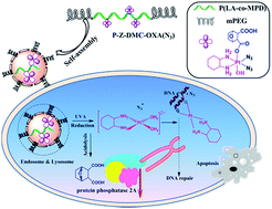
Taking advantage of the enhanced permeability and retention (EPR) effect of tumor, tumor-targeted drug delivery systems, e.g. micelles, liposomes, polymersomes and hydrogels, have been extensively developed to decrease side effects and prolong circulation time of small molecular drugs.20–22 Recently, we demonstrated a polymer–(multifunctional single-drug) conjugate strategy, in which three different drug components (cisplatin, azidyl radical and DMC) were rationally integrated.23 Comparing with other types of combination therapy based on nanoparticles, the polymer–(multifunctional single-drug) conjugate has several features: (i) simplified synthesis procedure by a single conjugation for batch production, (ii) delivery of multiple drugs with inter-synergisms to the targets of interest with definitive ratios and high drug loadings, (iii) ability to control drug exposure spatially and temporally via different responsiveness. The conjugate exhibited amplified signals of cancer treatment, especially in cisplatin-resistant cancer cells in vitro. However, improved results against tumor directly need to be demonstrated in vivo to verify our strategy. Herein, we constructed a multifunctional single-drug by integrating photo-activated oxaliplatin(IV) complex and DMC, which was further conjugated to polymer carrier to form multifunctional single-drug loaded nanoparticles. When the nanoparticles are internalized by cancer cells via endocytosis, active oxaliplatin(II) and azidyl radical can be released upon UVA irradiation to attack DNA, while DMC will be hydrolyzed subsequently from the polymer chain within intracellular environment to block DNA repair caused by the DNA-damaged chemotherapeutic drugs (oxaliplatin(II) and azidyl radical) through PP2A inhibition. Then the drugs would make concerted efforts to suppress tumor proliferation in vitro and in vivo (Scheme 1). The enhanced photo-chemotherapy efficacy with improved drug pharmacokinetics and reduced toxicity of the multifunctional single-drug loaded nanoparticles were also evaluated in vivo.


image file: c5ra26372g-s1.tif
Scheme 1 Possible pathways after multifunctional single-drug-loaded nanoparticles enter cancer cells.

Experimental section

Materials

Hydrogen peroxide (H2O2), N,N′-dicyclohexyl carbodiimide (DCC), N-hydroxybenzotriazole (HOBt), 1R,2R-cyclohexanediamine (DACH), Hoechst were purchased from Sigma-Aldrich. K2PtCl4 (purity 99%) was bought from Shandong Boyuan Chemical Company, China. Demethylcantharidin (DMC) (purity 99%) was bought from Nanjing Zelang Biomedical Company, China. Curcumin was purchased from Aladdin. All other commercially sourced chemicals and solvents were used without further purification.

General measurements

1H NMR spectra were measured by a Unity-300 MHz NMR spectrometer (Bruker) at room temperature. Fourier transform infrared (FT-IR) spectra were recorded on a Bruker Vertex 70 spectrometer. Mass spectroscopy (ESI-MS) measurements were performed on a Quattro Premier XE system (Waters) equipped with an electrospray interface (ESI). Inductively coupled plasma mass spectrometer (ICP-MS, Xseries II, Thermoscientific, USA) was used for quantitative determination of trace levels of platinum. Particle size and size distribution of nanoparticles were determined by DLS with a vertically polarized HeNe laser (DAWN EOS, Wyatt Technology, USA). The morphology of the nanoparticles was measured by TEM performed on a JEOL JEM-1011 electron microscope. Clinic parameters were measured by an automatic biochemical analyzer (Mindray BS-220, China). UV-visible absorption spectra were recorded on a Varian Cary 300 UV-visible spectrophotometer in 1 cm path-length cuvettes. Irradiations were carried out using a xenon lamp source (CEL-S500, AuLight, China) equipped with filters for parallel monochromatic lights at 365 nm. The power outage was measured using a power meter (FZ-A, AuLight, China).

Synthesis of Z-DMC-OXA(N3)

c,c,t-[Pt(DACH)(N3)2(OH)2] was prepared as previously reported.24 Z-DMC-OXA(N3) was synthesized as following procedures. Briefly, demethylcantharidin (100 mg, 0.6 mmol) was added to a solution of c,c,t-[Pt(DACH)(N3)2(OH)2] (254 mg, 0.6 mmol) in DMSO (10 mL) (Scheme S1) and the reaction mixture was stirred at 55 °C for 24 hours in the dark. Cooled diethyl ether (100 mL) was added to precipitate a light yellow solid, after washed for several times with acetone and diethyl ether, and dried. Z-DMC-OXA(N3) was isolated in 60% yield (212 mg). The product was characterized using IR (Fig. S1. iv), 1H NMR (Fig. S2) and ESI-MS (Fig. S3C). 1H NMR: (300 MHz, d6-DMSO, 25 °C): δH 1.03–2.2 ppm (10H, m, cyclohexane CH, CH2), 1.47 ppm (m, 4H; CH2CH2), 2.87 ppm (s, 2H; CHCH), 4.57 ppm (d, J = 4.5, 1H, OCH) and 4.68 ppm (d, J = 2.7, 1H; OCH), 8.11–6.35 ppm (4H, m, NH2); IR: cm−1 3480 (br, –OH), 2065 (sh, N3), 1724 (sh, –CO–OH), 1628 (sh, –OOC–C–); ESI-MS: (negative mode) m/z = 594.4 [M − H] (100%). *Caution!* Although no problems were encountered during this work, heavy metal azides are known to be heat and shock-sensitive detonators. Therefore, it is essential that any platinum azide compounds are handled with care.

Synthesis of P-Z-DMC-OXA(N3)

mPEG-b-P(LA-co-MPD) was prepared as previously reported.25 Z-DMC-OXA(N3) was conjugated to mPEG-b-P(LA-co-MPD) using the DCC/HOBt method (Scheme S2). Briefly, Z-DMC-OXA(N3) (50 mg), DCC (50 mg) and HOBt (20 mg) were added to DMF (5 mL). After stirring for 30 min, mPEG-b-P(LA-co-MPD) (100 mg) in CH2Cl2 (20 mL) was added and the reaction mixture was kept stirring at room temperature for 24 h. The solution was filtered and the filtrate was precipitated in ether. The solid was re-dissolved and re-precipitated in ether twice, giving a light yellow powder, P-Z-DMC-OXA(N3) conjugate.

Preparation of P-Z-DMC-OXA(N3) nanoparticles

P-Z-DMC-OXA(N3) nanoparticles were prepared by a nano-precipitation method. In brief, P-Z-DMC-OXA(N3) (50 mg) was dissolved in a solution of DMF (5 mL) and then water (25 mL) was added dropwise into the flask under stirring to form a micellar solution. The solution was dialyzed against water to remove DMF and then freeze-dried. The nanoparticle size was obtained from DLS. The morphology and size were further characterized using TEM. The platinum content of the nanoparticles was determined by ICP-OES.

Curcumin loaded P-Z-DMC-OXA(N3) nanoparticles were (Cur@P-Z-DMC-OXA(N3) nanoparticles) also prepared by the same method. UV-vis spectrophotometer was used to determine the curcumin content.

Photo-responsiveness of Z-DMC-OXA(N3) and P-Z-DMC-OXA(N3) nanoparticles

Photo-sensitivity. Aqueous solutions of Z-DMC-OXA(N3) and P-Z-DMC-OXA(N3) nanoparticles were UVA irradiated (365 nm, 10 mW cm−2) for the indicated periods of time (0 min to 90 min) and the UV-vis spectra of the aqueous solutions were taken. For stability in the dark, aqueous solutions of Z-DMC-OXA(N3) and P-Z-DMC-OXA(N3) nanoparticles in distilled water were kept in the dark and UV-vis spectra were taken over days. The size change of P-Z-DMC-OXA(N3) nanoparticles in response to UVA irradiation (365 nm, 10 mW cm−2) were also followed by DLS measurements.
Platinum release profile. Lyophilized P-Z-DMC-OXA(N3) nanoparticles (5 mg) were hydrated in 1 mL of buffered solution (0.1 mM PBS, pH 7.4 and pH 5.0), placed into a pre-swollen dialysis bag (MWCO = 1000 Da) and immersed in 19 mL buffered solution. The dialysis was conducted at 37 °C in a shaking culture incubator. Samples were UVA irradiated (365 nm, 10 mW cm−2) for 20 min before the experiments, and then kept in the dark. 1 mL of sample solution was withdrawn at specified time intervals from the dialysate and measured for Pt using ICP-OES. After sampling, fresh PBS (1 mL) was added to the dialysate. The platinum released from the nanoparticles was expressed as a percentage of cumulative platinum outside the dialysis bag compared to the total platinum in the nanoparticles as a function of release time.

In vitro studies

Cell culture. HeLa cervical cancer cells and fibroblast L929 normal cells were purchased from the Institute of Biochemistry and Cell Biology, Chinese Academy of Sciences, Shanghai, China and grown in DMEM (Life Technologies) supplemented with 0.03% L-glutamine and 1% penicillin/streptomycin in 5% CO2 at 37 °C.
Cell viability assay. Cytotoxicity was assessed using a 3-(4,5-dimethylthiazol-2-yl)-2,5-diphenyltetrazolium bromide (MTT) assay. L929 and HeLa cells were seeded in 96-well plates and incubated in DMEM overnight. The medium was then replaced by oxaliplatin, DMC, Z-DMC-OXA(N3) and P-Z-DMC-OXA(N3) nanoparticles with a final Pt concentration (or DMC concentration) from 3.375 to 216 μM. A MTT assay was performed in the absence and presence of UVA irradiation (365 nm) during 72 h incubation time. For control samples, incubation was performed in the dark. For the samples with irradiation, cells were incubated for 4 h in the dark after administration before UVA irradiation (365 nm, 10 mW cm−2) for 30 min and then incubated in the dark for the rest of the time. After 72 hours, MTT solution (20 μL) in PBS (5 mg mL−1) was added and the plates were incubated for another 4 h at 37 °C, followed by removal of the culture medium containing MTT and addition of DMSO (150 μL) to each well to dissolve the formazan crystals formed. Finally, the plates were shaken for 10 min and the absorbance of the formazan product was measured at 490 nm using a microplate reader. Treatment procedures for each cell line were replicated three times.
Cellular uptake. HeLa cells were seeded on the coverslips in 6-well plates (2 × 10⁵ cells per well) and cultured in 2 mL of DMEM for 24 h, 2 mL of Cur@P-Z-DMC-OXA(N3) nanoparticles in DMEM (at a final curcumin concentration of 10 μg mL−1) were added, and cells were incubated for additional 0.5 or 4 h. After three washes with PBS, 4% paraformaldehyde was added for another 30 min at 37 °C. Finally nuclei were counterstained with the Hoechst (1 μg mL−1) for 15 min at room temperature. Cellular uptake was observed with an Olympus FV1000 confocal laser scanning microscope (CLSM) imaging system (Japan).

In vivo experiments

Animal use. Male Sprague Dawley (SD) rats (6–8 weeks old) and Kunming (KM) mice (4–6 weeks old) were purchased from Jilin University (Changchun, China). All mice and rats were maintained under required conditions and had free access to food and water throughout the experiments. All the in vivo study protocols were approved by the local institution review board and performed according to the Guidelines of the Committee on Animal Use and Care of Changchun Institute of Applied Chemistry, Chinese Academy of Sciences.
In vivo anticancer efficacy evaluation. H22 murine hepatocarcinoma xenografts tumor model were implanted subcutaneously at the flank of KM mice (1 × 10⁷ H22 cells in 0.1 mL). Tumor nodules were allowed to grow to a volume ∼ 100 mm3 before initiating treatment. Prior to treatment, all the mice were numbered using ear tags, and their weight and the initial tumor volume were measured and recorded. Saline (n = 20), oxaliplatin (3.5 mg kg−1, n = 20), Z-DMC-OXA(N3) (3.25 mg Pt per kg, n = 20) and P-Z-DMC-OXA(N3) nanoparticles (3.25 mg Pt per kg, n = 20) were administered intravenously at day 0, day 3 and day 6 in the dark, where n is the number of mice in each group. Mice were then split into two groups: one group was kept constantly in the dark and the other group was irradiated with UVA (365 nm, 10 mW cm−2) at the tumor site for 0.5 h on day 1, day 4 and day 7. After the light treatment, mice were returned to the dark. Tumor length and width were measured with calipers, and the tumor volume was calculated using the following equation: tumor volume = length × width × width/2. The weight and tumor volume of each mouse were measured every two days over four weeks.

After that, the blood sample of each mouse was collected from the retro-orbital plexus into a coagulation-promoting tube, and then centrifuged at 3000 rpm for 5 min to obtain plasma samples for measuring clinic parameters including urea nitrogen (UREA), creatinine (CREA), uric acid (UA), alanine aminotransferase (ALT), aspartate aminotransferase (AST) by an automatic biochemical analyzer.

TUNEL assay. At the end of tumor inhibition study, the tumors were excised from sacrificed mice and fixed in 10% formalin solution followed by paraffin embedding and TUNEL staining. Embedding and staining were done in Norman Bethune Health Science Center of Jilin University. Images were acquired by OLYMPUS CX31 microscope and analyzed by Image Analysis System 10.0.
Blood clearance. The blood persistence properties of Z-DMC-OXA(N3) and P-Z-DMC-OXA(N3) nanoparticles were determined using male SD rats weighing ∼300 g. The animals, three per group, were injected in the tail vein with 1 mL of Z-DMC-OXA(N3) and P-Z-DMC-OXA(N3) nanoparticles with a Pt content of 1 mg kg−1. At predetermined time intervals, blood samples were collected in pre-weighed heparinized tubes. The percentage of Pt was calculated by taking into consideration that blood constitutes 5.4% of body weight.

Results and discussion

Synthesis and characterization of multifunctional single-drug loaded nanoparticles

Multifunctional single-drug, Z-DMC-OXA(N3), was obtained by ring-opening reaction of DMC with photo-activated oxaliplatin(IV) complex, c,c,t-[Pt(DACH)(N3)2(OH)2]. One of axial positions of c,c,t-[Pt(DACH)(N3)2(OH)2] was modified with one molecule DMC. FTIR (Fig. S1), 1H NMR (Fig. S2) and ESI-MS (Fig. S3) confirmed the structure of Z-DMC-OXA(N3). Biodegradable amphiphilic block copolymer PEG-b-P(LA-co-MPD) with pendent hydroxyl groups was chosen as the carrier for the multifunctional single-drug. DCC/HOBt condensation method was used to conjugate Z-DMC-OXA(N3) to the carrier forming a polymer–(multifunctional single-drug) conjugate (P-Z-DMC-OXA(N3)) (Scheme S2). Here, DMC was modified as a part of the single-drug and also as a linker between the hydroxyl-functionalized mPEG-b-P(LA-co-MPD) and photo-activated oxaliplatin(IV) prodrug. We expected the photo-reduction release of oxaliplatin(II) complex could induce hydrolysis of DMC in low pH condition (lysosomes and endosomes) sequentially. Thus oxaliplatin(II) could cause DNA damage and DMC could inhibit DNA damage repair, finally they could play a synergetic role to restrain tumor proliferation.

The newly formed P-Z-DMC-OXA(N3) could self-assemble into nanoparticles due to its amphiphilic character. The morphology of P-Z-DMC-OXA(N3) nanoparticles was analyzed using transmission electron microscope (TEM). The nanoparticles were successfully formed in a spherical shape with no aggregation was observed, and the mean diameter was about 150 nm determined by DLS (Fig. 1). The platinum content was also determined to be 6.5 wt% by ICP-OES.


image file: c5ra26372g-f1.tif
Fig. 1 (a) TEM morphology and (b) DLS characterization of P-Z-DMC-OXA(N3) nanoparticles.

Photo-sensitivity of multifunctional single-drug loaded nanoparticles

Z-DMC-OXA(N3) and P-Z-DMC-OXA(N3) nanoparticles shared the same intense N3–Pt ligand-to-metal charge-transfer (LMCT) UV-vis absorbance, like the previously reported photo-activated Pt(IV) complexes.12 The photo-activated Pt(IV) structure could be readily activated to Pt(II) form by light to kill cancer cells. So the small multifunctional single-drug and multifunctional single-drug loaded nanoparticles were expected to display high photo-responsiveness upon UVA irradiation. To confirm this, the photo-activation of Z-DMC-OXA(N3) and P-Z-DMC-OXA(N3) nanoparticles was monitored through UV-vis spectra. Under UVA irradiation (365 nm, 10 mW cm−2), the maximum absorption peak at 258 nm dropped dramatically, demonstrating the breakdown of the N3–Pt bond (Fig. 2a and b). Although the destruction speed of N3–Pt bond in multifunctional single-drug loaded nanoparticles was slight slower than small multifunctional single-drug, it was believed that the polymer carrier did not affect the activation character upon UVA irradiation (Fig. 2c). In this condition, the oxaliplatin(IV) species was reduced to the biologically active oxaliplatin(II) form and dissociated from the carrier for effective photo-chemotherapy. What's more, normalized UV absorbance (258 nm) of aqueous solutions in the dark was unchanged for all samples up to at least one week, which indicated the high stability of multifunctional single-drug loaded nanoparticles, making nanoparticles more convenience for preservation and application (Fig. 2d). The size change of P-Z-DMC-OXA(N3) nanoparticles for various UVA irradiation intervals was monitored by DLS measurement (Fig. 3a). After 15 min UVA irradiation, the size distribution of P-Z-DMC-OXA(N3) nanoparticles was divided to two districts (∼80 nm and ∼200 nm), and the intensity of smaller size district was strengthened after 30 min. It is reasonable that platinum release gradually from P-Z-DMC-OXA(N3) nanoparticles after irradiation may shift the hydrophilic and hydrophobic balance of nanoparticles and induce the re-assembly of nanostructure. Because only small amount of platinum was left after 45 min irradiation, no obvious size distribution change of P-Z-DMC-OXA(N3) nanoparticles was observed between 45 min and 60 min, which was consistent with the UV-vis spectra results upon UVA irradiation.
image file: c5ra26372g-f2.tif
Fig. 2 UVA sensitivity and dark stability of Z-DMC-OXA(N3) and P-Z-DMC-OXA(N3) nanoparticles: (a) UV-vis spectra of Z-DMC-OXA(N3) and (b) P-Z-DMC-OXA(N3) nanoparticles upon UVA irradiation from 0 to 90 min, (c) normalized UV absorbance (258 nm) of Z-DMC-OXA(N3) and P-Z-DMC-OXA(N3) nanoparticles versus irradiation time, (d) stability of Z-DMC-OXA(N3) and P-Z-DMC-OXA(N3) nanoparticles in the dark over days.

image file: c5ra26372g-f3.tif
Fig. 3 (a) Size of P-Z-DMC-OXA(N3) nanoparticles varies with time upon UVA irradiation, (b) Pt release from P-Z-DMC-OXA(N3) nanoparticles at pH 5.0 and pH 7.4 upon UVA irradiation and in the dark.

It has been confirmed that cancer tissue has idiosyncratic microenvironments including reduced extracellular and intracellular pH. Platinum release profiles of P-Z-DMC-OXA(N3) nanoparticles were monitored using a dialysis technique (MWCO = 1000 Da) at pH 7.4 and 5.0 to mimic the different pH conditions in blood circulation and endosomes/lysosomes (Fig. 3b). The experiment was implemented with UVA irradiation (365 nm, 10 mW cm−2) for 20 min, and then kept in the dark. The accelerated platinum release from the nanoparticles was found to be both pH and UVA dependent. In the dark, as predicted, Pt release at pH 5.0 was faster than that at pH 7.4 due to the ester bonds acidolysis of P-Z-DMC-OXA(N3). More specifically, after 8 h, 32% of Pt(IV) was release in the dark at pH 7.4, while 50% release occurred at pH 5.0. In the presence of UVA irradiation, from the beginning to the end (24 hours), the release rate and amount of platinum showed little difference between pH 7.4 and 5.0. Furthermore, the platinum release was significantly accelerated when UVA was applied. For instance, platinum release exceeded 72 wt% in 8 h, and the accumulative amount was more than 75 wt% at pH 7.4 and 77 wt% at pH 5.0 after 24 h. It could be concluded that it was much more susceptible for platinum release to UVA irradiation than to pH changes. These results further verified the photo-responsiveness of the multifunctional single-drug loaded nanoparticles for triggered drug release.

For the release of DMC, we stated it in details in our previous work.23 The specific PP2A inhibition proved the DMC hydrolysis indirectly. When A549 cells incubated with polymer–(multifunctional single-drug) conjugate in the presence of UVA irradiation, the value of PP2A activity dropped down obviously compared with its original level. This might due to the dissociation of DMC from the polymer and we expected this platform could enhance tumor growth inhibition by inhibiting PP2A activity.

In vitro cell cytotoxicity evaluation of multifunctional single-drug loaded nanoparticles

The cell cytotoxicity against normal cells and cancer cells of the multifunctional single-drug loaded nanoparticles under dark and after light irradiation was assessed by MTT assays using oxaliplatin, DMC and small single-drug as control for 72 hour incubation (Fig. 4). Cell viability reached over 97% after 0.5 h of UVA irradiation in the absence of any drug (Fig. S5). Without light treatment, P-Z-DMC-OXA(N3) nanoparticles (IC50 = 140.53 μM) exhibited much lower cytotoxicity than oxaliplatin and DMC against L929 normal fibroblasts, while oxaliplatin killed both HeLa cervical cancer cells and L929 normal fibroblasts without selectivity (IC50 for HeLa, 8.04 μM; IC50 for L929, 30.93 μM). Upon light treatment, the IC50 value of P-Z-DMC-OXA(N3) nanoparticles against HeLa cancer cells decreased dramatically as anticipated, since the photo-activated oxaliplatin(IV) was reduced to the more toxic oxaliplatin(II) form under light. P-Z-DMC-OXA(N3) nanoparticles was found to be roughly 2.6-fold more cytotoxic upon UVA light irradiation than in the dark (Fig. S6), suggesting the cytotoxicity of P-Z-DMC-OXA(N3) nanoparticles could be well improved by the external light stimulus.
image file: c5ra26372g-f4.tif
Fig. 4 In vitro cytotoxicity curves of oxaliplatin, DMC, Z-DMC-OXA(N3) and P-Z-DMC-OXA(N3) nanoparticles against (a) L929 normal fibroblasts in the dark and (b) HeLa cancer cell in the dark and in the presence of UVA irradiation (365 nm, 10 mW cm−2) during 72 hour incubation.

To correlate the observed cytotoxicity with cellular uptake and release of drugs, the internalization and intracellular distribution of P-Z-DMC-OXA(N3) nanoparticles was observed by CLSM (Fig. 5). Curcumin was used as a model dye and loaded in the nanoparticles. A rapid uptake and internalization of the green fluorescence labeled nanoparticles was observed in HeLa cells within 0.5 h. After 4 h incubation, green fluorescence emerged in the nuclei. The result suggested that the multifunctional single-drug loaded nanoparticles can be easily uptaken with a higher intracellular concentration due to the well-known intracellular uptake mechanism via endocytosis of the macromolecular drugs rather than free diffusion of free drugs.


image file: c5ra26372g-f5.tif
Fig. 5 Cellular uptake and localization of Cur@P-Z-DMC-OXA(N3) in HeLa cells after incubation with the cells for 0.5 h and 4 h. CLSM images were taken from the Hoechst channel (blue, a) the Cur channel (green, b) and overlapped images of the Cur and Hoechst channel (c).

Enhanced photo-chemotherapy and decreased toxicity of multifunctional single-drug loaded nanoparticles in vivo

Finally, the anti-tumor efficacy of the multifunctional single-drug loaded nanoparticles was tested in vivo using a H22 murine hepatocarcinoma model.22 Saline, oxaliplatin, (3.25 mg Pt per kg), Z-DMC-OXA(N3) (3.25 mg Pt per kg) and P-Z-DMC-OXA(N3) nanoparticles (3.25 mg Pt per kg) were injected intravenously for three times on day 0, day 3 and day 6 in the dark. The saline group was regarded as a control group without any drug treatment. The mice were then split into two groups: one group was kept constantly under dark and the other group was irradiated with UVA (365 nm, 10 mW cm−2) at the tumor site for 30 min on day 1, day 4 and day 7. After UVA treatment, mice were returned to the dark. Tumor growth inhibition in the dark and with UVA activation is shown in Fig. 6a. In the dark, P-Z-DMC-OXA(N3) nanoparticles displayed the highest anti-tumor efficacy compared with Z-DMC-OXA(N3) and oxaliplatin. The average tumor volume in Z-DMC-OXA(N3) group increased to about 1000 mm3 after four weeks, while in the micelle group the tumor growth was well restricted without excessive. At day 29, the average tumor volume for oxaliplatin group was 570 mm3 while the P-Z-DMC-OXA(N3) nanoparticles group was about 130 mm3. Upon UVA irradiation, Z-DMC-OXA(N3) was only slightly more effective in inhibiting tumor growth than in the dark, likely due to the rapid blood clearance of small molecular drugs by the reticuloendothelial system (RES).26 Furthermore, the tumor size in the multifunctional single-drug loaded nanoparticles group decreased to about 40 mm3 after four weeks upon UVA irradiation. TUNEL staining was further conducted to assess the apoptosis level of tumor after different drug treatment (Fig. S7). Significantly higher levels of apoptotic cells after UVA irradiation were observed in Z-DMC-OXA(N3) and P-Z-DMC-OXA(N3) nanoparticles groups than that under dark. It could be concluded that the photo-reduction release of oxaliplatin(II) and subsequent release of DMC enhanced the tumor therapeutic effect. Interestingly, the nanoparticles were more cytotoxic than oxaliplatin both in the presence and absence of UVA. It is reasonable that polymer conjugation of multifunctional single-drug could prolong the blood circulation time and take advantage of the EPR effect of tumor to improve the antitumor effect.
image file: c5ra26372g-f6.tif
Fig. 6 (a) Relative tumor volume, (b) variation of body weight and (c) survival rate of KM mice treated with oxaliplatin (3.25 mg Pt per kg), Z-DMC-OXA(N3) (3.25 mg Pt per kg), P-Z-DMC-OXA(N3) nanoparticles (3.25 mg Pt per kg) and saline in the dark and upon UVA irradiation, (d) variation of Pt level in blood with time in SD rats after a single intravenous injection of Z-DMC-OXA(N3) and P-Z-DMC-OXA(N3) nanoparticles (1 mg Pt per kg).

Hence, we further tested the clearance behavior of the nanoparticles in blood. Platinum could be applied as the marker for the in vivo fate of the nanoparticles, and the variation of Pt levels in blood with time was shown in Fig. 6d. Z-DMC-OXA(N3) was quickly cleared from blood after intravenous administration. After 24 hours, the left platinum doge of P-Z-DMC-OXA(N3) nanoparticles in the blood was over 4-fold more than that of Z-DMC-OXA(N3). Multifunctional single-drug loaded nanoparticles resulted in a significant prolongation of Pt presence in blood, consistent with previous studies.27,28 The main pharmacokinetic parameters also showed the improved pharmacokinetics after polymeric conjugation of small multifunctional single-drug. The area under the curve (AUC) increased from 13[thin space (1/6-em)]944.64 h ng mL−1 to 57[thin space (1/6-em)]618.73 h ng mL−1, and the clearance (CL) was a 4-fold less for P-Z-DMC-OXA(N3) nanoparticles over Z-DMC-OXA(N3) (Table 1). All the results confirmed that P-Z-DMC-OXA(N3) nanoparticles could improve the drug retention in blood and increase the bioavailability of the drug in vivo, resulting in enhanced efficacy and improved tolerability.

Table 1 Pharmacokinetic parameters in blooda
Test article AUC0–24 h h ng mL−1 Cmax ng mL−1 Tmax h CL mL h−1 Vd mL kg−1
a AUC, area under curve; Cmax, maximum concentration observed; Tmax, time at maximum concentration; CL, clearance; Vd, volume of distribution.
Z-DMC-OXA(N3) 13[thin space (1/6-em)]944.64 1643.89 0.5 13.99 608.31
P-Z-DMC-OXA(N3) 57[thin space (1/6-em)]618.73 4766.15 0.5 3.37 209.81


The relative body weight change in tumor inhibition experiment was measured to determine the systemic toxicity. Meanwhile, the survival rate clarified the long-time toxicity of different groups. Oxaliplatin both in the dark and after UVA treatment caused a significant decrease in total body weight, which resulted in the sacrifice of treated mice at day 8 due to excessive weight loss (Fig. 6b). Z-DMC-OXA(N3) had sacrifice in different degrees both in the dark and under the UVA light during the whole month, whereas the survival rate of P-Z-DMC-OXA(N3) nanoparticles was always being 100% (Fig. 6c). In the dark, Z-DMC-OXA(N3) and P-Z-DMC-OXA(N3) nanoparticles had little effect on body weight, suggesting almost no systemic toxicity. However, upon UVA irradiation, Z-DMC-OXA(N3) and P-Z-DMC-OXA(N3) nanoparticles caused a slight body weight reduction compared to the saline control group. The weight loss was still significantly lower than the oxaliplatin treated mice, and after a few days the body weight was comparable to the initial body weight at day 0, suggesting that even with UVA irradiation, multifunctional single-drug and micellar formulations caused significantly less systemic toxicity than the clinically used oxaliplatin. Alterations of blood biochemistry parameters ALT, AST, CREA, UA and UREA also certified the reduced hepatotoxicity and nephrotoxicity of P-Z-DMC-OXA(N3) nanoparticles (Fig. 7). In groups treated with oxaliplatin and multifunctional single-drug in the absence and presence of UVA irradiation, ALT and AST showed a great increase compared with the control groups. Similar results for UA and UREA were also found. On the contrary, the multifunctional single-drug loaded nanoparticles exhibited little effect on the levels of blood biochemistry parameters. In short, these results demonstrated the decreased systemic toxicity of P-Z-DMC-OXA(N3) nanoparticles in vivo.


image file: c5ra26372g-f7.tif
Fig. 7 Alterations of ALT, AST, CREA, UA and UREA in KM mice after treatment with oxaliplatin (3.25 mg Pt per kg), Z-DMC-OXA(N3) (3.25 mg Pt per kg), P-Z-DMC-OXA(N3) nanoparticles (3.25 mg Pt per kg) and saline in the dark and upon UVA irradiation.

Conclusions

We synthesized a multifunctional single-drug by tethering DMC to photo-activated oxaliplatin(IV) prodrug, which was conjugated to polymer carrier to form polymer–(multifunctional single-drug) conjugate nanoparticles for photo-chemotherapy. The multifunctional single-drug loaded nanoparticles displayed high photo-sensitivity under UVA irradiation and maintained stability in the dark. Upon UVA irradiation, multifunctional single-drug loaded nanoparticles exhibited enhanced anti-cancer efficacy in vitro and in vivo. What's more, the nanoparticles could prolong drug residence in the blood and decrease the systemic toxicity compared to clinical used oxaliplatin(II). This enhanced cancer treatment strategy could have potential clinical use in the near future.

Acknowledgements

The work is supported by the National Natural Science Foundation of China (No. 51403198 and 51573069), the Ministry of Science and Technology of China (863 Project, No. SS2012AA020603), and Jilin Provincial Science and Technology Department (No. 20150520019JH).

Notes and references

  1. E. Wong and C. M. Giandomenico, Chem. Rev., 1999, 99, 2451–2466 CrossRef CAS PubMed.
  2. R. C. Todd and S. J. Lippard, Metallomics, 2009, 1, 280–291 RSC.
  3. J. J. Wilson and S. J. Lippard, Chem. Rev., 2014, 114, 4470–4495 CrossRef CAS PubMed.
  4. M. D. Hall and T. W. Hambley, Coord. Chem. Rev., 2002, 232, 49–67 CrossRef CAS.
  5. R. C. Dolman, G. B. Deacon and T. W. Hambley, J. Inorg. Biochem., 2002, 88, 260–267 CrossRef CAS PubMed.
  6. S. Choi, C. Filotto, M. Bisanzo, S. Delaney, D. Lagasee, J. L. Whitworth, A. Jusko, C. Li, N. A. Wood, J. Willingham, A. Schwenker and K. Spaulding, Inorg. Chem., 1998, 37, 2500–2504 CrossRef CAS.
  7. S. G. Awuah, Y. R. Zheng, P. M. Bruno, M. T. Hemann and S. J. Lippard, J. Am. Chem. Soc., 2015, 137, 14854–14857 CrossRef CAS PubMed.
  8. N. J. Wheate, S. Walker, G. E. Craig and R. Oun, Dalton Trans., 2010, 39, 8113–8127 RSC.
  9. T. Boulikas and M. Vougiouka, Oncol. Rep., 2003, 10, 1663–1682 CAS.
  10. S. He, Y. Cong, D. Zhou, J. Li, Z. Xie, X. Chen, X. Jing and Y. Huang, J. Mater. Chem. B, 2015, 3, 8203–8211 RSC.
  11. F. S. Mackay, J. A. Woods, P. Heringova, J. Kasparkova, A. M. Pizarro, S. A. Moggach, S. Parsons, V. Brabec and P. J. Sadler, Proc. Natl. Acad. Sci. U. S. A., 2007, 104, 20743–20748 CrossRef CAS PubMed.
  12. P. J. Bednarski, F. S. Mackay and P. J. Sadler, Anti-Cancer Agents Med. Chem., 2007, 7, 75–93 CrossRef CAS PubMed.
  13. Y. Min, J. Li, F. Liu, E. K. L. Yeow and B. Xing, Angew. Chem., Int. Ed., 2014, 53, 1012–1016 CrossRef CAS PubMed.
  14. X. Yang, H. Xiang, L. An, S. P. Yang and J. G. Liu, New J. Chem., 2015, 39, 800–804 RSC.
  15. X. Li, J. Mu, F. Liu, E. W. P. Tan, B. Khezri, R. D. Webster, E. K. L. Yeow and B. Xing, Bioconjugate Chem., 2015, 26, 955–961 CrossRef CAS PubMed.
  16. E. Gabano, M. Ravera and D. Osella, Dalton Trans., 2014, 43, 9813–9820 RSC.
  17. X. Xue, S. You, Q. Zhang, Y. Wu, G. Z. Zou, P. C. Wang, Y. L. Zhao, Y. Xu, L. Jia, X. Zhang and X. J. Liang, Mol. Pharmacol., 2012, 9, 634–644 CrossRef CAS PubMed.
  18. D. Zhou, Y. Cong, Y. Qi, S. He, H. Xiong, Y. Wu, Z. Xie, X. Chen, X. Jing and Y. Huang, Biomater. Sci., 2015, 3, 182–191 RSC.
  19. R. Qi, H. Xiao, S. Wu, Y. Li, Y. Zhang and X. Jing, J. Mater. Chem. B, 2015, 3, 176–179 RSC.
  20. H. Maeda, Adv. Enzyme Regul., 2001, 41, 189–207 CrossRef CAS PubMed.
  21. H. Maeda, T. Sawa and T. Konno, J. Controlled Release, 2001, 74, 47–61 CrossRef CAS PubMed.
  22. D. Zhou, H. Xiao, F. Meng, X. Li, Y. Li, X. Jing and Y. Huang, Adv. Healthcare Mater., 2013, 2, 822–827 CrossRef CAS PubMed.
  23. D. Zhou, S. He, Y. Cong, Z. Xie, X. Chen, X. Jing and Y. Huang, J. Mater. Chem. B, 2015, 3, 4913–4921 RSC.
  24. H. Xiao, G. T. Noble, J. F. Stefanick, R. Qi, T. Kiziltepe, X. Jing and B. Bilgicer, J. Controlled Release, 2014, 173, 11–17 CrossRef CAS PubMed.
  25. H. Kuang, S. Wu, F. Meng, Z. Xie, X. Jing and Y. Huang, J. Mater. Chem., 2012, 22, 24832–24840 RSC.
  26. A. S. Paraskar, S. Soni, K. T. Chin, P. Chaudhuri, K. W. Muto, J. Berkowitz, M. W. Handlogten, N. J. Alves, B. Bilgicer, D. M. Dinulescu, R. A. Mashelkar and S. Sengupta, Proc. Natl. Acad. Sci. U. S. A., 2010, 107, 12435–12440 CrossRef CAS PubMed.
  27. J. H. Xu, F. P. Gao, L. L. Li, H. L. Ma, Y. S. Fan, W. Liu, S. S. Guo, X. Z. Zhao and H. Wang, Microporous Mesoporous Mater., 2013, 182, 165–172 CrossRef CAS.
  28. L. Rao, L. L. Bu, J. H. Xu, B. Cai, G. T. Yu, X. Yu, Z. He, Q. Huang, A. Li, S. S. Guo, W. F. Zhang, W. Liu, Z. J. Sun, H. Wang, T. H. Wang and X. Z. Zhao, Small, 2015, 11, 6225–6236 CrossRef CAS PubMed.

Footnote

Electronic supplementary information (ESI) available. See DOI: 10.1039/c5ra26372g

This journal is © The Royal Society of Chemistry 2016
Click here to see how this site uses Cookies. View our privacy policy here.