DOI:
10.1039/C5RA26291G
(Paper)
RSC Adv., 2016,
6, 21308-21316
Simple and signal-off electrochemiluminescence immunosensor for alpha fetoprotein based on gold nanoparticle-modified graphite-like carbon nitride nanosheet nanohybrids
Received
9th December 2015
, Accepted 16th February 2016
First published on 16th February 2016
Abstract
A simple, signal-off electrochemiluminescence (ECL) immunosensor for sensitive and selective detection of alpha fetoprotein (AFP) was developed based on gold nanoparticle (AuNPs) modified graphite-like carbon nitride nanosheet (g-C3N4 NSs) nanohybrids (Au-g-C3N4 NHs). Compared to the g-C3N4 NS modified glassy carbon electrode (GCE), the ECL intensity is more obvious at the Au-g-C3N4 NH modified GCE due to the fact that AuNPs can promote electron transfer and electrocatalytic reduction of S2O82− to produce large amounts of hole donor (SO4˙−). Results demonstrated that the ECL signal of the g-C3N4 NSs decreased due to the interaction between antibody and antigen on the modified electrode. Thus, an ECL immunosensor for the detection of AFP was realized by monitoring the ECL intensity change of g-C3N4 NSs. The factors influencing the performance of the immunosensor were investigated in detail. Under optimal conditions, the proposed biosensor achieved a wide line from 0.001 to 5.0 ng mL−1 with a detection limit of 0.0005 ng mL−1. Furthermore, the ECL immunosensor was successfully applied to the determination of AFP in serum samples, providing a promising effective strategy for AFP detection.
1. Introduction
Sensitive and accurate detection of tumor markers is critical to early diagnosis, point-of-care and portable medical supervision, as well as screening for many serious diseases.1 The levels of tumor markers in serum, tissue, or saliva can be indicative of the physiological state of a cancer at a specific time.2 As a tumor marker, alpha fetoprotein (AFP) is a plasma protein produced by the yolk sac and the liver during fetal life.3 However, it may also be found at high levels in the sera of adults with certain malignancies. An elevated AFP concentration in adult plasma is widely considered as an early indication of hepatocellular carcinoma or endodermal sinus tumor.4 Thus, it is of great significance to develop rapid and sensitive analytical methods to identify and quantify of AFP for early discovery, early diagnosis and early treatment. Immunological method has become the predominant analytical tool for quantitative monitoring of low-abundance tumor marker by the sophisticated techniques involving enzyme-linked immunosorbent assay,5,6 surface plasmon resonance,7,8 electrochemistry,9,10 photoluminescence,1,11 fluorescence immunoassay12 and electrochemiluminescence (ECL).13 Among these methods, ECL as a powerful analytical technique with high sensitivity, low background signal, well reproducibility, easy controllability and wide dynamic response range has been widely applied in the detection of biomolecules, clinical diagnostics, environmental and food monitoring.14,15 Up to now, ECL emitters can be either dissolved in a homogenous phase or immobilized in the solid state for analytical applications.16 The type of solid-state ECL, which immobilizes an ECL emitter as a sensing substrate at the electrode surface, has attracted considerable attention due to its regenerability, simplicity, portability and low cost.
A series of materials including Ru complexes, luminol, metallic oxide semiconductors and quantum dots have been used to design ECL sensors.16 Of two-dimensional (2D) nanomaterials, graphitic-like carbon nitride (g-C3N4) with a stacked 2D structure, has recently been proved to be an effective ECL emitter.17 Compared with traditional luminophores, g-C3N4 presents many charming advantages, such as easy preparation, metal-free, nontoxic, low cost, excellent biocompatibility. More importantly, several reports demonstrated the ultrathin g-C3N4 nanosheets (g-C3N4 NSs) have better ECL efficiency and stability than bulk g-C3N4.18,19 So far, g-C3N4 NSs have been used as the emitter for the constructed of ECL signal-off or on type sensor for sensitive determination of dopamine,20 carcinoembryonic antigen,21 bisphenol A in the presence of persulfate (S2O82−) or triethylamine as coreactant. However, at present based on the ECL property of g-C3N4 NSs to determination AFP is scarce now.
Recent years, various nanomaterials have been widely employed to improve the performance of electrode interface.22,23 The small size, high surface-to-volume ratio, good biocompatibility and unusual target binding characteristics can markedly improve the sensitivity and specificity of analyte detection, making nanomaterials particularly appealing for use as chemical and biosensors.24–26 Metal nanoparticles, especially gold nanoparticles (AuNPs), have been commonly utilized as intermediator to immobilize antibody on the electrode.27 The advantages that AuNPs bring to biosensors are presented as follows but not limited to: enlarging the active areas, accelerating electron transfer between electrodes and detection species, and behaving as biocompatible scaffolds for biomolecule immobilization.28,29 As the conductivity of the g-C3N4 NSs is low, a conducting nanomaterial such as AuNPs needs to be introduced for accelerating the electron transfer process.30,31 Moreover, AuNPs could improve and stabilize the cathodic ECL intensity of g-C3N4 NSs and have the excellent property of electrocatalytic reduction of S2O82− into hole donor (SO4˙−).21 The latter studies highlight the potential of using AuNPs modified electrode in ECL immunosensor to detect AFP. Thus, the integration of AuNPs and g-C3N4 NSs would exhibit promising application prospect in the ECL immunosensor.
Herein, a solid-state ECL immunosensor was developed with AuNPs modified g-C3N4 NSs nanohybrids (named as Au-g-C3N4 NHs) as sensing substrate to detect AFP. On the basis of steric hindrance from bio-recognition reactions, has enabled the development of a signal-off ECL sensing strategy via the insulation of attached biomacromolecule.32–34 After immobilization of alpha fetoprotein antibody (anti-AFP) on the modified electrode, the detection of AFP was realized by monitoring the ECL intensity change of g-C3N4 NSs resulting from the antigen–antibody reaction. The proposed strategy for the immunosensor is unique and advantageous for the following reasons: (1) Au-g-C3N4 NHs was immobilized in the solid state, where the electrode has good stability and reproducibility; (2) the synthesis method of Au-g-C3N4 NHs is simple and the ECL immunosensor is easily fabricated due to excellent film-forming ability of g-C3N4 NSs and remarkable reactivity between the thiol (or primary amine) groups of biomolecules and AuNPs; (3) the proposed detection strategy is completely label-free because of the bio-recognition reactions of antigen–antibody. More details about preparation of the electrode we proposed, and its relevant ECL detection and analysis were presented.
2. Experimental
2.1. Materials and reagents
AFP and anti-AFP were purchased from Beijing Biosynthesis Biotechnology Co., Ltd. HAuCl4·4H2O (99.9%), sodium borohydride (NaBH4, 96.0%), trisodium citrate (99.9%), and bovine serum albumin (BSA) were purchased from Sigma-Aldrich. Potassium persulfate (K2S2O8) and carbamide were purchased from Sinopharm Group Chemical Reagent Co. Phosphate buffer saline (PBS) solution with various pH were prepared by mixing the stock solution of 1/15 M Na2HPO4 and KH2PO4 in appropriate ratios. The supporting electrolyte was 0.1 M KCl. PBS (pH 7.4) solution containing 0.1 M K2S2O8 was used as the electrolyte in ECL analysis. All aqueous solutions were prepared with ultrapure water (≥18 MΩ cm−1), which was obtained from a Milli-Q water purification system. PBS (pH 7.4) solution was used for preparation of the antibody and antigen solution, washing buffer solution, and blocking buffer solution which contained 2% (w/v) BSA.
2.2. Apparatus
Transmission electron microscopic (TEM) images were obtained from a Hitachi H-600 microscope (Japan). The Fourier transform infrared (FT-IR) spectra were recorded on a NEXUS-670 FT-IR Spectrometer (Nicolet, USA). The phase characterization was performed by X-ray diffraction (XRD) using a D8 advance diffractometer system equipped with Cu-Kα radiation (Bruker Co., Germany). The UV-visible spectrum was recorded by a Shimadzu UV-2550 UV-visible spectrophotometer. Electrochemical impedance spectroscopy (EIS) was measured in the frequency region of 105 to 10−1 Hz with a voltage excitation amplitude of 5 mV on the IM6ex electrochemical workstation (ZAHNER, Germany). ECL measurements were carried out on an ECL detection system (MPI-B Remex Electronic Instrument Co., Xi'an, China) using modified glassy carbon electrode (GCE, Φ = 4 mm) as the working electrode, Ag/AgCl electrode as the reference electrode, and platinum wire as the auxiliary electrode in PBS (pH 7.4) solution containing 0.1 M K2S2O8.
2.3. Preparation of the g-C3N4 NSs
First, the bulk g-C3N4 was prepared by polymerization of carbamide molecules under high temperature. In detail, 10.0 g of carbamide powder was heated in an alumina crucible in a muffle furnace at 550 °C for 2.5 h under air condition with a ramp rate of about 5 °C min−1 of the heating and then cooling to room temperature. The obtained yellow product was the g-C3N4 powder.35 Second, the g-C3N4 NSs were obtained by liquid exfoliating of as-prepared bulk g-C3N4 in ultrapure water. In detail, 100 mg of bulk g-C3N4 powder dispersed in 100 mL ultrapure water, and then ultrasound for about 18 h. The initial formed suspension was then centrifuged at about 8000 rpm to remove the residual unexfoliated g-C3N4 nanoparticles and then the supernatant was collected and concentrated on a rotary evaporator at 75 °C under reduced pressure and result in a milk-like suspension (g-C3N4 NSs) for further study.
2.4. Synthesis of Au-g-C3N4 NHs
AuNPs were synthesized according to the standard sodium citrate reduction method.36 Briefly, 0.04 g of HAuCl4·4H2O was added into 100 mL ultrapure water and heated to boil under stirring, then 10 mL trisodium citrate (38.8 mM) solution were mixed rapidly with the boiling solution to induce particle formation. During prolonged heating under reflux for 15 min, the color of the solution turned to ruby red. This AuNPs aqueous dispersion was used without further purification in the following procedure. The Au-g-C3N4 NHs was prepared by a facile chemisorption method. AuNPs aqueous dispersion and g-C3N4 NSs suspension were mixed in different volume ratio, and then stirred at room temperature overnight. At this point, the obtained nanohybrid materials were collected by centrifugation and washed three times with ultrapure water and then redispersed into 0.5 mL of ultrapure water.
2.5. Fabrication of the ECL immunosensor
The ECL immunosensor was fabricated on a GCE. Briefly, the GCE was polished successively with 0.3, 0.05 μm alumina slurry, followed by rinsing thoroughly with ultrapure water. The fabrication procedure of the immunosensor is shown in Scheme 1. First, 10 μL of Au-g-C3N4 NHs was applied onto the surface of the cleaned GCE with a microsyringe and allowed to dry at room temperature to obtain Au-g-C3N4 NHs modified GCE (Au-g-C3N4 NHs/GCE). After the Au-g-C3N4 NHs/GCE was washed with PBS (pH 7.4) solution to remove free Au-g-C3N4 NHs. Then, the anti-AFP were introduced onto the Au-g-C3N4 NHs/GCE by dropping 50 μL antibody (2.5 μg mL−1) dissolved in PBS (pH 7.4) solution onto the electrode and allowing it to incubate at 4 °C for 12 h. After being rinsed with PBS solution thoroughly to remove physically adsorbed anti-AFP and then incubated with 50 μL of 2% BSA solution at 37 °C for 60 min to block nonspecific binding sites. Subsequently, the electrode was rinsed with PBS solution and used as an ECL immunosensor, and incubated in 50 μL of different concentrations of AFP at 37 °C for 60 min. Finally the electrode was rinsed with PBS solution and ready to be used.
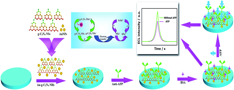 |
| | Scheme 1 Schematic representation of the fabricating procedures of the proposed ECL immunosensor. | |
3. Results and discussion
3.1. Characterization of g-C3N4 NSs and Au-g-C3N4 NHs
The phase structure of the as-synthesized g-C3N4 NSs sample was identified by XRD measurement. The XRD patterns of bulk g-C3N4 and g-C3N4 NSs are shown in Fig. 1A. In the case of bulk g-C3N4, the strong diffraction peak at 27.4° and the weak peak at 13.1° which correspond to the characteristic interlayer stacking reflection of conjugated aromatic systems and the inter-layer structural packing, respectively, which is in consistence with the reported g-C3N4.37–39 Remarkably enough, after exfoliation, the intensity of this two diffraction peaks significantly decreases, clearly demonstrating that the layered g-C3N4 has been successfully exfoliated as we expected. The chemical structure of the g-C3N4 NSs was further confirmed by the FT-IR spectra shown in Fig. 1B. The broad peaks around 3252 cm−1 originating from the N–H stretches can be clearly observed,40 suggesting the partial hydrogenation of some nitrogen atoms in the nanosheets. The band at 815 cm−1 belonged to the characteristic breathing mode of triazine ring and the set of peaks between 1647 and 1204 cm−1 were characteristic of aromatic carbon nitride heterocycles.41 The peak at 2150 cm−1 is attributed to cyano terminal groups C![[triple bond, length as m-dash]](https://www.rsc.org/images/entities/char_e002.gif) N.40 Clearly, the FT-IR spectrum of g-C3N4 NSs is similar to that of bulk g-C3N4, indicating that the exfoliated nanosheets keep the same chemical structure as their parent bulk g-C3N4. Combined with the results of the XRD and FT-IR analyses, it is seen that the g-C3N4 NSs has been successfully prepared.
N.40 Clearly, the FT-IR spectrum of g-C3N4 NSs is similar to that of bulk g-C3N4, indicating that the exfoliated nanosheets keep the same chemical structure as their parent bulk g-C3N4. Combined with the results of the XRD and FT-IR analyses, it is seen that the g-C3N4 NSs has been successfully prepared.
 |
| | Fig. 1 XRD patterns (A) and FT-IR spectra (B) of g-C3N4 NSs and bulk g-C3N4. The UV-vis spectrum (C) of g-C3N4 NSs (a), AuNPs (b), Au-g-C3N4 NHs (c). The TEM image of g-C3N4 NSs (D) and Au-g-C3N4 NHs (E). | |
UV-vis spectroscopy was used to confirm the combination of g-C3N4 NSs and AuNPs. As shown in Fig. 1C, the g-C3N4 NSs solution has an obvious absorption peak at 320 nm (curve a). The UV-vis absorption spectrum (curve b) of AuNPs shows a characteristic absorption for the surface plasmon resonance absorption at 520 nm. Au-g-C3N4 NHs (curve c) shows both the typical absorption features of g-C3N4 NSs and AuNPs and the absorption peaks had no shift, which indicated the intrinsic properties of g-C3N4 NSs and AuNPs have been maintained. The morphology of the g-C3N4 NSs and Au-g-C3N4 NHs were observed by TEM measurement, as shown in Fig. 1. From Fig. 1D, it is apparent that the g-C3N4 NSs shows a typical layered structure with sheet-like morphology, which means g-C3N4 NSs consisted of graphite planes stacking along the c-axis.42 In the TEM image of the Au-g-C3N4 NHs (Fig. 1E), we could find that the AuNPs were well-dispersed on the g-C3N4 NSs surface, again with little or no aggregation. This due to g-C3N4 NSs can be easily manipulated post-functionalization or elementally doped. The results above further confirming the formation of AuNPs on the g-C3N4 NSs.
3.2. The effect of AuNPs on the ECL behaviors of g-C3N4 NSs
In order to investigate the effect of AuNPs on the ECL behaviors of g-C3N4 NSs, the g-C3N4 NSs and Au-g-C3N4 NHs were modified onto the surface of GCE, respectively. The ECL was tested in PBS (pH 7.4) solution containing 0.1 M K2S2O8 at the potential range from 0 to −1.1 V with a scan rate of 100 mV s−1 and the results are shown in Fig. 2A. It is noted that the g-C3N4 NSs/GCE (curve b) has a strong ECL intensity in the S2O82− system, this is due to the strong high-energy annihilation between electrons (injected into the conduction band of g-C3N4 NSs by the anodic electrode) and holes (injected into the valence band of g-C3N4 NSs by the electrogenerated SO4˙− from S2O82−).43 Compared to the g-C3N4 NSs/GCE, the ECL intensity is more obvious at the Au-g-C3N4 NHs/GCE (curve c). The AuNPs electrocatalytic reduction of S2O82− produce more abundant hole-donor (SO4˙−) can be considered an essential reason for the enhanced ECL intensity. In addition, the effect of the AuNPs in the proportion of Au-g-C3N4 NHs on the ECL intensity was also investigated. As shown in Fig. 2B, when AuNPs increases from 0.37 wt% to 8.95 wt%, the ECL intensity gradually increases (0.37 wt% to 1.79 wt%) and then declines (1.79 wt% to 8.95 wt%). Therefore, 1.79 wt% AuNPs was employed for the determination to maximize the sensitivity.
 |
| | Fig. 2 (A) The ECL responses of (a) bare GCE, (b) g-C3N4 NSs/GCE, (c) Au-g-C3N4 NHs/GCE. (B) Effect of the AuNPs in the proportion of Au-g-C3N4 NHs on the ECL intensity. (a–d): 0.37 wt%; 1.79 wt%; 4.47 wt%; 8.95 wt%. Error bars represent standard deviation (n = 5). | |
3.3. Optimization of experimental conditions
To increase the sensitivity and selectivity of the biosensor, the optimization of the incubation time of the anti-AFP modified electrode in AFP solution was investigated. The AFP incubation time is an important parameter affecting the analytical performance of biosensor. At room temperature, the ECL signal for AFP (0.05 ng mL−1) decreased with the reaction time up to 60 min and then leveled off after 60 min (Fig. 3A), which showed the saturated bind between the anti-AFP and AFP. Therefore, 60 min of incubation time was used for the detection of AFP.
 |
| | Fig. 3 (A) Optimization of the incubation time of AFP. (B) Dependence of the ECL intensity of Au-g-C3N4 NHs/GCE on the concentration of S2O82− (M): (a) 0.03, (b) 0.05, (c) 0.1, (d) 0.15. | |
The ECL intensity is also affected by the concentration of coreactant. Fig. 3B shows that the ECL intensity increases with the concentration of S2O82− in the range of 0.03–0.15 M. Under the condition of the same technical parameters, when the concentration higher than 0.15 M the ECL intensity was over the test range since the ECL intensity obtained in 0.15 M S2O82− reaches the upper detection limit of the instrument. Therefore, the 0.1 M S2O82− was used to ensure a suitable sensitivity.
3.4. Characterization of the immunosensor
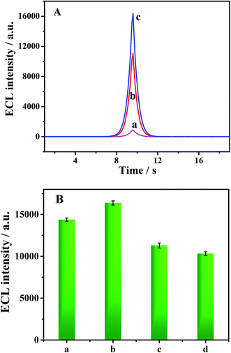
To probe the feature of the modified electrode surface, the modification of GCE surface was characterized by EIS and ECL signals at each immobilization steps were recorded to monitor the construction of the immunosensor. The impedance spectrum includes a semicircle portion and a linear portion. The semicircle portion observed at high frequencies corresponds to the charge-transfer limited process (Rct), a line portion observed at low frequencies by diffusion process. As shown in Fig. 4A, the bare GCE showed a very small semicircle domain with the Rct of 91.78 Ω, the characteristics of a mass diffusional limiting electron-transfer process (Fig. 4A, curve a), indicating a very fast charge-transfer process of [Fe(CN)6]3−/4−. After Au-g-C3N4 NHs film was deposited on the GCE, the value of Rct increased to 135.4 Ω (Fig. 4A, curve b), this is due to the g-C3N4 NSs is semiconductor, thus restricted the electron transfer of [Fe(CN)6]3−/4− to the GCE. Subsequent surface was modified with anti-AFP, the value of Rct increased to 293.9 Ω (Fig. 4A, curve c). These results attributed to the antibodies hindered the electron transfer of [Fe(CN)6]3−/4− on the interface of the modified electrode. After blocking the residual nonspecific binding sites on the fabricated immunosensor with BSA leaded to a more obvious increase of Rct to 566.3 Ω (Fig. 4A, curve d), which was attributed to the fact that BSA (acted as an inert electron layer) could hinder the electrochemical probe from getting close to the electrode interface. Finally, owing to the antigen–antibody complex blocking layer, the AFP immobilization on the electrode greatly increased of Rct to 814.8 Ω (Fig. 4A, curve e). To give more detailed information about the impedance of the modified electrode, the Randles circuit (the inset plot of Fig. 4A) is further chosen to fit the impedance data obtained in the experiments. Fig. 4B illustrates the ECL intensity of different modified electrodes in PBS (pH 7.4) solution containing 0.1 M K2S2O8. It was clearly observed that the successive modification steps, i.e., the conjugation of anti-AFP (Fig. 4B, curve b), BSA (Fig. 4B, curve c), and AFP (Fig. 4B, curve d), result in the gradual decreases in ECL intensity. Compared with the Au-g-C3N4 NHs/GCE, the ECL intensity of anti-AFP/Au-g-C3N4 NHs/GCE, BSA/anti-AFP/Au-g-C3N4 NHs/GCE and AFP/BSA/anti-AFP/Au-g-C3N4 NHs/GCE were decreased by 21.98%, 28.93% and 63.33%, respectively. This could be explained by the fact that the immobilization of these non-conductive bioactive substances on the Au-g-C3N4 NHs/GCE surface forming a barrier for electron transfer and hindered the diffusion of ECL coreactant to the electrode surface. These results were in accordance with those obtained from EIS results. All these experimental results demonstrated that the immunosensor has been successfully fabricated.
 |
| | Fig. 4 (A) The EIS for (a) bare GCE, (b) Au-g-C3N4 NHs/GCE, (c) anti-AFP/Au-g-C3N4 NHs/GCE, (d) BSA/anti-AFP/Au-g-C3N4 NHs/GCE and (e) AFP/BSA/anti-AFP/Au-g-C3N4 NHs/GCE in 0.1 M KCl solution containing 1.0 mM [Fe(CN)6]3−/4− with the frequencies swept from 105 Hz to 10−1 Hz. Inset is the Randles circuit model for the modified electrodes in the cell. (B) The ECL responses of (a) Au-g-C3N4 NHs/GCE, (b) anti-AFP/Au-g-C3N4 NHs/GCE, (c) BSA/anti-AFP/Au-g-C3N4 NHs/GCE and (d) AFP/BSA/anti-AFP/Au-g-C3N4 NHs/GCE at the AFP concentration of 0.1 ng mL−1. | |
3.5. ECL detection of AFP
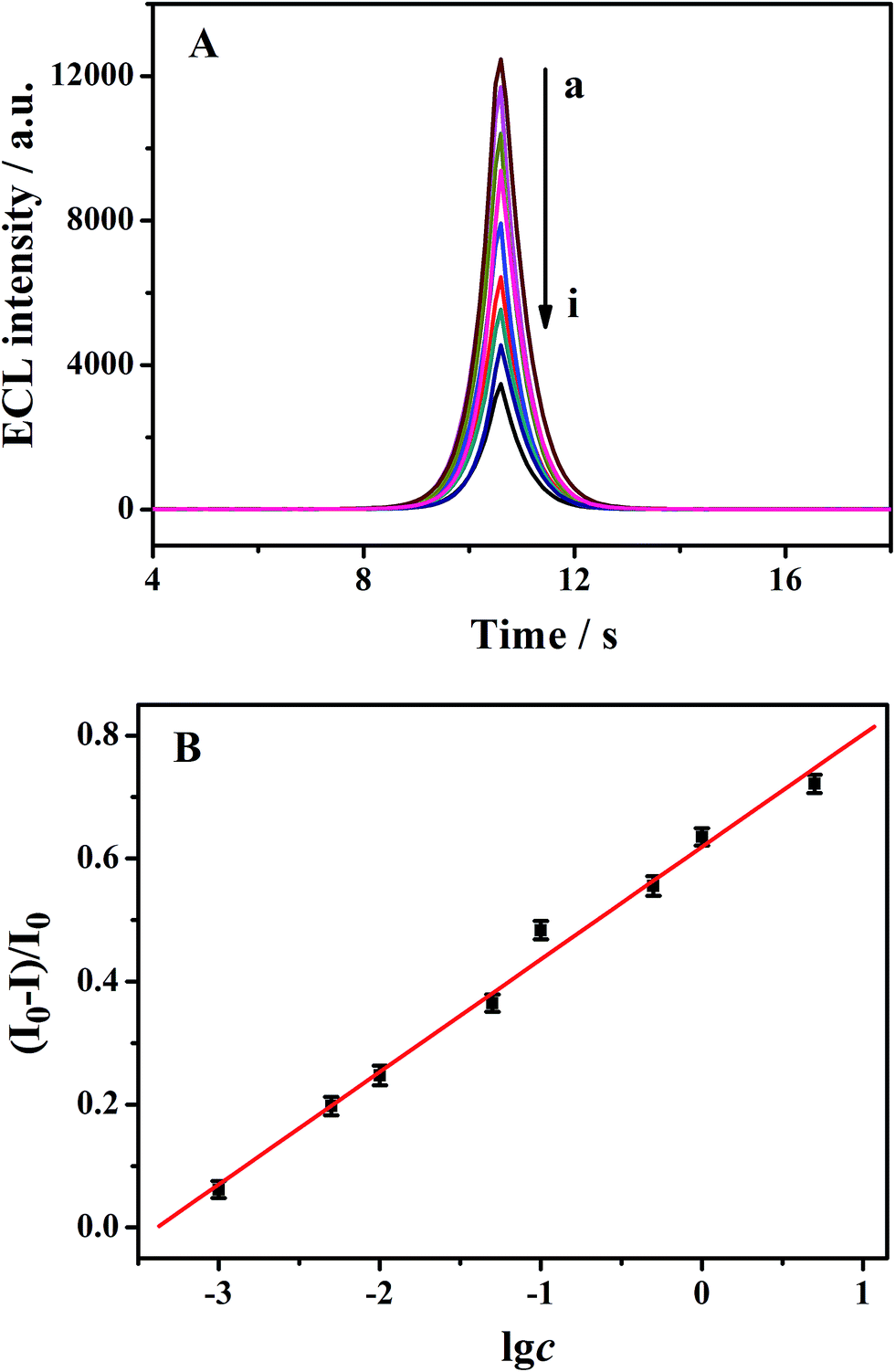
Under the optimal conditions, the ECL immunosensor was applied to quantify the concentration of target. AFP standards with concentrations from 0.001 to 5.0 ng mL−1 were tested, with five measurements each in parallel. As shown in Fig. 5A, the ECL signal decreases gradually with an increase in the concentrations of AFP. The inhibition ratio (I0 − I)/I0 of ECL intensity was found to be proportional to the logarithm of AFP concentration in range from 0.001 to 5.0 ng mL−1 (where I0 and I represented the ECL intensity of BSA/anti-AFP/Au-g-C3N4 NHs/GCE incubated without AFP and with 50 μL of different concentrations of AFP, respectively). The corresponding linear function is (I0 − I)/I0 = 0.6191 + 0.1830![[thin space (1/6-em)]](https://www.rsc.org/images/entities/char_2009.gif) lgc (ng mL−1, R2 = 0.9953) (Fig. 5B). The detection limit (S/N = 3) also evaluated to be as low as 0.0005 ng mL−1 for AFP. Additionally, the analytical performance of the ECL immunosensor for AFP detection was compared with those of other reported methods (Table 1). The comparison indicates that the ECL aptasensor is one of the most excellent sensors for AFP detection.
lgc (ng mL−1, R2 = 0.9953) (Fig. 5B). The detection limit (S/N = 3) also evaluated to be as low as 0.0005 ng mL−1 for AFP. Additionally, the analytical performance of the ECL immunosensor for AFP detection was compared with those of other reported methods (Table 1). The comparison indicates that the ECL aptasensor is one of the most excellent sensors for AFP detection.
 |
| | Fig. 5 (A) ECL responses of the immunosensors with increasing concentration of AFP (ng mL−1) from (a) to (i): (a) 0, (b) 0.001, (c) 0.005, (d) 0.01, (e) 0.05, (f) 0.1, (g) 0.5, (h) 1.0 and (i) 5.0. (B) Calibration curve of the immunosensor for detection of AFP in the range between 0.001 and 5.0 ng mL−1. Error bars represent standard deviation (n = 5). | |
Table 1 Comparison of different methods for the detection of AFP
| Method |
Linear range (ng mL−1) |
Detection limit (ng mL−1) |
Reference |
| Electrochemiluminescence |
0.05–50 |
0.03 |
44 |
| Horseradish peroxidase based immunosensor |
0.02–4.0 |
0.01 |
45 |
| Label-free electrochemical immunosensor |
0.02–50 |
0.01 |
46 |
| Sandwich-type electrochemical immunosensor |
0.01–60 |
0.0016 |
47 |
| Sandwich-type immunosensor |
0.01–40 |
0.0023 |
48 |
| Label-free electrochemical immunosensor |
0.01–12 |
0.005 |
49 |
| Photoelectrochemical immunosensor |
0.05–50 |
0.04 |
50 |
| Electrochemiluminescence |
0.001–5 |
0.0005 |
This work |
3.6. Selectivity, reproducibility and stability of the ECL immunosensor
To evaluate the selectivity of the ECL immunosensor, we challenged the immunosensor with other possible interferences such as human IgG (HIgG), luteotropic hormone (LH), L-cysteine (L-Cys), and L-glutamate (L-Glu) and carcinoembryonic antigen (CEA) in the same conditions. And the results are exhibited in Fig. 6. When the immunosensor was incubated with 20 ng mL−1 HIgG, LH, L-Cys, L-Glu and CEA solution, respectively, no apparent signal change was observed compared to the blank test (no target molecular) in the same testing conditions. However, when AFP was coexistence with the interferences, the signal change was almost the same as that with only AFP. All these results indicating that the proposed immunosensor has a good selectivity for AFP.
 |
| | Fig. 6 Selectivity of the proposed ECL immunosensor. The mixture is containing HIgG (20 ng mL−1), LH (20 ng mL−1), L-Cys (20 ng mL−1), CEA (20 ng mL−1) and AFP (1.0 ng mL−1). Error bars represent standard deviation (n = 5). | |
To evaluate the reproducibility of the immunosensor, we investigated the immunosensor through the coefficient of variation of the intra- and interassays. When the immunosensor was investigated by using five electrodes to test the same concentration of AFP (0.1 ng mL−1) at the same conditions, the relative standard deviation (RSD) was 3.7%. When a same electrode was used to test repetitively for five times at a certain concentration of AFP (0.1 ng mL−1), the RSD was 4.8%. In addition, when the immunosensor was stored in PBS (pH 7.4) solution at 4 °C, over 90% of the initial ECL intensity remained after a storage period of two weeks. These results showed acceptable reproducibility and good stability of our proposed method.
3.7. Application of the immunosensor
As the practical applications, the developed immunosensor was explored to detect AFP in real serum sample. We evaluated the recovery in different concentrations of AFP solutions diluted in the serum sample (Table 2). The serum sample was diluted with PBS (pH 7.4). The obtained results showed satisfactory recoveries in the range of 100.2% to 105.3%. The results suggested that the biosensor has a promising potential application for the detection of AFP in clinical analysis.
Table 2 Determination of AFP added in serum sample (n = 3) with the proposed immunosensor
| Serum sample |
Concentration of AFP added (ng mL−1) |
Concentration obtained with immunosensor (ng mL−1) |
Recovery (%) |
RSD (%) |
| 1 |
0.01 |
0.01053 |
105.3 |
2.8 |
| 2 |
0.1 |
0.1047 |
104.7 |
3.6 |
| 3 |
0.5 |
0.5012 |
100.2 |
4.1 |
| 4 |
1.0 |
1.005 |
100.5 |
5.3 |
| 5 |
2.0 |
2.017 |
100.9 |
3.9 |
4. Conclusions
This work demonstrated a facile pathway to fabricate a solid-state ECL immunosensor utilizes Au-g-C3N4 NHs as the substrate material. The introduction of the AuNPs, on the one hand, as antibody capture substrate can be combined with antibodies and immobilization antibody on the surface of electrode, on the other hand, can promote electron transfer and electrocatalytic reduction of S2O82− to produce large amounts of hole donor (SO4˙−), effectively enhance the ECL intensity. The fabrication of the ECL immunosensor is easy, inexpensive and completely label-free. The resulting immunosensor for quantitative detection of AFP possesses high sensitivity, selectivity, good reproducibility, and long-term stability. This proposed method can be expanded readily for detecting other cancer biomarkers. In view of the above advantages, we anticipate that this high sensitive and selective method has potential to be applied in clinical applications.
Acknowledgements
This work was supported by the National Natural Science Foundation of China (No. 21205048), the Natural Science Foundation of Shandong Province, China (No. ZR2013BM003, No. ZR2009BM034) and the Science and Technology Development Plan Project of Shandong Province (No. 2012G0022116).
References
- Y. J. Li, M. J. Ma and J. J. Zhu, Dual-Signal Amplification Strategy for Ultrasensitive Photoelectrochemical Immunosensing of α-Fetoprotein, Anal. Chem., 2012, 84, 10492–10499 CrossRef CAS PubMed.
- W. B. Li, X. Q. Jiang, J. C. Xue, Z. K. Zhou and J. H. Zhou, Antibody modified gold nano-mushroom arrays for rapid detection of alpha-fetoprotein, Biosens. Bioelectron., 2015, 68, 468–474 CrossRef CAS PubMed.
- D. Du, Z. X. Zou, Y. S. Shin, J. Wang, H. Wu, M. H. Engelhard, J. Liu, I. A. Aksay and Y. H. Lin, Sensitive Immunosensor for Cancer Biomarker Based on Dual Signal Amplification Strategy of Graphene Sheets and Multienzyme Functionalized Carbon Nanospheres, Anal. Chem., 2010, 82, 2989–2995 CrossRef CAS PubMed.
- M. Giannetto, L. Elviri, M. Careri, A. Mangia and G. Mori, A voltammetric immunosensor based on nanobiocomposite materials for the determination of alpha-fetoprotein in serum, Biosens. Bioelectron., 2011, 26, 2232–2236 CrossRef CAS PubMed.
- W. Q. Lai, D. P. Tang, J. Y. Zhuang, G. N. Chen and H. H. Yang, Magnetic Bead-Based Enzyme-Chromogenic Substrate System for Ultrasensitive Colorimetric Immunoassay Accompanying Cascade Reaction for Enzymatic Formation of Squaric Acid-Iron(III) Chelate, Anal. Chem., 2014, 86, 5061–5068 CrossRef CAS PubMed.
- J. A. Brochot and I. W. Siddiqi, Peroxidase-catalysed rupture of the C-F bond as and indicator reaction for enzyme immunoassay: Kinetic determination of human immunoglobulin G, α-fetoprotein and placental lactogen, Anal. Chim. Acta, 1989, 224, 329–337 CrossRef CAS.
- Y. Teramura and H. Iwata, Label-free immunosensing for α-fetoprotein in human plasma using surface plasmon resonance, Anal. Biochem., 2007, 365, 201–207 CrossRef CAS PubMed.
- S. Scarano, M. Mascini, A. P. F. Turner and M. Minunni, Surface plasmon resonance imaging for affinity-based biosensors, Biosens. Bioelectron., 2010, 25, 957–966 CrossRef CAS PubMed.
- J. J. Miao, X. B. Wang, L. D. Lu, P. Y. Zhu, C. Mao, H. L. Zhao, Y. C. Song and J. Shen, Electrochemical immunosensor based on hyperbranched structure for carcinoembryonic antigen detection, Biosens. Bioelectron., 2014, 58, 9–16 CrossRef CAS PubMed.
- X. L. Shao, H. Gu, Z. Wang, X. L. Chai, Y. Tian and G. Y. Shi, Highly Selective Electrochemical Strategy for Monitoring of Cerebral Cu2+ Based on a Carbon Dot-TPEA Hybridized Surface, Anal. Chem., 2013, 85, 418–425 CrossRef CAS PubMed.
- X. R. Zhang, S. G. Li, X. Jin and S. S. Zhang, A new photoelectrochemical aptasensor for the detection of thrombin based on functionalized graphene and CdSe nanoparticles multilayers, Chem. Commun., 2011, 47, 4929–4931 RSC.
- J. L. Yuan, G. L. Wang, K. Majima and K. Matsumoto, Synthesis of a Terbium Fluorescent Chelate and Its Application to Time-Resolved Fluoroimmunoassay, Anal. Chem., 2001, 73, 1869–1876 CrossRef CAS PubMed.
- L. C. Chen, X. T. Zeng, A. R. Ferhan, Y. W. Chi, D.-H. Kim and G. N. Chen, Signal-on electrochemiluminescent aptasensors based on target controlled permeable films, Chem. Commun., 2015, 51, 1035–1038 RSC.
- Y. L. Cao, R. Yuan, Y. Q. Chai, L. Mao, H. Niu, H. J. Liu and Y. Zhuo, Ultrasensitive luminol electrochemiluminescence for protein detection based on in situ generated hydrogen peroxide as coreactant with glucose oxidase anchored AuNPs@MWCNTs labeling, Biosens. Bioelectron., 2012, 31, 305–309 CrossRef CAS PubMed.
- L. H. Chen, D. J. Huang, S. Y. Ren, Y. W. Chi and G. N. Chen, Carbon Dioxide Gas Sensor Based on Ionic Liquid-Induced Electrochemiluminescence, Anal. Chem., 2011, 83, 6862–6867 CrossRef CAS PubMed.
- S. Y. Deng and H. X. Ju, Electrogenerated chemiluminescence of nanomaterials for bioanalysis, Analyst, 2013, 138, 43–61 RSC.
- C. M. Cheng, Y. Huang, J. Wang, B. Z. Zheng, H. Y. Yuan and D. Xiao, Anodic Electrogenerated Chemiluminescence Behavior of Graphite-Like Carbon Nitride and Its Sensing for Rutin, Anal. Chem., 2013, 85, 2601–2605 CrossRef CAS PubMed.
- N. Y. Cheng, J. Q. Tian, Q. Liu, C. J. Ge, A. H. Qusti, A. M. Asiri, A. O. Al-Youbi and X. P. Sun, Au-Nanoparticle-Loaded Graphitic Carbon Nitride Nanosheets: Green Photocatalytic Synthesis and Application toward the Degradation of Organic Pollutants, ACS Appl. Mater. Interfaces, 2013, 5, 6815–6819 CAS.
- J. Q. Tian, Q. Liu, A. M. Asiri, A. O. Al-Youbi and X. P. Sun, Ultrathin Graphitic Carbon Nitride Nanosheet: A Highly Efficient Fluorosensor for Rapid, Ultrasensitive Detection of Cu2+, Anal. Chem., 2013, 85, 5595–5599 CrossRef CAS PubMed.
- Y. T. Liu, Q. B. Wang, J. P. Lei, Q. Hao, W. Wang and H. X. Ju, Anodic electrochemiluminescence of graphitic-phase C3N4 nanosheets for sensitive biosensing, Talanta, 2014, 122, 130–134 CrossRef CAS PubMed.
- L. C. Chen, X. T. Zeng, P. Si, Y. M. Chen, Y. W. Chi, D. H. Kim and G. N. Chen, Gold Nanoparticle-Graphite-Like C3N4 Nanosheet Nanohybrids Used for Electrochemiluminescent Immunosensor, Anal. Chem., 2014, 86, 4188–4195 CrossRef CAS PubMed.
- Q. F. Li, L. X. Zeng, J. C. Wang, D. P. Tang, B. Q. Liu, G. N. Chen and M. D. Wei, Magnetic Mesoporous Organic-Inorganic NiCo2O4 Hybrid Nanomaterials for Electrochemical Immunosensors, ACS Appl. Mater. Interfaces, 2011, 3, 1366–1373 CAS.
- F. Peng, Y. Y. Su, Y. L. Zhong, C. H. Fan, S.-T. Lee and Y. He, Silicon Nanomaterials Platform for Bioimaging Biosensing, and Cancer Therapy, Acc. Chem. Res., 2014, 47, 612–623 CrossRef CAS PubMed.
- X. R. Xia, N. A. Monteiro-Riviere, S. Mathur, X. F. Song, L. S. Xiao, S. J. Oldenberg, B. Fadeel and J. E. Riviere, Mapping the Surface Adsorption Forces of Nanomaterials in Biological Systems, ACS Nano, 2011, 5, 9074–9081 CrossRef CAS PubMed.
- G. Aragay, F. Pino and A. MerkocI, Nanomaterials for Sensing and Destroying Pesticides, Chem. Rev., 2012, 112, 5317–5338 CrossRef CAS PubMed.
- O. N. Oliveira Jr, R. M. Iost, J. R. Siqueira Jr, F. N. Crespilho and L. Caseli, Nanomaterials for Diagnosis: Challenges and Applications in Smart Devices Based on Molecular Recognition, ACS Appl. Mater. Interfaces, 2014, 6, 14745–14766 Search PubMed.
- Y. Zhu, P. Chandra and Y.-B. Shim, Ultrasensitive and Selective Electrochemical Diagnosis of Breast Cancer Based on a Hydrazine-Au Nanoparticle-Aptamer Bioconjugate, Anal. Chem., 2013, 85, 1058–1064 CrossRef CAS PubMed.
- G. Z. Liu, E. Luais and J. J. Gooding, The Fabrication of Stable Gold Nanoparticle-Modified Interfaces for Electrochemistry, Langmuir, 2011, 27, 4176–4183 CrossRef CAS PubMed.
- J. Wang, L. L. Yang, S. Boriskina, B. Yan and B. M. Reinhard, Spectroscopic Ultra-Trace Detection of Nitroaromatic Gas Vapor on Rationally Designed Two-Dimensional Nanoparticle Cluster Arrays, Anal. Chem., 2011, 83, 2243–2249 CrossRef CAS PubMed.
- B. Jeong, R. Akter, O. H. Han, C. K. Rhee and M. Aminur Rahman, Increased Electrocatalyzed Performance through Dendrimer-Encapsulated Gold Nanoparticles and Carbon Nanotube-Assisted Multiple Bienzymatic Labels: Highly Sensitive Electrochemical Immunosensor for Protein Detection, Anal. Chem., 2013, 85, 1784–1791 CrossRef CAS PubMed.
- F. Yang, J. Han, Y. Zhuo, Z. H. Yang, Y. Q. Chai and R. Yuan, Highly sensitive impedimetric immunosensor based on single-walled carbon nanohorns as labels and bienzyme biocatalyzed precipitation as enhancer for cancer biomarker detection, Biosens. Bioelectron., 2014, 55, 360–365 CrossRef CAS PubMed.
- L.-L. Li, K.-P. Liu, G.-H. Yang, C.-M. Wang, J.-R. Zhang and J.-J. Zhu, Fabrication of Graphene-Quantum Dots Composites for Sensitive Electrogenerated Chemiluminescence Immunosensing, Adv. Funct. Mater., 2011, 21, 869–878 CrossRef CAS.
- G. F. Jie, J. J. Zhang, D. C. Wang, C. Cheng, H.-Y. Chen and J.-J. Zhu, Electrochemiluminescence Immunosensor Based on CdSe Nanocomposites, Anal. Chem., 2008, 80, 4033–4039 CrossRef CAS PubMed.
- L. C. Chen, D. J. Huang, S. Y. Ren, T. Q. Dong, Y. W. Chi and G. N. Chen, Preparation of graphite-like carbon nitride nanoflake film with strong fluorescent and electrochemiluminescent activity, Nanoscale, 2013, 5, 225–230 RSC.
- X. D. Zhang, X. Xie, H. Wang, J. J. Zhang, B. C. Pan and Y. Xie, Enhanced photoresponsive ultrathin graphitic-phase C3N4 nanosheets for bioimaging, J. Am. Chem. Soc., 2013, 135, 18–21 CrossRef CAS PubMed.
- G. Frens, Controlled Nucleation for the Regulation of the Particle Size in Monodisperse Gold Suspensions, Nature, 1973, 241, 20–22 CAS.
- C. M. Cheng, Y. Huang, X. Q. Tian, B. Z. Zheng, Y. Li, H. Y. Yuan, D. Xiao, S. P. Xie and M. M. F. Choi, Electrogenerated Chemiluminescence Behavior of Graphite-like Carbon Nitride and Its Application in Selective Sensing Cu2+, Anal. Chem., 2012, 84, 4754–4759 CrossRef CAS PubMed.
- X. C. Wang, K. Maeda, A. Thomas, K. Takanabe, G. Xin, J. M. Carlsson, K. Domen and M. Antonietti, A metal-free polymeric photocatalyst for hydrogen production from water under visible light, Nat. Mater., 2009, 8, 76–80 CrossRef CAS PubMed.
- S. B. Yang, Y. J. Gong, J. S. Zhang, L. Zhan, L. L. Ma, Z. Y. Fang, R. Vajtai, X. C. Wang and P. M. Ajayan, Exfoliated Graphitic Carbon Nitride Nanosheets as Efficient Catalysts for Hydrogen Evolution Under Visible Light, Adv. Mater., 2013, 25, 2452–2456 CrossRef CAS PubMed.
- Y. G. Li, J. Zhang, Q. S. Wang, Y. X. Jin, D. H. Huang, Q. L. Cui and G. T. Zou, Nitrogen-Rich Carbon Nitride Hollow Vessels: Synthesis, Characterization, and Their Properties, J. Phys. Chem. B, 2010, 114, 9429–9434 CrossRef CAS PubMed.
- B. V. Lotsch, M. Doblinger, J. Sehnert, L. Seyfarth, J. Senker, O. Oeckler and W. Schnick, Unmasking Melon by a Complementary Approach Employing Electron Diffraction, Solid-State NMR Spectroscopy, and Theoretical Calculations-Structural Characterization of a Carbon Nitride Polymer, Chem.–Eur. J., 2007, 13, 4969–4980 CrossRef CAS PubMed.
- G. Liao, S. Chen, X. Quan, H. Yu and H. Zhao, Graphene oxide modified g-C3N4 hybrid with enhanced photocatalytic capability under visible light irradiation, J. Mater. Chem., 2012, 22, 2721–2726 RSC.
- W. J. Miao, Electrogenerated Chemiluminescence and Its Biorelated Applications, Chem. Rev., 2008, 108, 2506–2553 CrossRef CAS PubMed.
- S. R. Yuan, R. Yuan, Y. Q. Chai, L. Mao, X. Yang, Y. L. Yuan and H. Niu, Sandwich-type electrochemiluminescence immunosensor based on Ru-silica@Au composite nanoparticles labeled anti-AFP, Talanta, 2010, 82, 1468–1471 CrossRef CAS PubMed.
- J. Tang, B. L. Su, D. P. Tang and G. N. Chen, Conductive carbon nanoparticles-based electrochemical immunosensor with enhanced sensitivity for α-fetoprotein using irregular-shaped gold nanoparticles-labeled enzyme-linked antibodies as signal improvement, Biosens. Bioelectron., 2010, 25, 2657–2662 CrossRef CAS PubMed.
- J. F. Liu, G. H. Lin, C. Xiao, Y. Xue, K. Yang, H. X. Ren, W. S. Lu, H. Zhao, X. J. Li and Z. B. Yuan, Sensitive electrochemical immunosensor for α-fetoprotein based on graphene/SnO2/Au nanocomposite, Biosens. Bioelectron., 2015, 71, 82–87 CrossRef CAS PubMed.
- Z. H. Yang, Y. Q. Chai, R. Yuan, Y. Zhuo, Y. Li, J. Han and N. Liao, Hollow platinum decorated Fe3O4 nanoparticles as peroxidase mimetic couple with glucose oxidase for pseudobienzyme electrochemical immunosensor, Sens. Actuators, B, 2014, 193, 461–466 CrossRef CAS.
- G. K. Parshetti, F. Lin and R. Doong, Sensitive amperometric immunosensor for α-fetoprotein detection based on multifunctional dumbbell-like Au-Fe3O4 heterostructures, Sens. Actuators, B, 2013, 186, 34–43 CrossRef CAS.
- T. T. Qi, J. F. Liao, Y. S. Li, J. R. Peng, W. T. Li, B. Y. Chu, H. Li, Y. Q. Wei and Z. Y. Qian, Label-free alpha fetoprotein immunosensor established by the facile synthesis of a palladium-graphene nanocomposite, Biosens. Bioelectron., 2014, 61, 245–250 CrossRef CAS PubMed.
- G. L. Wang, J. J. Xu, H. Y. Chen and S. Z. Fu, Label-free photoelectrochemical immunoassay for α-fetoprotein detection based on TiO2/CdS hybrid, Biosens. Bioelectron., 2009, 25, 791–796 CrossRef CAS PubMed.
|
| This journal is © The Royal Society of Chemistry 2016 |
Click here to see how this site uses Cookies. View our privacy policy here.