M. Tagayaab
aDepartment of Materials Science and Technology, Nagaoka University of Technology, 1603-1 Kamitomioka, Nagaoka, Niigata 940-2188, Japan. E-mail: tagaya@mst.nagaokaut.ac.jp
bTop Runner Incubation Center for Academica-Industry Fusion, Nagaoka University of Technology, 1603-1 Kamitomioka, Nagaoka, Niigata 940-2188, Japan
First published on 20th January 2016
The effective segregation of chitosan (Chi) molecules in a silica-surfactant nanostructure formation process was investigated to find unique self-assembled nanostructures of Chi. The formation process induced the well-defined segregation nanofiber networks to exhibit osteoblast-like cell adhesion and spreading, suggesting the unique Chi segregation nanostructures cytocompatibility.
The supramolecular-templating method using structure-directing agent (SDA) such as surfactant can easily composite with silica to produce ordered nanostructures in a large area. By this method, the self-assembly film and nanostructure formation of chemical entities is at the basis of many biological or biomimetic processes and is increasingly utilized in the nanostructure materials syntheses.4 Self-assembled ionic and nonionic surfactants have been successfully utilized for preparing ordered inorganic–organic hybrids with mesostructures as well as for their outstanding properties,5 and the hybrids have then been employed for biomedical applications.6 It is suggested that these hybrids can be widely applicable for cell culture plates, contact lenses, and teeth and bone cements. However, the hybrids still have some problems of poor mechanical and viscoelastic properties such as high brittleness and low strength. They also react with the surrounding biological solutions and cells too fast in vitro and in vivo,7 which deteriorates their long-term stability. Thus, silica-surfactant hybrids with a flexible and biocompatible chitosan (Chi) molecule,8 which is used as a commercial and low-cost biopolymer derived from the shells of crustaceans and the cell walls of fungi and yeast, as well as in squid pens,9 is one effective strategy for enhancing their mechanical properties as well as their cytocompatibility. However, the Chi molecules exhibit low compatibility with silicate materials because of strong Chi self-aggregation forces among the hydrophilic hydroxyl and α-glycoside groups in the molecular structure. Thus, it is proposed in this study that segregated Chi nanostructures can be assisted by the surfactant-silica self-assembly process for their utilization.
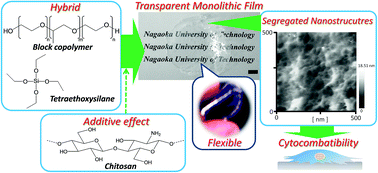
In this study, effective Chi-segregation nanodomains were prepared via silica-surfactant hybrid film formation, which was synthesized based on a room-temperature sol–gel process using poly(ethylene oxide)–poly(propylene oxide)–poly(ethylene oxide) (PEO–PPO–PEO) triblock copolymer (TBC) as the SDA.5j The hybrid films with different Chi concentrations were synthesized to clarify the additive effect of the Chi molecules on the self-assembly process at the film surface, as shown in Fig. 1.
The synthetic process of the films followed the modified procedure from previous reports,5h,j whereby 0.94 g of TBC (F127, PEO106–PPO70–PEO106, Mw = 12600) was dissolved in 6.8 mL of ethanol, and stirred. 2.0 mL of an hydrochloric acid aqueous solution (pH = 1.4) was added and stirred. Then, the Chi flakes (Funakosi Co. Ltd., Chitosan 7B KH001001) were added into the solution at the weight concentration of 0.0, 1.0, 5.0, 10 wt%. Then, 2.0 mL of tetraethylorthosilicate (TEOS) was added to the solution, and stirred at 40 °C for 2 h. The solution was transferred to Teflon vessels (bottom area: 7.9 cm2) at a density of 0.42 mL cm−2, and stored for 3 days for the gelation, and then they were dried at 65 °C for 18 h. The films were peeled from the vessels and were abbreviated as 0w-, 1w-, 5w- and 10w-films prepared at the concentration of 0.0, 1.0, 5.0, and 10 wt%, respectively.
The films were characterized by a UV-visible spectrophotometer and X-ray diffraction (XRD) pattern. The total visible transmittance was calculated by averaging the values in the wavelength range between 400 and 800 nm. The surface nanostructures and viscoelastic properties were analyzed by an atomic force microscope (AFM: Nanocute, SII Investments, Inc.) in areas of 0.5 × 0.5 and 1.0 × 1.0 μm2. The surface roughness (Rrms) was calculated by the root mean squares in the height images. The viscoelastic properties were calculated from the force curves, with the detailed measurement procedure being described in the ESI, Experimental Procedure S1 and Scheme S1.†
The osteoblast-like MC3T3-E1 cells were cultured in a cell culture flask containing 15 mL of the fetal bovine serum (FBS) dispersed in alpha-minimum essential medium (αMEM) at 10 vol% (10%FBS/αMEM). The cells were incubated at 37 °C in a humidified atmosphere of CO2, and subcultured every 7 days with trypsin-EDTA. After being washed with 15 mL of phosphate buffered saline (PBS) and treated with 1 mL of the trypsin-EDTA, the cells were dispersed in 15 mL of PBS and then in 15 mL of the 10%FBS/αMEM. The number of cells in the suspension was seeded at the density of 3.0 × 10³ cells cm−2 and then cultured for 36 h. After the culture, the cells were rinsed by PBS. Then, the cells adhered only on the film surfaces were characterized by light microscopy. The density and area of the adherent cell were counted and calculated from the 2-D images (n = 50). The cell viability with the culture time was measured as described in the ESI, Experimental Procedure S2 in detail.† As compared to the reference, commercially-available tissue culture poly(styrene) (TCPS) was used. The statistical analysis was examined by the Student's t test.
The films with different Chi concentrations were placed in a transparent and self-standing state with a length of up to several centimetres (Fig. 1), whereas TEOS alone was in a fragmented state. The monolithic films had thicknesses of several hundred micrometers (150–200 μm), and the total transmittance was in the order of 0w- (81% T) > 1w- (64% T) > 5w- (54% T) > 10w- (19% T) films, thus it was found that the Chi addition reduces the visible-light transmittance and scattering. The hand-bending state image (5w-film) shown in Fig. 1 indicates the flexibility of the films. Therefore, nanocomposite structures with silica frameworks combined with the surfactant functional groups were able to homogeneously stabilize the Chi molecules on the film surfaces.
Fig. 2 shows the AFM topographic and phase-shift images of the films at an observation area of 1 × 1 μm2. The 0w-film showed a nanoparticulate surface morphology that was a flat surface structure at an Rrms value of 3.3 ± 1.9 nm. In contrast, the network fibrous nanostructures of Chi molecules were observed in the films containing Chi at Rrms values of 3.3 ± 1.3 nm, 3.4 ± 0.91 nm and 4.4 ± 2.2 nm for the 1w-, 5w- and 10w-films. Only by the Chi molecules being cast on the 0w-film after the silica–surfactant hybridization, did the Chi molecules form homogeneously-packed structures without nanostructures (ESI, Fig. S1†). All the samples from the XRD patterns exhibited the broad patterns only from the 100 plane (d100 = 7.4–7.6 nm) and the pattern shapes were almost the same, which also corresponded to the worm-like mesostructures of our previous report.5j Therefore, the segregated nanofiber networks were successfully formed by the silica-surfactant nanocomposite process.
Fig. 3(a–d) shows the force curves for the film surfaces. The five points measured for the force curves are marked in the AFM topographic images (0.5 × 0.5 μm2) in the ESI, Fig. S2.† Typical tip–sample interactive behaviors were observed depending on the film surfaces. During the initial stage, there was a measurable attraction at separation distances below 75 nm. A slight attractive force between the tip and the sample during the approaching process was then observed, which could be attributed to the interactions between the tip and surface molecules. This force strongly appears in the 0w-film, suggesting that only the silica–surfactant composite strongly adsorbs on the SiO2 probe tip surface.5j Furthermore, the starting distance increased with increasing Chi concentrations, suggesting an increase in the Chi segregation layer thickness.
![]() | ||
Fig. 3 Force curves (•: approaching, ○: retracting) of the (a) 0w-, (b) 1w-, (c) 5w- and (d) 10w-films. (Inset): the adhesive areas magnified in the retracting curves. (e) The S (close circles) and Fad (open circles) value changes with the chitosan concentration. The dotted lines indicate the decrease of the slopes. Calculation of the S and Fad values is described in the ESI, Experimental Procedure S1 and Scheme S1.† |
A repulsion force between the tip and the sample is evident from the slopes, starting at the distance of around a few nanometers on 0w- and 1w-films and several tens of nanometers on 5w- and 10w-films. This force is attributed to the overlap of the electric double layers around the two surfaces between the tip and sample surfaces. The S (i.e., saturated slope) values of the film surfaces containing Chi (1w: 6.6 ± 0.43, 5w: 7.0 ± 0.21, 10w: 13.0 ± 0.41 nN nm−1) are higher than that of the 0w-film surface (5.6 ± 0.38 nN nm−1) in Fig. 3(e), and the 10w-film surface is the highest of all the surfaces, indicating the importance of the Chi segregation for the viscoelastic properties of the films. In particular, the 10w film shows the two-step changes, wherein the lower and higher S values can be attributed to the self-aggregation layer among Chi molecules and the Chi–silica–surfactant interactive layer, respectively, indicating the existence of the separated Chi layer. Furthermore, the E* values of the films were 138 ± 9.5, 165 ± 10, 175 ± 5.3, and 325 ± 10.3 MPa for the 0w-, 1w-, 5w- and 10w-films, respectively.
At a separation distance in the retracting process, the tip jumps inward and subsequently a maximum Fad was observed (Fig. 3(a–d), insets). It is reasonable to conclude that the film surfaces differently interact with the fragmented surfaces after the insert of the tip. The Fad of the 0w-film is the highest of all the surfaces, suggesting the favorable interactions (e.g. hydrogen bonding, electrostatic interactions) of the SiO2 tip surfaces with the sample surfaces of both the TBC and silica. In contrast, the segregated Chi nanofibers were difficult to interact with the tip surfaces due to the dominant self-aggregation among the Chi molecules.
In this study, the utilization of the insufficiency behavior of natural products with the formation of siliceous compounds, which is thought to be scientific interest,12 justifies the search for bioinspired synthesis that could lead to novel silica-based hybrids with simple hierarchical structures or, at least, that could allow their unique production under soft-conditions. In fact, inspired by nature, a variety of chemical reagents have been used to check in vitro silicification processes.13 In the material preparation, the additives (e.g., Chi), which is used to mimic active natural molecules, can play different roles, e.g. as aggregation promoting reagents or SDA. In this study, a possible segregation mechanism was proposed, as shown in Scheme 1. The Chi addition effectively induces suitable segregation to form nanofiber networks on the films, which was driven by the well-known silica–surfactant nanostructure formation based on the self-assembly process. As a result, a suitable segregation with the nanostructures occurred on the hybrids. Furthermore, this procedural approach can lead to bulk biosilica monolithic films or, alternatively, to similar highly-ordered mesoporous architectures when the SDA addition is well-controlled.
Fig. 4(a–d) shows light-microscope images of the cells adhered on the films. Taking into account the transparent and mechanical properties described above for the biomedical applications, 0w-, 1w- and 5w-films were used in the following. The cells adhered on the films modified with Chi nanofibers clearly had expanded fibrous pseudopods and showed anisotropic cellular morphologies (Fig. 4(a and b)), whereas cells on the 0w-film and TCPS surfaces had smaller isotropic morphologies. At a culture time of 36 h, the number of the expanded fibrous pseudopods per one cell was ordered 5w (7.8 ± 1.6 pieces per cell) > 1w (5.5 ± 1.1 pieces per cell) > 0w (5.1 ± 1.3 pieces per cell) > TCPS (5.2 ± 1.7 pieces per cell), suggesting the effective interfacial cell film binding at 5w. As shown in Fig. 4(c), the cell viability with the 0w-film showed the lowest viability due to the influence of the hydrophilic PEO group. With increasing the Chi amount, the cell viability significantly increased. This indicated that the Chi molecules were functionalized on the surfaces and exhibited a good cytocompatibility, and 1w- and 5w-films showed the higher cell viability. Moreover, the density and area of the adherent cell were in the order of (1w, 5w) > 0w > TCPS (Fig. 4(d and e)). Herein, it has been reported that the cell survival was affected by the adherent cell shape.10 The osteoblast-like cells on Ta, Cr, and hydroxyapatite had different adhesive areas,11 suggesting that the difference in the surface properties affected the cell adhesive areas. In totally considering these results, the Chi segregation films (1w-, 5w-films) of this study would provide good cell activity and long cell survival. The different structures could be attributed to the cell adhesion points with the sample surfaces, so that the cytoskeleton changes and extracellular matrix arrangements at the interfaces caused the binding behavior and morphologies.11c Thus, the Chi nanostructures on the silica–TBC effectively promoted the osteoblast-like cell adhesion. The detailed nanostructural effect on the cell functions will be reported by our laboratory.
It has been reported that the bone healing process could be improved by controlling the composition and structure of the bone substitute nanomaterials composited with biocompatible polymers.14 The Chi nanocomposite film surfaces of this study significantly exhibited higher cytocompatibility at the initial cell culture stage. Therefore, the recognition of cells on the Chi nanostructural surfaces caused the adhesion behaviors, and the interfacial viscoelastic layers by Chi nanofibers also effectively determined the cellular shapes (anisotropy or not) and the functions, although the pre-adsorbed protein states are also important.11c
Footnote |
† Electronic supplementary information (ESI) available. See DOI: 10.1039/c5ra26241k |
This journal is © The Royal Society of Chemistry 2016 |