
Fabrication, hydrolysis and cell cultivation of microspheres from cellulose-graft-poly(L-lactide) copolymers
Lili Yangab,
Jinming Zhanga,
Jiasong Hea,
Jun Zhang*a and
Zhihua Gan*c
aBeijing National Laboratory for Molecular Sciences, CAS Key Laboratory of Engineering Plastics, Institute of Chemistry, Chinese Academy of Sciences (CAS), Beijing 100190, China. E-mail: jzhang@iccas.ac.cn
bState Key Laboratory of Petroleum Resources and Prospecting, China University of Petroleum (Beijing), Beijing 102249, China
cState Key Laboratory of Organic–Inorganic Composites, Beijing Laboratory of Biomaterials, College of Life Science and Technology, Beijing University of Chemical Technology, Beijing 100029, China. E-mail: zhgan@mail.buct.edu.cn
First published on 1st February 2016
Abstract
Porous microspheres of cellulose-graft-poly(L-lactide) (cellulose-g-PLLA) copolymers were fabricated by a water/oil/water (W/O/W) emulsion evaporation method. Their morphology, hydrophilicity and amount of hydroxyl group content (–OH content) on the surface were adjustable with the change of molar substitution of PLLA (MSPLLA). After a hydrolysis post-processing, surface physical properties of microspheres were regulated further. The influence of physical properties of microspheres on cell cultivation was investigated by setting hepatocellular liver carcinoma cell line (HepG-2) as the example. Compared with the cells on PLLA microspheres, HepG-2 cells with more pseudopods spread well on the surface of cellulose-g-PLLA microspheres. Particularly, the cellulose-g-PLLA microspheres with MSPLLA of 11.5 were the most appropriate candidate for cell adhesion and proliferation. Therefore, a moderate PLLA content of cellulose-g-PLLA copolymers was beneficial to the cell cultivation. This work indicated that combining cellulose with PLLA was an available route to developing cellulose-based materials for cell cultivation.
Introduction
In recent decades, cell cultivation has attracted extensive interest in tissue engineering for repair, restoration or regeneration via the combination of cells, biomaterials and growth factors. As a major family of cell species, anchorage-dependent cells (ADCs) must rely on appropriate substrates that function to maintain the cell population and also ensure the intact phenotype. Among the numerous strategies for propagating ADCs, the use of microspheres as microcarriers is a promising approach that originated from a setup by van Wezel in 1967 to mass-produce viral vaccines and biological cells by using mammalian cells. Given its superiority in both cell line expansion and conveyance, this approach has latterly been developed and adopted for tissue engineering use.1 For this purpose, microspheres with diameters typically ranging from 100 μm to 400 μm were fabricated with natural and synthetic polymers such as dextran,2 collagen,3 poly(lactic acid) (PLA) and poly(ε-caprolactone) (PCL), enabling vast and conveyable cell-loading interfaces to be generated. Considering that the chemical composition of microcarriers plays an important role in the expansion rate and differentiation type of cells, a lot of effort has been devoted to fabricating novel microspheres with suitable solid-microstructural and surface properties in order to favor attachment and proliferation of ADCs.Natural polymers have been widely used for biomedical applications because of their good biocompatibility. Microspheres which are fabricated by natural polymers, such as collagen,4 gelatin,5 alginate6 and chitosan,7 are often formulated via water-based solutions. Cellulose, which is the most abundant natural polysaccharide, has also been widely applied in biomedical fields such as wound closure and healing,8 drug delivery,9 vascular grafting and tissue engineering. However, cellulose is insoluble in water and common organic solvents owing to the vast intermolecular and intramolecular hydrogen bonds. Moreover, it usually requires material soluble in water or organic solvents to fabricate microspheres via common methods including emulsion solvent extraction/evaporation, polymer extrusion, spray-drying and coacervation or precipitation.10 Obviously, it is impossible to prepare cellulose-based microspheres via the normal techniques. Although the ionic liquids (ILs) such as AmimCl11 is able to dissolve cellulose, it is hard to be used to fabricate cellulose-based microspheres due to its non-volatility and high viscosity. However, in consideration of the abundant hydroxyl groups of cellulose which are the useful sites of further functionalization for protein adsorption and promoting cell attachment, it would be of great interest to fabricate microspheres of cellulose-based polymers by a facile technique.
The synthetic aliphatic polyesters such as PLA have been widely utilized to fabricate microspheres12,13 via emulsion solvent evaporation method. However, the application of these polyesters is limited by their surface and bulk properties, such as hydrophobicity, lack of cell recognition sites. To overcome these shortcomings, the hydrolysis as a common chemical way has been applied to endow PLLA with hydrophilicity.14,15 During this process, carboxyl and hydroxyl groups can be created on the surface of the microspheres due to the break of ester bond of polyester chains. By controlling the degree of hydrolysis, the physical and chemical properties of microspheres including size, surface morphology,16 surface energy,17 chemical composition,18 hydrophilicity19 and charge20 can be well controlled, which show great influence on cell attachment and growth on those microspheres.
In consideration of the good solubility of synthetic aliphatic polyesters in common organic solvents, the modification of cellulose via copolymerization with aliphatic polyesters has attracted great interests in order to increase the solubility and processability of cellulose.21,22 The cellulose-graft-poly(L-lactide) (cellulose-g-PLLA) has been widely studied in our research group.23,24 Not only the mechanical properties but also the solubility in common organic solvents has been improved substantially for cellulose. Furthermore, cellulose in the copolymers can enhance the wettability of PLLA, which might benefit the attachment and proliferation of cells. Therefore, the successful synthesis of cellulose-g-PLLA copolymer and its solubility in organic solvents make it possible to fabricate cellulose-based microspheres via a emulsion solvent evaporation technique.
Although much effort has been made to develop new materials for the fabrication of microspheres, to our knowledge, there are seldom literature reported about the fabrication of microspheres by using cellulose so far.25 Herein, based on our previous work that the modification of cellulose by grafting PLLA can increase the solubility of cellulose in dichloromethane,26 we reported the fabrication of microspheres by using cellulose-g-PLLA copolymers via water/oil/water (W/O/W) emulsion solvent evaporation technique. After then, the surface properties of the microspheres were changed by the controlled hydrolysis in alkali aqueous solution. Finally, the influence of surface morphology and property on the morphology, attachment and proliferation of cells on microspheres was investigated in details.
Experimental section
Materials
Microcrystalline cellulose with a degree of polymerization 220 was purchased from Beijing Fengli Jingqiu Commerce and Trade Company (China), and dried in vacuum at 50 °C for 12 h before use. L-Lactide (LLA) was supplied by Corbion Purac (Netherlands). Poly(L-lactide) (PLLA) was got from J & K Scientific (China). 4-Dimethylaminopyridine (DMAP) was obtained from Haili Chemical Industry Company (China). Ionic liquid 1-allyl-3-methylimidazolium chloride (AmimCl) was synthesized according to the literature.27 Polyvinyl alcohol (PVA) was purchased from Sigma-Aldrich (USA). All other chemicals used were of analytical grade and purchased from Beijing Chemical Reagent Factory (China) and used without further purification.Hepatocellular liver carcinoma cell line (HepG-2) was supplied by Chinese Academy of Medical Sciences & Peking Union Medical College. Dulbecco's modified eagle's medium (DMEM), phosphate buffered saline (PBS), fetal bovine serum (FBS) and penicillin–streptomycin were obtained from Hyclone (USA). Fluorescein diacetate (FDA), propidium iodide (PI) and cell counting kit-8 (CCK-8) were supplied by Sigma-Aldrich (USA).
Synthesis of cellulose-g-PLLA copolymers in AmimCl
A typical polymerization procedure was used as follows. Dried cellulose (1.7 g) was dispersed in AmimCl (43.0 g) in a Schlenk tube with a magnetic stirrer. After stirred at 80 °C for 2–3 h to get a clear homogenous solution, LLA (14.5 g) and DMAP (1.9 g) were added into the tube in sequence. Then the exhausting–refilling process was repeated three times. After reaction at 80 °C for 24 h, the tube was cooled to room temperature, and the mixture was precipitated in deionized water. And then, the product was redissolved in DMSO and then precipitated in an excess of water to obtain the final cellulose-g-PLLA copolymer. The purified copolymer was dried in a vacuum oven until reaching a constant weight. The name of samples is labeled as cellulose-g-PLLAMS. MS represents the molar substitution of PLLA, which is defined as the average number of introduced LA units per anhydroglucose residue of cellulose calculated from 1H NMR.Preparation and hydrolysis of microspheres
The cellulose-g-PLLA microspheres were prepared by a double emulsion solvent evaporation technique (W1/O/W2). Briefly, cellulose-g-PLLA (0.125 g) was dispersed in dichloromethane (3 mL) and stirred until the copolymer dissolved completely. The PVA aqueous solution (1 mL, 0.1 w/v%) was added as the inner aqueous phase and emulsified in the organic phase. The primary emulsion was dripped into PVA outer aqueous solution (200 mL, 0.25%) under agitation at a rate of 500 rpm by a mechanical stirrer to generate a W1/O/W2 emulsion. After stirring for 4 h, the hardened microspheres were harvested and rinsed with distilled water several times. The microspheres with a diameter of 100–250 μm were separated by standard sieves and used for all the following experiments. As control, the PLLA microspheres were prepared by the same technique except that cellulose-g-PLLA was replaced by neat PLLA.For hydrolysis, a certain amount of microspheres was immersed into NaOH aqueous solution (10 mL, 0.05 w/v%). After 1 h, the alkali solution was removed and the microspheres were rinsed thoroughly by a large amount of distilled water until a constant pH 7.0 was reached.
Preparation and hydrolysis of PLLA and cellulose-g-PLLA films
The films for wettability measurement were prepared by compression molding at 190 °C for PLLA and at 150–170 °C for cellulose-g-PLLA. The hydrolysis of films was performed at the same condition of microspheres.Cell experiments
HepG-2 cells were cultured in culture flasks (10 cm2) for 2–3 days in DMEM supplemented with 10% FBS, 100 μg mL−1 penicillin and 0.1 mg mL−1 streptomycin at 37 °C in a 5% CO2 incubator. The microspheres were sterilized by irradiation at ambient temperature for 8 h under 10 kGy doses with 60Co as radiation source.Cell morphology was observed by SEM. The microspheres dispersed in 50 μL water were placed into a 96-well plate and the water was removed afterwards. Then 200 μL HepG-2 cell suspension was added into the wells (1 × 10⁵ cells per well). After incubation at 37 °C in a humidified atmosphere with 5% CO2 for 1 day, the samples were fixed with 1 mL glutaraldehyde for one night at 4 °C. Subsequently the samples were washed with PBS for three times and dehydrated with 40%, 50% and 60% ethanol solution for 1 minute in turn. The microspheres were dried by lyophilization.
The cell density was observed by fluorescence microscopy. The microspheres dispersed in 50 μL water were added into a 96-well plate. 200 μL HepG-2 cells were seeded in the wells (5 × 10⁴ cells per well). The cell-seeded microspheres were replenished with fresh medium every day. The cells were harvested at 37 °C and 5% CO2 on 1, 3, 5 day respectively. The culture medium was aspirated and the samples were washed with PBS. After then, FDA/PI solution (50 μL) was added into the wells. After 3 min, the solution was removed and the samples were washed with PBS later.
The cell viability was assayed by CCK-8 reagent. After the microspheres in 50 μL water were added into a 96-well plate above all respectively, the 200 μL culture medium containing HepG-2 cells was added into the wells at a density of 2 × 10³ cells per well. Each kind of microspheres was repeated for six times. As controls, the cells with only 200 μL culture medium were measured. The cells were incubated at 37 °C in a humidified atmosphere with 5% CO2 for 1, 3, 5 days respectively. After incubation, 20 μL CCK-8 was filled into each well and then the samples were incubated at 37 °C and 5% CO2 for 2 hours. 100 μL liquid was drawn to a new plate and the plate was read with a spectrophotometer to detect the absorbance of the samples at wavelength of 450 nm which reflects the number of viable cells. The cell viability was calculated according to the following equation:
| Cell viability (%) = (Asample/Acontrol) × 100% |
Measurements
For NMR, the 1H NMR spectra of cellulose-g-PLLA copolymers were measured with a Bruker AV 400 NMR apparatus. The DMSO-d6 was used as solvent in which a drop of trifluoroacetic acid-d1 was added to shift active hydrogen to low field.For contact angle measurement, the surface wettability was measured on a Dataphysics OCA20 contact-angle system at ambient temperature. The average contact angle was obtained by measuring more than five different positions of the same sample.
For SEM measurement, the shape and morphology of microspheres were observed by JSM-6700F scanning electron microscopy. The specimens were sputter coated with platinum by Bal Tec before the observation.
For fluorescence microscopy, the fluorescence photographs of viable and dead cells adhered on microspheres were acquired by Nikon eclipse Ti.
For spectrophotometer, the viability of HepG-2 cells on microspheres was measured by Thermo Multiskan MK3. The wavelength was set at 450 nm.
Results and discussion
Synthesis and characterization of cellulose-g-PLLA copolymers
As shown in Scheme 1, the cellulose-g-PLLA copolymer was synthesized in AmimCl by grafting the PLLA from hydroxyl groups of cellulose through ring-opening polymerization of LLA with DMAP as catalyst. The cellulose-g-PLLA copolymers with different molar substitutions of PLLA (MSPLLA) were synthesized by changing molar feed ratios of LLA monomer to cellulose. | ||
| Scheme 1 Graft copolymerization of cellulose with L-lactide (LLA). | ||
Fig. 1 shows the 1H NMR spectra of cellulose-g-PLLA copolymers. The chemical shifts at 5.14 and 1.45 ppm were attributed to methine protons and methyl protons of internal lactyl respectively, which are also the characteristic peaks of PLLA. Compared with neat PLLA, the chemical shifts at 4.19 and 1.27 ppm were assigned to methine protons and methyl protons of terminal lactyl. Besides, the signal peaks in the range from 2.80 to 5.00 ppm except for the peak belonged to methine protons (4.19 ppm) are the characteristic peaks of the protons of cellulose backbone chains. The above observation clearly reveals that PLLA has been grafted onto cellulose successfully.
 | ||
| Fig. 1 1H NMR spectra of (a) cellulose-g-PLLA and (b) neat PLLA. | ||
Preparation and hydrolysis of microspheres
For the cellulose-g-PLLA copolymers with various compositions (Table 1), the solubility of the copolymers in dichloromethane improves with PLLA content, i.e. MSPLLA. It was found in this work that the cellulose-g-PLLA11.5 is soluble completely, while cellulose-g-PLLA6.7 swells in dichloromethane. Due to the different solubility of these copolymers in dichloromethane, the surface morphology of microspheres fabricated from these copolymers is affected significantly, as shown in Fig. 2. There are a lot of winkles on the surface of neat PLLA microspheres, which attribute to the spherulites of PLLA during emulsion solvent evaporation process, as is shown in Fig. 3c. But for cellulose-g-PLLA11.5 microspheres, smooth surface with porous structure was observed in Fig. 2b and b1. Compared with cellulose-g-PLLA11.5, cellulose-g-PLLA6.7 microspheres (Fig. 2a and a1) showed surface more rough and more holes, which was attributed to the more poor solubility in dichloromethane for cellulose-g-PLLA6.7 with lower content of PLLA component.| Sample | MSPLLAb | MStc | Grafting content (%) | Solubility | ||
|---|---|---|---|---|---|---|
| Water | DCM | DMSO | ||||
| a +: soluble; −: insoluble; ±: swollen.b MSPLLA: molar substitution of PLLA, which is defined as the average number of introduced LA units per anhydroglucose residue of cellulose, and calculated from 1H NMR.c MSt: molar substitution of PLLA in theory. | ||||||
| Cellulose-g-PLLA2.9 | 2.9 | 4.0 | 56.3 | + | − | + |
| Cellulose-g-PLLA6.7 | 6.7 | 8.0 | 77.2 | − | ± | + |
| Cellulose-g-PLLA11.5 | 11.5 | 16.0 | 85.4 | − | + | + |
 | ||
| Fig. 2 SEM images of microspheres. (a)–(c) are surface morphology of cellulose-g-PLLA6.7, cellulose-g-PLLA11.5 and neat PLLA microspheres before hydrolysis. (d)–(f) are surface morphology after hydrolysis of (a)–(c), respectively. (a1)–(c1) are cross-section morphology of cellulose-g-PLLA6.7, cellulose-g-PLLA11.5 and neat PLLA microspheres. The scale is 50 μm. | ||
 | ||
| Fig. 3 XRD spectra of microspheres. (a)–(c) are the microspheres composed of cellulose-g-PLLA6.7, cellulose-g-PLLA11.5, and PLLA, respectively. | ||
Hydrolysis has been used as a chemical etching technique for biodegradable polymers to modify the surface property for promoting cell growth.28,29 Fig. 2d–f showed the surface morphologies of microspheres from PLLA and cellulose-g-PLLA copolymers with different compositions after hydrolysis in NaOH aqueous solution. It was found that there is little change on the surface of PLLA microspheres after hydrolysis (Fig. 2f), while the size and amount of holes on the surface of cellulose-g-PLLA microspheres increased after hydrolysis compared with that of the original ones (Fig. 2d and e). Especially, the holes on surface of cellulose-g-PLLA6.7 microspheres after hydrolysis are larger than that of cellulose-g-PLLA11.5 microspheres.
The morphological changes of above microspheres after hydrolysis are due to the selective degradation of PLLA component in NaOH aqueous solution. The little change of neat PLLA microspheres is due to the slow hydrolysis rate of PLLA. This is because PLLA chains in microspheres pack compactly in crystalline state. However, the water is easier to penetrate into microspheres and break the ester bond of PLLA chains with the help of cellulose which reduces the crystallizability and induces the porous structure of cellulose-g-PLLA microspheres. And, the hydrolysis rate increases with increasing the cellulose content. Therefore, combined with the control of cellulose-g-PLLA composition, the control of hydrolysis degree is another efficient method to guide the design of microspheres with different porous structures.
Fig. 4 shows the water contact angles of the films of cellulose-g-PLLA copolymers and neat PLLA before and after hydrolysis. It was found the contact angle of neat PLLA is 90°. After copolymerized with cellulose, the contact angle of cellulose-g-PLLA11.5 film with less cellulose component is similar to that of PLLA around 90°, while that of cellulose-g-PLLA6.7 film with more cellulose component shows declined to 75°, indicating the increase of hydrophilicity. The further investigation indicated that the cellulose content in copolymers shows significant influence on the hydrolysis of films in NaOH aqueous solution. For PLLA film, the contact angle decreases just from 90° to 80°. While for cellulose-g-PLLA11.5 and cellulose-g-PLLA6.7 films, the contact angles decrease from 90° to 30° and from 75° to 10°, respectively. These results suggest that the hydrolysis results in surface with more hydrophilicity, which is owing to the increased amount of hydroxyl groups and carboxylic groups from broken ester bonds of PLLA chains. And it can be also inferred that the contact angle of film decreased more and faster as cellulose content increased in the copolymer. In consideration of the similar surface properties of cellulose-g-PLLA films and microspheres, the hydrophilicity is also an important role in determining the morphological change of microspheres after hydrolysis in alkali solution.
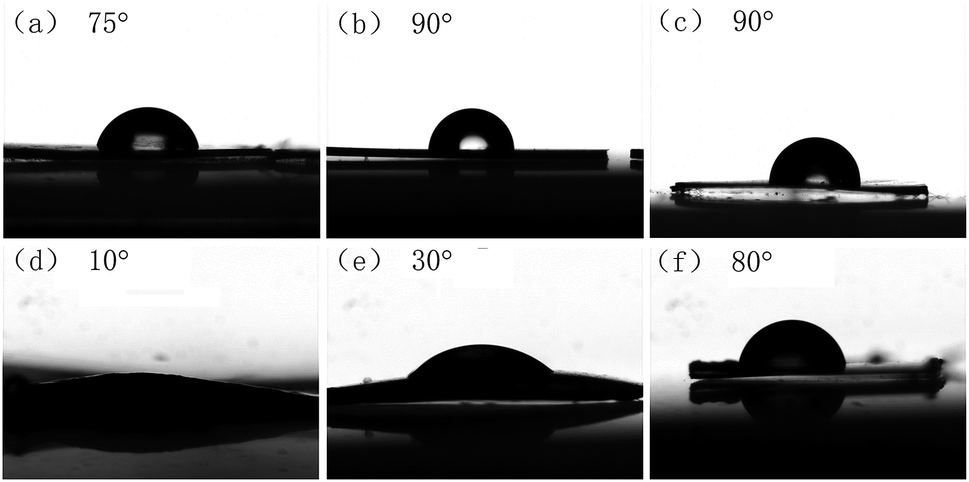 | ||
| Fig. 4 Contact angle of polymer films. (a)–(c) are the films of cellulose-g-PLLA6.7, cellulose-g-PLLA11.5 and PLLA before hydrolysis. (d)–(f) are the films of (a)–(c) after hydrolysis, respectively. | ||
Fig. 5 shows the XPS analysis on cellulose-g-PLLA and neat PLLA microspheres before and after hydrolysis. The microspheres show three peaks with different C 1s binding energies in spectra, i.e. 284.9 eV for alkyl –C–HX, 286.7 eV for ether –C–O, and 289.7 eV for carboxylic –C(![[double bond, length as m-dash]](https://www.rsc.org/images/entities/char_e001.gif) O)–O. Before hydrolysis, PLLA has the most carboxylic groups, while cellulose-g-PLLA6.7 copolymer has the least carboxyl groups corresponding to the lowest PLLA content. Owing to the hydrophilicity, the cellulose-g-PLLA copolymers were easily to be attacked by NaOH aqueous solution compared with neat PLLA microspheres. For this reason, the peak areas of hydroxyl and ester of cellulose-g-PLLA6.7 microspheres after hydrolysis show the most obvious increase among three samples, while no detectable changes in surface composition were found for PLLA microspheres. For cellulose-g-PLLA11.5 after hydrolysis, there is a shift on the ratio to hydroxyl. The resultant XPS spectra after hydrolysis indicated that the cellulose-g-PLLA microspheres exhibit more enriched concentration of ether and carboxylic groups on the surface of microspheres compared with untreated ones on the basis of corresponding peak areas. Actually, the increased amount of carboxylic and hydroxyl groups could be regarded as the hydrolysis degree of microsphere surface. Therefore, the XPS results clearly indicated that the microspheres of cellulose-g-PLLA with higher cellulose composition showed easier hydrolysis in alkali solution.
O)–O. Before hydrolysis, PLLA has the most carboxylic groups, while cellulose-g-PLLA6.7 copolymer has the least carboxyl groups corresponding to the lowest PLLA content. Owing to the hydrophilicity, the cellulose-g-PLLA copolymers were easily to be attacked by NaOH aqueous solution compared with neat PLLA microspheres. For this reason, the peak areas of hydroxyl and ester of cellulose-g-PLLA6.7 microspheres after hydrolysis show the most obvious increase among three samples, while no detectable changes in surface composition were found for PLLA microspheres. For cellulose-g-PLLA11.5 after hydrolysis, there is a shift on the ratio to hydroxyl. The resultant XPS spectra after hydrolysis indicated that the cellulose-g-PLLA microspheres exhibit more enriched concentration of ether and carboxylic groups on the surface of microspheres compared with untreated ones on the basis of corresponding peak areas. Actually, the increased amount of carboxylic and hydroxyl groups could be regarded as the hydrolysis degree of microsphere surface. Therefore, the XPS results clearly indicated that the microspheres of cellulose-g-PLLA with higher cellulose composition showed easier hydrolysis in alkali solution.
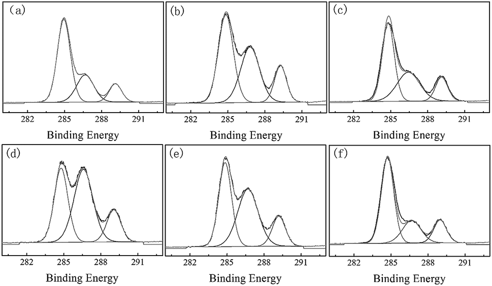 | ||
| Fig. 5 High resolution XPS C 1s spectra of microspheres. (a)–(c) are the microspheres composed of cellulose-g-PLLA6.7, cellulose-g-PLLA11.5 and neat PLLA. (d)–(f) are the hydrolyzed microspheres of (a)–(c), respectively. | ||
In vitro HepG-2 cells growth on the microspheres
Cell morphology on microspheres revealed the dependence of cell growth on surface properties of microspheres resulted from the different chemical structures of cellulose-g-PLLA and neat PLLA. Fig. 6 showed the SEM images of HepG-2 cells on the microspheres before and after hydrolysis. As shown in Fig. 6a, a1, b, b1, c and c1 for the microspheres before hydrolysis, cells adhered onto all microspheres, but showed different morphologies. Cells flattened to form monolayers with few pseudopod on PLLA microspheres. However, more cells with a number of pseudopod were observed on microspheres prepared by cellulose-g-PLLA11.5 and cellulose-g-PLLA6.7. It can be found that cell adhesion was significantly enhanced on cellulose-g-PLLA microspheres, even though different opinions were reported regarding whether modified biodegradable polyesters can encourage cell adhesion and growth.30,31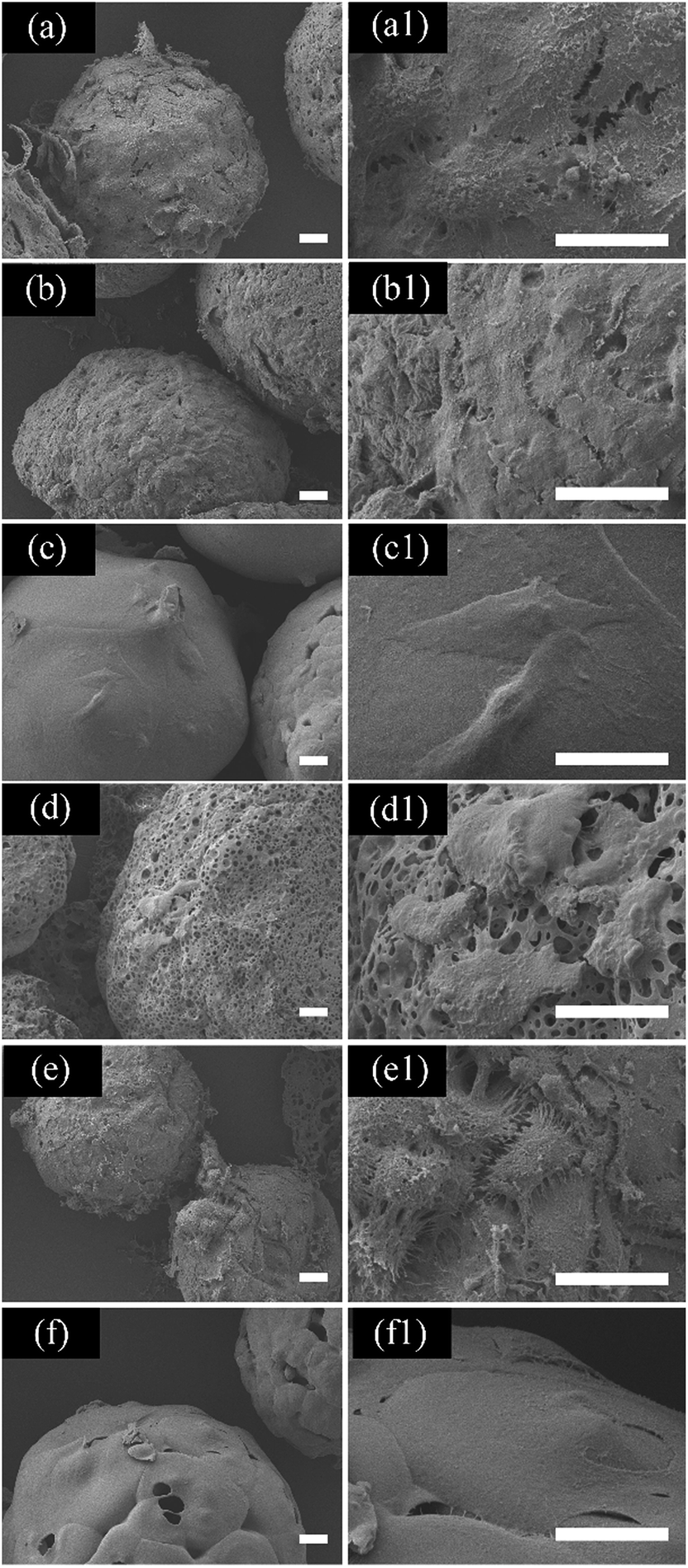 | ||
| Fig. 6 SEM images of HepG-2 cells growth on microspheres fabricated by PLLA and cellulose-g-PLLA copolymers. (a)–(c) are the microspheres composed of cellulose-g-PLLA6.7, cellulose-g-PLLA11.5 and neat PLLA. (a1)–(c1) Higher magnification of (a)–(c). (d)–(f) are the hydrolyzed microspheres of (a)–(c), respectively. (d1)–(f1) Higher magnification of (d)–(f). The scale is 20 μm. | ||
Furthermore as shown in Fig. 6d, d1, e, e1, f and f1 for cell growth on microspheres after hydrolysis, HepG-2 cells did not show different morphologies on PLLA microspheres before and after hydrolysis, but showed more pseudopod on cellulose-g-PLLA11.5 microspheres after hydrolysis. However, the cells did not grow well on cellulose-g-PLLA6.7 microspheres after hydrolysis. Only a small amount of cells adhered without pseudopod onto the hydrolyzed cellulose-g-PLLA6.7 microspheres after hydrolysis. Therefore, it is suggested that the microspheres with suitable cellulose content on the surface (e.g. cellulose-g-PLLA11.5) will endow the microspheres with suitable hydrophilicity, crystallinity and morphology, which are favorable for cell adhesion and growth.
Fig. 7 was the live/dead assay of HepG-2 cells in culture on microspheres for different time observed by fluorescence microscopy. Viable cells are stained green, and dead cells are stained red. The cell density observed by SEM and fluorescence microscopy in Fig. 6 and 7 was different. As shown in Fig. 7a1–f1 on the first day, the spindle-shaped cells were observed on both the untreated and treated PLLA microspheres, while only few cells clustered together as rounded ones adhered to unhydrolyzed and hydrolyzed cellulose-g-PLLA microspheres except for cellulose-g-PLLA11.5 microspheres before hydrolysis. From the viewpoint of chemical structure of microspheres, it could be concluded that the moderate cellulose can provide hydrophilic surface for enhancing cell growth, however, too much cellulose might decrease the cytocompatibility.
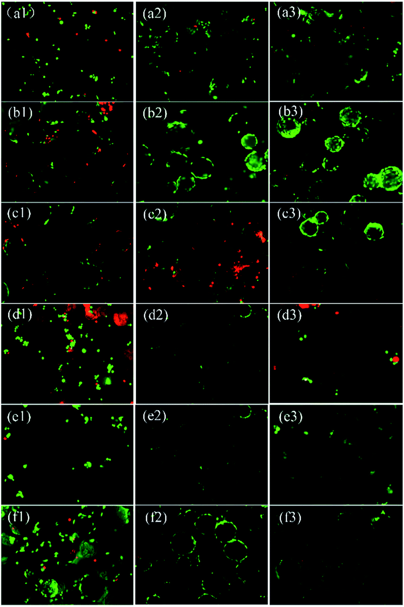 | ||
| Fig. 7 Viability of cells on microspheres. (a) and (d) are cellulose-g-PLLA6.7 microspheres before and after hydrolysis treatment, respectively. (b) and (e) are cellulose-g-PLLA11.5 microspheres before and after hydrolysis, respectively. (c) and (f) are PLLA microspheres before and after hydrolysis, respectively. The numbers “1, 2 and 3” after “a, b and c” represent cultivation of cells on microspheres for 1, 3 and 5 days. The bar is 100 μm in the picture. | ||
When the time came to the third day, the number of cells on untreated microspheres increased as a function of time and displayed characteristic flattened spindle. On the contrary, the number of cells decreased on the hydrolyzed cellulose-g-PLLA microspheres. With further elongating cultivation time to fifth day, even though the number of cells on cellulose-g-PLLA6.7 microspheres did not change, the cells on cellulose-g-PLLA11.5 microspheres show good conditions even better than those on PLLA microspheres. It might be the hydrophilicity resulted from the suitable cellulose content in cellulose-g-PLLA11.5 microspheres more suitable for cell growth.
As shown in Fig. 8, the cell viability on all microspheres increased with time. On the first day, the HepG-2 cells have similar viability on all microspheres. On the third day, the cells on cellulose-g-PLLA6.7 have the lowest viability among all the microspheres either before or after hydrolysis, while the number of cells on PLLA microspheres was the largest. The number of cells on cellulose-g-PLLA11.5 microspheres was ranged in the middle. This result revealed the importance of cellulose content in cellulose-g-PLLA copolymers for cell growth. When the time came to the fifth day, the microspheres fabricated by PLLA or cellulose-g-PLLA11.5 before hydrolysis showed good biocompatibility to cells, and the latter was even better. These results indicate that moderate cellulose in the cellulose-g-PLLA copolymers can lead a positive influence on the biocompatibility of microspheres, while too much cellulose would have a negative influence.
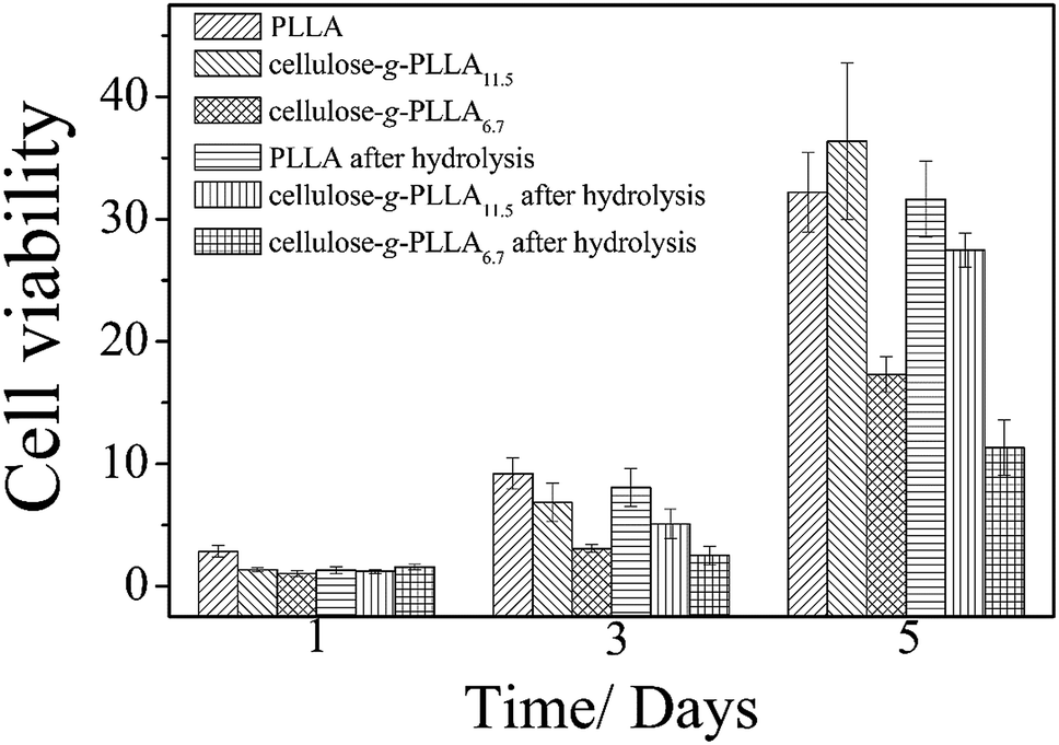 | ||
| Fig. 8 HepG-2 cell population on PLLA and cellulose-g-PLLA microspheres before and after hydrolysis for 1, 3 and 5 days of cell cultivation. | ||
Conclusions
In this work, cellulose-g-PLLA copolymers were synthesized by ring-opening polymerization homogeneously in an ionic liquid AmimCl. With the solubilization function in organic solvent of PLLA for cellulose, the microspheres composed of cellulose-g-PLLA copolymers were fabricated by the normal emulsion solvent evaporation method. Due to existence of cellulose in copolymer, the cellulose-g-PLLA microspheres hydrolyzed faster than the pure PLLA. And the morphology, the hydrophilicity, and the composition of microspheres can be tuned by hydrolysis. For the microspheres of copolymers with moderate cellulose content (e.g. cellulose-g-PLLA11.5), cell growth could be promoted. The results of this work will guide the research in the future about the modification of cellulose through copolymerization with PLLA, and processability of cellulose-based scaffolds for the application in tissue engineering.Acknowledgements
This work was supported by the National Science Foundation of China (No. 51425307 and No. 50973124).References
- N. R. Mercier, H. R. Costantino, M. A. Tracy and L. J. Bonassar, Biomaterials, 2005, 26, 1945–1952 CrossRef CAS PubMed.
- J. A. Cadee, L. A. Brouwer, W. den Otter, W. E. Hennink and M. J. A. van Luyn, J. Biomed. Mater. Res., 2001, 56, 600–609 CrossRef CAS PubMed.
- Y. Y. Li, H. W. Cheng, K. M. C. Cheung, D. Chan and B. P. Chan, Acta Biomater., 2014, 10, 1919–1929 CrossRef CAS PubMed.
- Y. Yao, F. Phan and Y. B. C. Li, Stem Cell Res. Ther., 2013, 4, 109 CrossRef PubMed.
- Y. Jin, I.-Y. Kim, I.-D. Kim, H.-K. Lee, J.-Y. Park, P.-L. Han, K. K. Kim, H. Choi and J.-K. Lee, Acta Biomater., 2014, 10, 3126–3135 CrossRef CAS PubMed.
- A. Moshaverinia, S. Ansari, C. Chen, X. Xu, K. Akiyama, M. L. Snead, H. H. Zadeh and S. Shi, Biomaterials, 2013, 34, 6572–6579 CrossRef CAS PubMed.
- E. Y. Chung, H. M. Kim, G. H. Lee, B. K. Kwak, J. S. Jung, H. J. Kuh and J. Lee, Carbohydr. Polym., 2012, 90, 1725–1731 CrossRef CAS PubMed.
- S. U. Park, B. K. Lee, M. S. Kim, K. K. Park, W. J. Sung, H. Y. Kim, D. G. Han, J. S. Shim, Y. J. Lee, S. H. Kim, I. H. Kim and D. H. Park, Int. Wound J., 2014, 11, 35–43 CrossRef PubMed.
- G. F. Picheth, M. R. Sierakowski, M. A. Woehl, L. Ono, A. R. Cofre, L. P. Vanin, R. Pontarolo and R. A. De Freitas, J. Pharm. Sci., 2014, 103, 3958–3965 CrossRef CAS PubMed.
- N. K. Varde and D. W. Pack, Expert Opin. Biol. Ther., 2004, 4, 35–51 CrossRef CAS PubMed.
- T. Setoguchi, K. Yamamoto and J.-I. Kadokawa, Polymer, 2012, 53, 4977–4982 CrossRef CAS.
- Y. Mao, Y. S. Dong, P. H. Lin, C. L. Chu, X. B. Sheng and C. Guo, Mater. Sci. Eng., C, 2011, 31, 9–13 CrossRef CAS.
- P. B. O'Donnell and J. W. McGinity, Adv. Drug Delivery Rev., 1997, 28, 25–42 CrossRef.
- B. Asplund, J. Sperens, T. Mathisen and J. Hilborn, J. Biomater. Sci., Polym. Ed., 2006, 17, 615–630 CrossRef CAS PubMed.
- S. P. Sun, M. Wei, J. R. Olson and M. T. Shaw, ACS Appl. Mater. Interfaces, 2009, 1, 1572–1578 CAS.
- S. J. Hong, H. S. Yu and H. W. Kim, Macromol. Biosci., 2009, 9, 639–645 CrossRef CAS PubMed.
- R. Daw, S. Candan, A. J. Beck, A. J. Devlin, I. M. Brook, S. MacNeil, R. A. Dawson and R. D. Short, Biomaterials, 1988, 19, 1717–1725 CrossRef.
- H. W. Kim, H. J. Gu and H. H. Lee, Tissue Eng., 2007, 13, 965–973 CrossRef CAS PubMed.
- K. Webb, V. Hlady and P. A. Tresco, J. Biomed. Mater. Res., 1998, 41, 422–430 CrossRef CAS PubMed.
- N. Faucheux, R. Schweiss, K. Lützow, C. Werner and T. Groth, Biomaterials, 2004, 25, 2721–2730 CrossRef CAS PubMed.
- Y. Teramoto and Y. Nishio, Polymer, 2003, 44, 2701–2709 CrossRef CAS.
- M. Krouit, J. Bras and M. N. Belgacem, Eur. Polym. J., 2008, 44, 4074–4081 CrossRef CAS.
- Y. Luan, J. Wu, M. Zhan, J. Zhang, J. Zhang and J. He, Cellulose, 2012, 20, 327–337 CrossRef.
- C. Yan, J. Zhang, Y. Lv, J. Yu, J. Wu, J. Zhang and J. He, Biomacromolecules, 2009, 10, 2013–2018 CrossRef CAS PubMed.
- H. L. Cai, S. Sharma, W. Y. Liu, W. Mu, W. Liu, X. D. Zhang and Y. L. Deng, Biomacromolecules, 2014, 15, 2540–2547 CrossRef CAS PubMed.
- C. H. Yan, J. Wu, J. M. Zhang, J. S. He and J. Zhang, Polym. Degrad. Stab., 2015, 118, 130–136 CrossRef CAS.
- H. Zhang, J. Wu, J. Zhang and J. He, Macromolecules, 2005, 38, 8272–8277 CrossRef CAS.
- J. M. Anderson and M. S. Shive, Adv. Drug Delivery Rev., 1997, 28, 5–24 CrossRef CAS.
- S. W. Zielhuis, J. F. W. Nijsen, G. C. Krijger, A. D. van het Schip and W. E. Hennink, Biomacromolecules, 2006, 7, 2217–2223 CrossRef CAS PubMed.
- X. P. Bi, Z. W. You, J. Gao, X. Q. Fan and Y. D. Wang, Acta Biomater., 2014, 10, 2814–2823 CrossRef CAS PubMed.
- A. R. Amini, J. S. Wanllace and S. P. Nukavarapu, J. Long-Term Eff. Med. Implants, 2011, 21, 93–122 CrossRef CAS PubMed.
| This journal is © The Royal Society of Chemistry 2016 |