Interactions governing the entrapment of anticancer drugs by low-molecular-weight hydrogelator for drug delivery applications

Siddhi Guptaa, Manish Singhb, Amarendar Reddy M.a, Prabhu S. Yavvaria, Aasheesh Srivastava*a and Avinash Bajaj*b
aDepartment of Chemistry, Indian Institute of Science Education and Research, Bhopal, India. E-mail: asri@iiserb.ac.in
bLaboratory of Nanotechnology and Chemical Biology, Regional Centre for Biotechnology, India. E-mail: bajaj@rcb.res.in

Received 4th December 2015 , Accepted 5th January 2016

First published on 12th January 2016


Abstract

We present the effect of size, charge, and hydrophobicity of anticancer drugs on their drug encapsulation efficacy in an L-alanine-based small-molecule hydrogelator. Entrapment of various anticancer drugs in a hydrogel was depicted and correlated towards interactions between gelator and drug molecules. Hydrogel showed the highest entrapment for 5-fluorouracil, which was as high as ∼1.2 mg mL−1 in 1.5% (w/v) hydrogel; however, with small polar anticancer drugs such as cisplatin and carboplatin, poor encapsulation was observed. Hydrogel was also able to entrap and retain hydrophobic drugs, such as docetaxel and tamoxifen, with a high drug-loading efficiency. The hydrogel-entrapped drugs were then characterized by rheology and SEM studies to understand the effect of the drugs on hydrogel assembly. Drug release and anticancer activity studies showed slow and sustained release of drugs from hydrogels, making them suitable for exploring with regard to future cancer therapeutic applications.

Introduction

Current cancer chemotherapy regimens include the use of biomaterials based on polymers, liposomes or protein-based drug nanoparticles that help in the controlled release of drugs into the blood circulation for enhanced bio-distribution and improved pharmacokinetics.1 These cancer chemotherapeutic approaches have strong limitations, such as several systemic toxic effects and poor targeting at tumor sites,2 which calls for improved localized injectable therapies that can inevitably surpass such limitations by enhancing localized drug concentrations and minimizing systemic toxicity, coupled with reduced dosage through slow and sustained release.3

Hydrogels provide suitable alternatives to existing clinically available biomaterials due to their ability to encapsulate and allow slow and sustained release of drugs.4 The injectability of these hydrogels at tumor sites provides the additional benefits of releasing drugs at tumor sites and avoiding systemic toxicity of drugs.5 In this regard, low molecular weight hydrogelators (LMHGs) gained considerable attention due to their small molecular weight and their ability to self-assemble into mechanically robust gels that can easily encapsulate drugs and subsequently release them in a sustained manner.6 Many LMHGs based on peptides,7 semi-synthetic molecules,8 carbohydrates9 and lipid-based10 carriers have been reported. Unlike conventional polymeric carriers, these LMHGs (MW < 1000 Da) can be easily synthesized and have known biodegradation pathways causing fewer side-effects.11 These LMHGs are based on secondary interactions such as hydrogen bonds, van der Waals forces, π–π stacking and electrostatic interactions, rendering their self-assemblies structurally reversible.12 These interactions also help the hydrogels in retaining drugs with variable charges and hydrophobicity. Sutton et al. demonstrated the gelation behavior of Fmoc–phenylalanine and Fmoc–tyrosine gels in pH-sensitive Fickian drug release.13 Banerjee and co-workers have developed many peptide-based gelators and explored their thixotropic behavior, drug encapsulation and release studies.14 A peptide amphiphilic system for cisplatin delivery was developed, where a peptide molecule having the MMP-2 sensitive sequence, GTAGLIGQRGDS, was used.15

As the majority of commercial anti-cancer drugs are highly hydrophobic, there is an imminent need to design hydrogels that can entrap higher amounts of such drugs.16 In many instances, LMHGs are amphiphilic,17 where gel formation is assisted by H-bonding as well as hydrophobic interactions. Although gels formed from hydrophobic molecules possess higher strength, very few systems have been explored so far for their biomedical utility.18 The challenges of homogenous dispersion of gelator molecules in water prior to gelation limits homogenous supramolecular assembly and thereby gelation. Gao et al. made the first attempts in forming hydrogels using hydrophobic interactions, where gelation was induced by phosphatase-assisted flipping of a hydrophilic precursor to a hydrophobic molecule.19 Using a similar strategy, Yang et al. developed a taxol-folic acid derivative that underwent self-assembly by coupling with a motif, GpYk, followed by a phosphatase-catalysed reaction.20 Cheetham et al. adopted a unique approach by developing structures by assembly of the anticancer drug, camptothecin, itself.21

Although hydrogelator–drug conjugates display a clear advantage in terms of slower and controlled drug release, the need for reactive functional groups to form reversible linkages during conjugation limits the drug conjugation strategy to lower numbers of molecules. Moreover, the conjugation always involves tedious synthetic procedures that can be unfruitful in terms of cost-effectiveness and accessibility to non-specialists. Therefore, direct physical entrapment of drug molecules in LMHGs still stands as a sound and popular strategy for entrapping drugs. It offers the advantage of entrapping a wide range of payload molecules individually or in combination, which is of special relevance in cancer therapy.22

Recently, we reported injectable hydrogels prepared from an L-alanine derivative (ALA-HYD) that could encapsulate and release doxorubicin, a potent cancer chemotherapeutic drug. Significant reductions in tumor volumes were observed when the hydrogel-entrapped drug was injected at tumor sites in mice.23 Formed at an optimum gelator concentration of 1.5% (w/v), these hydrogen-bond-based hydrogels exhibited fairly good mechanical strength and thixotropic behaviour. As anticancer drugs vary from each other in terms of their aqueous solubility, hydrophobicity and charge, it is impossible to develop a universal hydrogelator system for all kinds of anticancer drugs. To best of our knowledge, no study has been performed to systematically investigate the therapeutic potential of different anticancer drugs to be encapsulated in a given LMHG.24

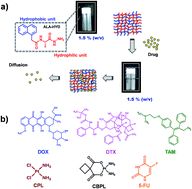
Therefore, to address this issue we studied encapsulation efficacy, drug release, mechanical strength, injectability and anticancer potential of ALA-HYD hydrogel using six anticancer drugs differing in charge and hydrophobicity viz. docetaxel (DTX), tamoxifen (TAM), cisplatin (CPL), carboplatin (CBPL) and 5-fluorouracil (5-FU) along with doxorubicin (DOX) (Fig. 1). Each drug has a different mode of action towards cancer cells, as DTX is known for its activity in inhibiting microtubule depolymerization25 and TAM acts as an anti-estrogen in mammary tissues.26 The platinum drugs, CPL and CBPL both bind with DNA to form intra-strand crosslinks, thus affecting replication,27 whereas 5-FU generally inhibits the nucleotide synthetic enzyme, thymidylate synthase.28 In this manuscript, the drug encapsulation efficiency, release and characteristic anti-cancer potency of ALA-HYD gelator for different anticancer drugs differing in their size, hydrophobicity and H-bonding ability were investigated.


image file: c5ra25847b-f1.tif
Fig. 1 (a) Schematic of present study depicting drug encapsulation in L-alanine-based hydrogelator, ALA-HYD, having both hydrophilic and hydrophobic moieties in its fibrillar network and drug-release by diffusion; (b) chemical structures of six different anticancer drugs, doxorubicin (DOX), docetaxel (DTX), tamoxifen (TAM), cisplatin (CPL), carboplatin (CBPL) and 5-fluorouracil (5-FU) used in this study.

Results and discussion

The initial studies involved determining the encapsulation efficacy of 1.5% ALA-HYD hydrogel for different anticancer drugs. Measured amounts of drug stock solutions were added to a hot, aqueous super-saturated solution (sol) of gelator and addition was continued until the integrity of the gel was retained. Fig. 2 shows inverted-vial images of hydrogel formation entrapping different drug molecules, where the ability of the gelator to gelate the solvent decreased with increasing amount of drug added to the sol prior to gelation. Encapsulation efficacies differed with each drug, with the least entrapment being achieved with cisplatin (80 μg) and the highest (640 μg) amount of entrapment with 5-FU under these conditions (Table 1).
image file: c5ra25847b-f2.tif
Fig. 2 Digital images of inverted vials with hydrogels showing gelation and maximum encapsulation efficiency wrt to drugs DOX, DTX, TAM, 5-FU, CPL and CBPL. Encapsulation efficiency is indicated as the amount of drug (μg) per 500 μL of 1.5% (w/v) ALA-HYD hydrogel.
Table 1 Maximum encapsulation efficacy of ALA-HYD for different anticancer drugs and their injectability
Drug Maximum encapsulation (μg per 500 μL) Maximum injectability (μg per 500 μL)
Cisplatin (CPL) 80 80
Carboplatin (CBPL) 120 80
Docetaxel (DTX) 160 80
Doxorubicin (DOX) 200 120
Tamoxifen (TAM) 240 120
5-Fluorouracil (5-FU) 640 640


Differential encapsulation efficacies for anticancer drugs might be due to differential non-covalent inter- and intra-molecular interactions, such as hydrogen bonding, van der Waals and π–π stacking between drug and gelator molecules. Self-assembly is a prevalent phenomenon that leads to gelation for LMHGs in the absence of chemical cross-linkers. Therefore, significant differences in encapsulation efficacy can be directly correlated with degrees of hydrophobicity and molecular structures of drugs. 5-FU, being the smallest moiety and having a somewhat planar architecture, has the greatest ability to become encapsulated. The presence of amide bonds might provide hydrogen-bonded water solubility, whereas a fluorine group provides weak hydrophobic interactions. A balance of these two forces, along with π–π stacking as another possible intermolecular interaction between drug, solvent and gelator, led to gelation and high drug encapsulation. Furthermore, due to its small size, 5-FU is expected to cause the least perturbation to the gelator assembly, leading to stable gels even at high drug loadings.

The order of encapsulation among DOX, DTX and TAM anticancer drugs was TAM > DOX > DTX (Fig. 2, Table 1) as TAM presents the maximum aromatic planar architecture for aromatic interactions between itself and the gelator molecule, whereas DTX is highly hydrophobic in nature with minimum ability for π–π interactions. Very surprisingly, the hydrogel showed a minimum encapsulation efficacy for CPL and CBPL in spite of the small size and polar nature of these drugs, suggesting that the presence of free amine groups on CPL and CBPL disrupt the H-bonding between gelator molecules at high concentrations. The absence of any aromatic interactions between these molecules and ALA-HYD further alleviates the problem of their becoming encapsulated in the hydrogel.

The effect of drug entrapment on the injectability of hydrogels for different anticancer drugs was also checked simultaneously. Injectability of the gel followed a regressive pattern with increasing concentration of the drug entrapped, with the only exception being 5-FU, where the gel was injectable even at its maximum encapsulation. A maximum of 80 μg of each drug could be incorporated into the gel (per 500 μL) while maintaining injectability. We then performed rheological studies, including both amplitude sweep and frequency sweep experiments on hydrogel-encapsulated drugs, as shown in Fig. 3.


image file: c5ra25847b-f3.tif
Fig. 3 Rheological studies showing (a) frequency sweep, (b) amplitude sweep and (c) storage modulus (G′) variation in the free gel and drug-loaded gels tested at a capacity of 80 μg per 500 μL of gel volume. These studies show the mechanical stability of gels upon entrapment with (G′ > G′′) and their varied strengths as an outcome of the gel–drug interactions with their strengths varying with the drug entrapped.

The G′ (storage modulus) and G′′ (loss modulus) values clearly indicate the formation of mechanically stable gels (G′ > G′′). Hydrogels entrapping DOX had the highest mechanical strength, with the G′ value even higher than pristine gel, which might be due to the formation of imine bonds between DOX and the gelator.23 However, gel-entrapped TAM exhibited the lowest G′ amongst the systems tested, which was almost half of the pristine gel. The lower mechanical strength of gel-entrapped TAM might be due to the presence of a tertiary ammonium unit in the drug interfering with self-assembly of the gelator, resulting in a significant decrease in gel strength. It is noteworthy that self-assembly of the gelator is resilient enough to accommodate a large hydrophobic molecule like DTX without a significant decrease in gel-strength. Not surprisingly, gels could readily accommodate 5-FU without much difficulty and the resulting gels were as strong as the pristine ones. Gel-entrapped CPBL was mechanically less stable compared to gel-entrapped CPL due to the molecular complexity of CBPL over CPL and the inability of carboxylates over chloride ligands to become accommodated in gelator molecules.

SEM studies indicated that there are not many perceptible differences in the nanoscale morphology of hydrogels except in gel-entrapped CPL (Fig. 4). Gel-entrapped CPL showed hollow, rod-like structures, whereas other gel-encapsulated drugs showed long, entangled fibrous morphologies similar to pristine gels. The higher hydrogen-bonding ability of –NH2 moieties of CPL with gelator molecules and the higher planarity of the drug facilitates its easy incorporation into an intra-fibrillar network, leading to altered morphology of the gel. In contrast, the non-planarity of CBPL diminishes gel–drug interactions and we find no perceivable change in architecture with gel-entrapped CBPL.


image file: c5ra25847b-f4.tif
Fig. 4 Scanning electron micrographs of gels with different drugs at 80 μg encapsulation. Scale bar is 200 nm for all the gels except CPL, which is shown at a scale of 1 μm.

We then performed drug-release studies of these hydrogels at 37 °C (Fig. 5) at physiological pH. DOX-loaded hydrogels showed the maximum and fastest release where 80% of the drug was released over 96 h, whereas 5-FU hydrogels exhibited the slowest release, with around 70% release in 96 h. 5-FU showed maximum incorporation into the gel, being a structurally smaller moiety compared to the other drugs (Table 1) and such a slow and sustained release pattern suggested favorable interactions and compatibility between 5-FU and gelator molecules. Interestingly, the mechanical strength of 5-FU-loaded gels was also comparable with that of the free gel. DTX and TAM-loaded hydrogels showed a similar trend with 60% release after 48 h. We could not, however, conduct release studies with CPL and CBPL-loaded gels as it was difficult to detect the released amounts with HPLC (being below the detection limit of the instrument). This could be due to strong interactions between –NH2 groups of drugs and gelator molecules, which might have led to a much slower release, which may also account for these exhibiting the least incorporation into the gel structure compared to the other four drugs.


image file: c5ra25847b-f5.tif
Fig. 5 Cumulative drug release profiles from hydrogels entrapping 80 μg of DOX, DTX, TAM and 5-FU over a course of 96 h, showing the DOX being released at a faster rate and 5-FU with slow and consistent release, compared to all other drugs.

The gel network provides a sustained release of drugs, which helps the drugs to be retained at the tumor site for a longer time, thereby increasing their anti-cancer potency for longer time periods. The sustained release also helps in preventing the recurrence of tumors. The anticancer potential of hydrogel-encapsulated drugs against the murine breast cancer 4T1 cell line was investigated. 4T1 cells were cultured on the lower compartment of a transwell plate and drug-loaded hybrid gels were cast on the transwell. We performed an MTT assay to study cell proliferation in 4T1 cells in the presence of free drugs and also the drugs entrapped in gel. Highly entrapped drugs, 5-FU, CPL and CBPL showed almost two fold difference in the cell viabilities of gel-entrapped and free drug. Comparison of cell death between drug and hydrogel-drug treated cells (Fig. 6) suggested that drug-loaded hydrogels are less toxic towards the cells compared to free drugs, which further proves the benefits of controlled release of the drugs from the hydrogel.


image file: c5ra25847b-f6.tif
Fig. 6 Cytotoxicity towards 4T1 cell lines of free drug as well as drug entrapped in hydrogels, indicating reduced toxicity of the drugs entrapped in hydrogel, in comparison to those added directly to the cells, owing to the controlled release of drug from the gel network. Mean ± SD, n = 3.

Conclusions

In summary, we studied the ability of L-alanine-derived hydrogelator, ALA-HYD, to physically entrap various anticancer drugs. ALA-HYD could entrap all the drugs with different capacities by mere physical mixing, without use of any covalent or coupling strategies. All six drugs formed injectable hydrogel systems at different loading capacities, indicating optimum mechanical strength. These gel–drug systems revealed nano-fibrous morphologies and the potential to show therapeutic applicability. CPL and CBPL showed the least incorporation into gel networks owing to their ability to form stronger interactions with gel molecules, thereby disrupting physical interactions among gelator molecules. In vitro release of drugs via a process of diffusion at 37 °C showed desirable slow and sustained release of drugs in each case, which was evident from an increased cell survival compared to treatment with free DOX and 5-FU. This system, therefore, opens up further possibilities for dual encapsulation of drugs in several combinations (hydrophilic–hydrophilic or hydrophilic–hydrophobic) and for testing their efficacy against tumors. The present study not only introduces a suitable carrier molecule for hydrophobic drugs but increases the scope for further encapsulating a suitable combination of two drugs in the same gel so as to increase efficiency.

Experimental section

Materials and methods

Anticancer drugs doxorubicin, docetaxel, tamoxifen, 5-fluorouracil, cisplatin, carboplatin, sodium dodecyl sulfate (SDS) and Tween-80 were purchased from Sigma Aldrich. HPLC grade methanol and acetonitrile were from Spectrochem. Gelator compound ALA-HYD was synthesized as described earlier.23

Preparation of hydrogel

The hydrogels were prepared with a gelator concentration of 1.5% (w/v) in aqueous medium with the method reported earlier.23 Gelation was checked by inverting the glass vials.

Drug encapsulation and injectability studies

Drug stocks were prepared in their respective solvents, i.e. hydrophilic drugs, namely DOX and 5-FU, were dissolved in water, whereas hydrophobic drugs, i.e. TAM and DTX, were dissolved in DMSO. Methanol was used for CPL and CBPL. From these drug stocks, desired amounts of drugs were added to gel solution to find out the ‘maximum encapsulation capacity’ and the ‘maximum injectability limit’ for these drugs individually. The ‘maximum encapsulation capacity’ is simply the maximum amount of drug that could be taken up by the gel without disrupting the structure, while the ‘maximum injectability’ limit was the maximum amount of drug that could be encapsulated in the gel while retaining the injectability. An inverted vial method was used to demonstrate gelation, which was further confirmed by rheology. Gelation was checked a minimum of three times for each drug[thin space (1/6-em)]:[thin space (1/6-em)]gel ratio. Injectability was tested using a surgical 20′′ needle generally used for in vivo experiments. As a control, gel was prepared by adding DMSO of a volume equal to the highest amount of drug solution added to ensure that the loss of gel integrity was solely due to the drug and not due to the DMSO.

Rheology studies

For the strain sweep experiments, a 25 mm diameter, 1° angle cone was used at the top and a flat plate was used at the bottom. Samples were placed on the bottom plate. A 50 μm gap distance was maintained between the cone and the plate. All the rheological studies were performed at 25 °C on the hydrogel-entrapped drugs (80 μg and at the injectability limit of the drugs in a 15 mg mL−1 gelator). Strain-sweep tests were performed from 0.01 to 100% strain at constant frequency = 1 rad s−1. Frequency sweep experiments were carried out from 0.1 to 100 Hz at a constant strain of 0.1%.

Scanning electron microscopy (SEM)

Drug-entrapping hydrogels (80 μg of drugs at their injectability limit in 15 mg mL−1 gelator) were dried inside a vacuum desiccator for 60 h. These dried samples were spread on carbon tape and gold-coated for 120 s. The images were taken at 5 kV accelerating voltage on a Carl Zeiss (Ultraplus) field emission scanning electron microscope.

Drug release studies

We initially prepared drug-release media for each drug for carrying out an in vitro release assay. Hydrophilic drugs were tested in phosphate buffer saline (PBS) along with a pH of 7.4. For DTX, 0.01% Tween-80 was added to the buffer to enhance its solubility, whereas for TAM, 0.1% SDS was mixed with PBS in a ratio of 14[thin space (1/6-em)]:[thin space (1/6-em)]1 v/v. The drug-loaded hydrogels were immersed in their respective release media and aliquots were taken out at stipulated time intervals of 24, 48, 72 and 96 h followed by replacement with fresh media. For each drug, the release assay was carried out three times.

High-pressure liquid chromatography (HPLC) studies

Released drugs were quantified using HPLC with the help of a standard curve using a Waters HPLC (Germany) equipped with a UV/Visible detector and a TSK gel ODS 100V 5 μm column. For all the systems, HPLC acetonitrile, methanol and Milli Q double-filtered water were used as the mobile phases. UV detection at respective wavelengths was carried out in each case.

In vitro cytotoxicity (MTT assay) studies

Mouse mammary gland cell line (4T1) was cultured in RPMI 1640 medium (Sigma-Aldrich), supplemented with 10% heat-inactivated fetal calf serum (FCS; Invitrogen), 1% (v/v) penn–strep, at 37 °C, under 5% CO2 in a humidified atmosphere. We seeded ∼1.0 × 10⁴ cells per well in the lower compartment of a 24-well Corning transwell plate. Cells were seeded 1 day before the experiment for proper attachment and treated with various anticancer drugs at concentrations near to their IC50 values reported in the literature29 (DOX-0.25 μg mL−1, 5-FU-2.3 μg mL−1, CPL-2.8 μg mL−1, CBPL-36 μg mL−1, DTX-0.6 μg mL−1 and TAM-6 μg mL−1) and drugs entrapped in hydrogels (same concentrations) were cast in transwell filters. MTT assay was performed after 48 h. The % cell viability was reported by comparing the absorbance of blank cells (w/o gel) as 100% viability.

Acknowledgements

We thank RCB and IISER for intramural funding and the Department of Biotechnology, Govt. of India for funding. A. B. thanks DST for Ramanujan fellowship. S. G. thanks DBT for fellowship. A. R. M. thanks IISER Bhopal for institute fellowship. Y. P. S. thanks UGC for senior research fellowship.

References

  1. C. Chun, S. M. Lee, C. W. Kim, K. Y. Hong, S. Y. Kim, H. K. Yang and S. C. Song, Biomaterials, 2009, 30, 4752 CrossRef CAS PubMed.
  2. K. J. Skilling, F. Citossi, T. D. Bradshaw, M. Ashford, B. Kellam and M. Marlow, Soft Matter, 2014, 10, 237 RSC.
  3. H. Wang and Z. Yang, Soft Matter, 2012, 8, 2344 RSC.
  4. M. Kwak, K. Hur, J. E. Yu, T. S. Han, K. Yanagihara, W. H. Kim, S. M. Lee, S. C. Song and H. K. Yang, Invest. New Drugs, 2010, 28, 284 CrossRef CAS PubMed.
  5. B. Ding, Y. Li, M. Qin, Y. Ding, Y. Caoa and W. Wang, Soft Matter, 2013, 9, 4672 RSC.
  6. D. Ma, H. B. Zhang, K. Tua and L. M. Zhang, Soft Matter, 2012, 8, 3665 RSC.
  7. Y. Yang, M. Nakazawa, M. Suzuki, H. Shirai and K. Hanabusa, J. Mater. Chem., 2007, 17, 2936–2943 RSC; S. Zhang, Nat. Biotechnol., 2003, 21, 1171–1178 CrossRef CAS PubMed; H. A. Behanna, J. J. J. M. Donners, A. C. Gordon and S. I. Stupp, J. Am. Chem. Soc., 2005, 127, 1193–1200 CrossRef PubMed.
  8. L. Frkanec and M. Zinić, Chem. Commun., 2010, 46, 522–537 RSC.
  9. H. Komatsu, S. Matsumoto, S. Tamaru, K. Kaneko, M. Ikeda and I. Hamachi, J. Am. Chem. Soc., 2009, 131, 5580–5585 CrossRef CAS PubMed; Z. Yang, G. Liang, M. Ma, A. S. Abbah, W. W. Lu and B. Xu, Chem. Commun., 2007, 843–845 RSC.
  10. H. Svobodová, V. Noponen, E. Kolehmainen and E. Sievänen, RSC Adv., 2012, 2, 4985 RSC.
  11. A. M. Reddy and A. Srivastava, Soft Matter, 2014, 10, 4863 RSC.
  12. R. H. Zha, S. Sur and S. I. Stupp, Adv. Healthcare Mater., 2013, 2, 126 CrossRef CAS PubMed.
  13. S. Sutton, N. L. Campbell, A. I. Cooper, M. Kirkland, W. J. Frith and D. J. Adams, Langmuir, 2009, 25, 10285 CrossRef CAS PubMed.
  14. J. Naskar, G. Palui and A. Banerjee, J. Phys. Chem. B, 2009, 113, 11787 CrossRef CAS PubMed; B. Adhikari and A. Banerjee, Soft Matter, 2011, 7, 9259 RSC; J. Nanda, A. Biswas and A. Banerjee, Soft Matter, 2013, 9, 4198 RSC; A. Baral, S. Roy, A. Dehsorkhi, I. W. Hamley, S. Mohapatra, S. Ghosh and A. Banerjee, Langmuir, 2014, 30, 929 CrossRef PubMed.
  15. J. K. Kim, J. Anderson, H. W. Jun, M. A. Repka and S. Jo, Mol. Pharm., 2009, 6, 978 CrossRef CAS PubMed.
  16. F. Zhao, M. Lung Ma and B. Xu, Chem. Soc. Rev., 2009, 38, 883 RSC.
  17. P. K. Vemula, N. Wiradharma, J. A. Ankrum, O. R Miranda, G. John and J. M. Karp, Curr. Opin. Biotechnol., 2013, 24, 1174 CrossRef CAS PubMed.
  18. M. Suzuki, M. Yumoto, H. Shirai and K. Hanabusa, Chem.–Eur. J., 2008, 14, 2133–2144 CrossRef CAS PubMed; S. Nandi, H.-J. Altenbach, B. Jakob, K. Lange, R. Ihizane, M. P. Schneider, U. Gün and A. Mayer, Org. Lett., 2012, 14, 3826–3829 CrossRef PubMed; P. S. Yavvari, A. Reddy M and A. Srivastava, RSC Adv., 2013, 3, 17244–17253 RSC; N. Mohmeyer and H.-W. Schmidt, Chem.–Eur. J., 2005, 11, 863–872 CrossRef PubMed.
  19. J. Gao, H. M. Wang, L. Wang, J. Y. Wang, D. L. Kong and Z. M. Yang, J. Am. Chem. Soc., 2009, 131, 11286 CrossRef CAS PubMed.
  20. C. Yang, D. Li, Q. F. Zhao, L. Wang, L. Wang and Z. Yang, Org. Biomol. Chem., 2013, 11, 6946 CAS.
  21. A. G. Cheetham, P. Zhang, Y. Lin, L. L. Lock and H. Cui, J. Am. Chem. Soc., 2013, 135, 2907 CrossRef CAS PubMed.
  22. R. Tian, J. Chen and R. Niu, Nanoscale, 2014, 6, 3474 RSC.
  23. M. Singh, S. Kundu, A. Reddy, V. Sreekanth, R. K. Motiani, S. Sengupta, A. Srivastava and A. Bajaj, Nanoscale, 2014, 6, 12849 RSC.
  24. V. J. Venditto and F. C. Szoka Jr, Adv. Drug Delivery Rev., 2013, 65, 80 CrossRef CAS PubMed.
  25. K. Fuzitaka, N. Hattori, T. Senoo, H. Iwamoto, S. Ohshimo, M. Kanehara, N. Ishikawa, Y. Haruta, H. Murai and N. Kohno, Oncol. Lett., 2011, 2, 167 Search PubMed.
  26. W. Bursch, A. Ellinger, H. Kienzl, L. Torok, S. Pandey, M. Sikorska, R. Walker and R. S. Hermann, Carcinogenesis, 1996, 17, 1595 CrossRef CAS PubMed.
  27. M. H. Jamal, W. C. Chng, K. Yusoff and N. Shafee, Cancer Cell Int., 2012, 12, 1 CrossRef PubMed.
  28. S. Violette, L. Poulain, E. Dussaulx, D. Pepin, A. M. Faussat, J. Chambaz, J. M. Lacorte, C. Staedel and T. Lesuffleur, Int. J. Cancer, 2002, 98, 498 CrossRef CAS PubMed.
  29. C. Urueña, J. Mancipe, J. Hernandez, D. Castañeda, L. Pombo, A. Gomez, A. Asea and S. Fiorentino, BMC Complementary Altern. Med., 2013, 13, 74 CrossRef CAS PubMed; L. Bao, A. Haque, K. Jackson, S. Hazari, K. Moroz, R. Jetly and S. Dash, Am. J. Pathol., 2011, 178, 838–852 CrossRef PubMed; D. L. Morse, Mol. Cancer Ther., 2005, 4, 1495–1504 CrossRef PubMed; D. Lu, X. Wen, J. Liang, X. Zhang, Z. Gu and Y. Fan, Chin. J. Polym. Sci., 2008, 26, 369–374 CrossRef; X. Sun, R. Jiang, A. Przepiorski, S. Reddy, K. P. Palmano and G. W. Krissansen, BMC Cancer, 2012, 12, 591 CrossRef PubMed; P. Sengupta, S. Basu, S. Soni, A. Pandey, B. Roy, M. S. Oh, K. T. Chin, A. S. Paraskar, S. Sarangi, Y. Connor, V. S. Sabbisetti, J. Kopparam, A. Kulkarni, K. Muto, C. Amarasiriwardena, I. Jayawardene, N. Lupoli, D. M. Dinulescu, J. V. Bonventre, R. A. Mashelkar and S. Sengupta, Proc. Natl. Acad. Sci. U. S. A., 2012, 109, 11294–11299 CrossRef PubMed.

This journal is © The Royal Society of Chemistry 2016