Electronic structure and spectral properties of aurones as visible range fluorescent probes: a DFT/TDDFT study

Yunsheng Xue*, Yunyan Dou, Lin An, Youguang Zheng, Ling Zhang and Yi Liu*
Jiangsu Key Laboratory of New Drug Research and Clinical Pharmacy, School of Pharmacy, Xuzhou Medical College, No. 209, Tongshan Road, Xuzhou, Jiangsu 221004, China. E-mail: xueyunsheng@xzmc.edu.cn; liuyi@xzmc.edu.cn; Tel: +86-516-83262137 Tel: +86-516-83262136

Received 3rd December 2015 , Accepted 8th January 2016

First published on 12th January 2016


Abstract

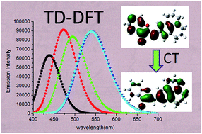
The absorption and emission spectra of aurone and its derivatives 1–4 have been investigated with density functional theory (DFT) and time-dependent density functional theory (TD-DFT). The performance of ten xc-functionals including BLYP, B3LYP, PBE0, BHHLYP, BMK, M06, M06-2X, M06-HF, LC-BLYP and CAM-B3LYP in combination with different basis sets has been analyzed. It turns out that within the selected TDDFT framework, B3LYP and PBE0 emerge as the most efficient functionals for the aurones studied. The experimentally determined spectral properties and substitution effects are well reproduced by calculations, which allowed a detailed assignment and interpretation of the spectra. The results reveal that the lowest energy transitions predominantly correspond to the π → π* transitions between the HOMO and LUMO with charge transfer (CT) character.


1 Introduction

Aurones (2-benzylidenebenzofuran-3(2H)-ones) are a group of lesser known flavonoids that are structural isomers of flavones (Fig. 1). They are found in vegetables and especially in fruits and flowers where they contribute to their coloration.1,2 Aurones are biosynthesized from chalcones by the key enzyme aureusidin synthase.3 In addition to their pigmentation role, aurones exhibit a wide variety of biological activities, including antifungal,4 antibacterial,5 insect antifeedant,6 antioxidant,7 and anticancer activities.8,9 Moreover, they have also been described as inhibitors of tyrosinase,10,11 acetylcholinesterase,12 hepatitis C virus RNA-dependent RNA polymerase,13 histone deacetylase,14 monoamine oxidase,15 and ABCG2,16 and as inducers of NAD(P) H:quinone oxidoreductase 1.17 In particular, as an important class of organic heterocyclic dyes, aurones exhibit unique photochemical and photophysical properties, which render them useful in a variety of applications such as fluorescent labels and probes in biology and medicine.18,19
image file: c5ra25733f-f1.tif
Fig. 1 General structure of aurone, chalcone and flavone molecular scaffolds.

Shanker and coworkers20 recently synthesized four amine-substituted aurone derivatives (Fig. 2) to discover fluorescent probes for biomolecules from new chromophores. The effect of substituents on the absorption and emission properties of the aurone chromophore has been explored and the results shown promising results in the potential application as fluorescent probes. In view of the considerable importance of these compounds and to further develop applications of these systems as fluorescent probes, it is essential to understand fundamentally about the structural and electronic basis of the optical properties of these chromophores.


image file: c5ra25733f-f2.tif
Fig. 2 Structure and atom numbering of the four studied aurone derivatives.

Nowadays, quantum chemical calculations have been proven to be an important tool for investigation of the relationships between structures and spectral properties of the organic molecules and for the interpretation of experimental results arising from industrial interest and applications. In recent years, TD-DFT has been widely used in electronic transition energy predictions.21–51 It appears as one of the most successful methods in terms of the balance between the accuracy and the computational cost. Indeed, recent benchmarks performed by several groups have concluded that TD-DFT's average accuracy for low-lying excited-states is in the 0.1–0.3 eV range.52–55

Recently, Sigalas and coworkers have systematically studied the radical scavenging activity of a series of aurones with hydroxyl substitutions at DFT-B3LYP level.56,57 The vibrational and electronic absorption spectra of 4′-nitro-aurone have been investigated theoretically (DFT/TD-DFT) and experimentally by Chaitanya et al.58 Using the B3LYP/6-311++G (2d,2p) method, Chandraju and Fun have studied the FTIR and NMR spectra as well as some physical properties of 5,6-dimethyl-4′-nitro-aurone.59 Despite this, to the best of our knowledge, there has been no report on the use of TD-DFT for the systematic investigation of spectral property of the aurones investigated.

In continuation of our theoretical research on spectral properties of molecules with unique optical properties,60–63 we decided to study the electronic structure and absorption and emission spectra of aurone and its four aurone derivatives 1–4 (Fig. 2) by DFT and TD-DFT. The aim of the present study is to determine the structural and electronic properties of title compounds and to gain insights into the nature of spectra feature observed in the experiments. The relationship between structure and spectra as well as the effects of substitution on electronic spectra are discussed. From a theoretical point of view, it is valuable to use a computational design to predict the spectral properties before designing and synthesizing new chromophores as fluorescent probes.

2 Computational methods

All calculations have been performed with the Gaussian 09 suite of program.64 The ground-state (S0) geometry of each molecule has been fully optimized with default thresholds on residual forces and displacements. Subsequently, vibrational frequencies were calculated analytically at the same level of theory as in the previous step to verify the optimized structure. After the ground-state geometry optimization, the absorption spectrum of each molecule has been computed with TD-DFT formalism. The first excited-state (S1) geometrical optimization was performed using the TD-DFT method. Fluorescence emission energies were computed from TD-DFT calculations based on the optimized geometry of the lowest excited state.

The performance of different DFT functionals in combination with different basis sets was explored. The vertical absorption energy have been computed employing a wide panel of exchange–correlation (xc) functionals, including pure GGA functional (BLYP),65 global hybrid functionals (B3LYP,65,66 PBE0,67,68 BHHLYP69), global hybrid meta GGA functionals (BMK,70 M06, M06-2X and M06-HF),71 and recent long-range corrected and range-separated hybrids functionals (LC-BLYP65,72 (ω = 0.47 bohr−1) and CAM-B3LYP73). To account for the solvation, the TD-DFT calculations have been combined with polarizable continuum model (PCM),74,75 which has emerged as the effective tools to treat bulk solvent effects for both the ground- and excited-states.

3 Results and discussions

3.1 Ground and excited state geometries

The ground-state (S0) geometry of the five aurones was optimized at the PBE0/6-311G(d,p) approach which has been applied successfully in the study of chalcones60–63 and indigoids.54 This computational model was further evaluated by comparing gas-phase optimized bond length and bond angle values with experimental ones (XRD) reported in the literature for Z-2-p-methoxyaurone.76 Some selected parameters are listed in Table S1 in ESI. It can be seen from Table S1 that the PBE0/6-311G(d,p) method is reasonably accurate in predicting geometrical parameters. The computed mean absolute error (MAE) for the bond lengths and angles is only 0.008 Å and 0.46°, respectively. As shown in Table S2 in ESI, the differences between gas-phase and solvated (ethanol) geometries are very small. For instance, the MAE of bond lengths and bond angles for aurone are 0.001 Å and 0.1°, respectively. Therefore, we have selected the gas-phase PBE0/6-311G(d,p) for determining the ground-state structural parameters.

Both E- and Z-isomers of aurones can be found in nature, although the latter is much more abundant. According to our calculation, Z-isomers are more stable than E-isomers with energy differences of 3.07 kcal mol−1 (aurone), 2.43 kcal mol−1 (1), 2.55 kcal mol−1 (2), 2.32 kcal mol−1 (3) and 2.09 kcal mol−1 (4). The thermodynamic stability of both isomers is similar, which can be one of the reasons for their simultaneous rise in nature. Our DFT calculations resulted in a planar geometry for Z- and E-isomers of all five aurones. Comparing the calculated bond lengths and angles of the two isomers for aurone (Table S3 in ESI), we find that the corresponding values are not very much different, with the largest differences being less than 0.02 Å for bond lengths and 2.1° for bond angles. This indicates that isomerization has little effect on these geometrical parameters.

As observed in Table S4 in ESI, the corresponding geometrical parameters of Z-aurones are very similar with each other. This indicates that the introduction of substituent has little effect on these geometric parameters. Moreover, the linkage between the benzofuranone system and phenyl ring with average bond lengths of C2–C3 (1.4846 Å), C2–C10 (1.3432 Å), C10–C1′ (1.4415 Å) and C2–O1 (1.3820 Å), suggests that these atoms are highly conjugated, leading to a π-bridge for the charge transfer (CT) between the benzofuranone system and phenyl ring B. According to the calculations, the key backbone of all aurones has a planar structure, while aurone 3 and 4 are not completely planar due to the deviation of amine substituents.

Similar to the ground state, the excited state geometry of the five aurones optimized using the TD-PBE0 method also adopt a planar structure, with the exception of some deviation from the plane in the amine substituents. It was noted that some notable difference exists in bond lengths and angles between the ground and excited state, especially for the bond angles. Comparison of the key structural parameters list in Table S5 in ESI and Table S4 shows that the bond lengths of excited state are prone to equalization, implying higher degree of conjugation than ground state geometry. The largest change of bond length presents in the central C2[double bond, length as m-dash]C10 bond and the C3[double bond, length as m-dash]O11 bond. For instance, upon excitation to the S1 state, the length of C2[double bond, length as m-dash]C10 and C3[double bond, length as m-dash]O11 bonds in aurone elongated by about 0.06 and 0.04 Å respectively, while that of the neighboring single bonds (C2–O1, C2–C3 and C10–C1′) shortened by average 0.03 Å. Compared to bond length, bond angles change more distinctly than that of the ground state. Moreover, the bond angles in coumaranone moiety changes more obviously than other parts with the largest deviation of about 2° compared to that of ground state. All of these changes in geometrical parameters serve to rationalize the observed large Stokes shifts.

3.2 Absorption spectra

On the basis of above geometrical study, a systemic evaluation of the performance of computation model on the TD-DFT step was performed based on the calculation of vertical absorption energy λmax of Z-aurone. First, we evaluated the impact of the selected atomic basis set (BS) because excited-state properties are often significantly BS-dependent. Table 1 gives the λmax,ab computed using different basis sets for a given geometry.
Table 1 The λmax,ab (in nm) of Z-aurone obtained with PCM (ethanol)-TD-PBE0/X based on the geometries optimized at the PBE0/6-311G(d,p) level in gas phasea
Basis sets (X) λmax,ab
a The experimental value (in ethanol) is 377 nm.20b Optimization using PCM model.
6-31G 356.35
6-31G(d) 359.08
6-311G(d,p) 365.57
6-311G(2d,2p) 366.54
6-31+G(d) 370.64(370.78)b
6-311+G(d,p) 372.49
6-311++G(2d,2p) 373.43


As shown in Table 1, the 6-311++G (2d,2p) basis set gives the best result relative to experimental value (377 nm). On the contrary, the 6-31G gives the poorest result. The extension of basis set from 6-31G to 6-311G (2d,2p) or from 6-31+G (d) to 6-311++G (2d,2p) leads to an increase in λmax,ab. Adding extra polarization function to 6-311G (d,p), increases the λmax,ab by only 1 nm, suggesting that the second set of polarization function is unnecessary. Adding diffuse function to 6-31G (d) or 6-311G (d,p) changes λmax,ab by +11 nm or +7 nm, respectively, indicating diffuse function is necessary for TD-DFT calculation. From 6-31+G (d) to 6-311++G (2d,2p), the change of λmax,ab is only 3 nm, while the computational time increases significantly. Therefore we have selected the 6-31+G (d) for TD-DFT calculation because it provides results close to 6-311++G (2d,2p) with much lower computational costs. When solvent effect was added in the geometrical optimization, almost no change of λmax,ab was observed with respect to the data in gas phase (370.64 vs. 370.78 nm), indicating that the solvent effect has very small impact on the geometrical structure of the aurones.

Next, we investigated the impact of functional on the excitation energies. The results are collected in Table 2. Ten functionals were used for the TD-DFT calculations, including BLYP, B3LYP, PBE0, BMK, BHHLYP, M06, M06-2X, M06-HF, LC-BLYP and CAM-B3LYP. The accuracy of the calculated values was assessed against the experimental data.

Table 2 Comparison between computed and experimental λmax,ab (in nm) for Z-aurone. The theoretical values are calculated at the PCM (ethanol)-X/6-31+G(d)//PBE0/6-311G(d,p) level
Functional λmax,ab f
a Ref. 20.
BLYP 433.42 0.3322
B3LYP 379.97 0.5058
PBE0 370.64 0.5396
M06 372.08 0.5650
BMK 345.53 0.6263
BHHLYP 329.78 0.7366
M06-2X 337.76 0.6404
M06-HF 308.08 0.7230
LC-BLYP 311.77 0.7175
CAM-B3LYP 341.06 0.6387
Exp.a 377  


As shown in Table 2, B3LYP functional gives the most accurate λmax,ab value, with discrepancy of only 3 nm relative to the experimental data. Hybrid functionals PBE0 and M06 also give a reasonable accurate result; the calculated λmax are about 6 and 5 nm smaller than the experimental one, respectively. The pure functional BLYP presents too large λmax,ab (+56 nm), while long-range corrected functionals (LC-BLYP and CAM-B3LYP) and other global hybrids seriously underestimate the λmax,ab.

Beside aurone itself, we have also applied the same set of functionals for the four aurone derivatives 1–4 (Table 3) in order to provide complete information. For clarity, the calculated λmax,ab are graphically displayed in Fig. 3. As can be seen from Table 3 and Fig. 3, the conventional global hybrid B3LYP gives the most accurate result compared to experimental values, with mean absolute error (MAE) of only 18.9 nm. The hybrids PBE0 and M06 functionals predict λmax,ab with accuracy similar to that obtained at B3LYP level, with MAE of 25.4 and 27.7 nm, respectively. The similar accuracy between PBE0 and M06 can be rationalized by the comparable amount of HF-like exchange (25 versus 27%), which is the dominating factor for the performance of standard hybrid functional.77 On the contrary, poor results were obtained using all other functionals, especially for LC-BLYP and M06-HF functionals. LC-BLYP and M06-HF dramatically underestimate the λmax,ab with a MAE of up to 102 and 100 nm with respect to experimental values. In fact, it has been shown that M06-HF significantly overestimates the vertical transition energies for π → π* states.78

Table 3 Comparison of calculated and experimental absorption maxima λmax,ab (nm) in ethanol for aurone derivatives 1–4 at the PCM-X/6-31+G(d)//PBE0/6-311G(d,p) levela
Functional 1 2 3 4 MAEb
λmax,ab f λmax,ab f λmax,ab f λmax,ab f
a From ref. 20 in ethanol.b Mean absolute error of λmax,ab.
BLYP 459.97 0.7917 479.68 0.7126 548.20 0.7857 541.10 0.7929 46.7
B3LYP 404.08 0.9366 421.59 0.8667 478.63 0.9612 474.06 0.971 18.9
PBE0 393.84 0.9726 413.98 0.9191 463.63 1.0159 460.04 1.0256 25.4
M06 393.17 0.9629 408.92 0.8946 459.99 0.9921 456.02 1.0017 27.7
BMK 366.42 1.0442 382.06 0.9823 430.71 1.1106 429.22 1.1114 55.0
BHHLYP 348.63 1.1197 361.88 1.0442 402.22 1.1717 401.52 1.1640 76.9
M06-2X 357.51 1.0431 373.27 0.9829 417.17 1.1182 419.17 1.1075 64.8
M06-HF 324.51 1.1081 338.82 1.0456 376.36 1.2017 382.17 1.1697 99.8
LC-BLYP 326.07 1.1009 337.60 1.0671 369.26 1.2355 374.08 1.2032 102.0
CAM-B3LYP 358.49 1.0388 371.94 0.9962 412.53 1.1431 414.5 1.1265 66.1
Exp. 395   441   501   515    


image file: c5ra25733f-f3.tif
Fig. 3 Comparison between the calculated and experimental λmax,ab.

As can be seen from Fig. 3, the λmax,ab decrease gradually with increasing HF exchange percentage from B3LYP (20%) to M06-HF (100%) method, although the trend between M06 and PBE0 are sometimes an exception. On the other hand, the oscillator strengths also increase gradually with increasing HF exchange percentage. These phenomena were also observed in previous studies, i.e., increasing the HF exchange ratio in the global hybrids yields smaller absorption maxima.54,79–81

As shown in Tables 2 and 3 and Fig. 3, all functionals provide almost the same trend of the change of excitation energies for all compounds, which is in good agreement with experimental results. It was noted that the experimental observed weak red shift (only 14 nm) of λmax,ab from 3 to 4 was not correctly reproduced by computation using pure GGA and conventional hybrid functionals. This can be attributed to the charge transfer character of the main electronic transitions in aurones 3 and 4. Indeed, the two hybrid meta-GGA functionals (M06-2X, M06-HF) and two long-range corrected functionals (LC-BLYP and CAM-B3LYP) reproduced this trend well. Despite the somewhat poor performance in description of the weak spectral shift, hybrid functionals PBE0 and B3LYP behave well in predicting the absorption maximum with MAE of only 25 nm (0.15 eV) and 19 nm (0.11 eV), respectively, in agreement with the level of accuracy expected at these levels of theory.22 Concerning the oscillator strengths (f), the highest oscillator strength is found for 4 and the lowest for aurone, which is consistent with the experimental results. As a whole, considering the accuracy and consistency, the following discussion are mainly based on the results obtained at the PCM-TD-PBE0/6-31+G(d) level.

Structurally, aurone and its derivatives 1–4 have the classic donor–acceptor π conjugate (D–π–A) characteristic with the electron deficient coumaranone moiety (A) linked to the electron donating ring B (D). As shown in Table 4, introduction of electron-donating amine substituents results in an obvious red-shift of λmax,ab. The amine substituent (2) extends the λmax,ab of the parent compound by about 43 nm into the visible region of the spectrum. The amide group (1) evokes the least red shift (only 23 nm) compared to parent aurone due to the fact that the nitrogen atom is bonded to the electron withdrawing carbonyl. Aurone 3 has considerably larger red shift relative to 2 (50 nm in ethanol). This might be due to the rigid ring structure in 3, which limits the free rotation of the N–C bond in amine, and then enhances the electron-donating ability of the amine.82

Table 4 Calculated absorption wavelength λab (eV, nm), main configuration and oscillator strength (f) for the studied aurones at the PCM-TD-PBE0/6-31+G(d) level
Molecule Exp.a λab (eV/nm) f Configuration
a Ref. 20, in methanol.b Ref. 83, in ethanol.
Aurone 379 (4.06)b 3.35/370.6 (S1) 0.5396 H → L (81%)
316 (4.27)b 3.92/316.3 (S3) 0.3463 H−1 → L (90%)
279 (3.85)b 4.56/272.1 (S5) 0.0965 H−4 → L (88%)
251 (4.10)b 5.01/247.6 (S6) 0.2325 H → L+1 (88%)
1 395 (4.09) 3.15/393.8 (S1) 0.9762 H → L (83%)
3.75/330.8 (S3) 0.1433 H−1 → L (92%)
4.55/272.4 (S5) 0.1462 H−4 → L (88%)
4.71/263.3 (S7) 0.2854 H → L+1 (87%)
2 442 (4.33) 2.99/414.0 (S1) 0.9191 H → L (82%)
3.77/328.5 (S3) 0.0391 H−1 → L (90%)
4.58/270.9 (S5) 0.1443 H → L+1 (76%)
4.65/266.6 (S6) 0.1733 H−4 → L (80%)
4.75/261.1 (S7) 0.1595 H → L+2 (64%)
3 503 (4.15) 2.67/463.6 (S1) 1.0159 H → L (82%)
3.73/332.6 (S3) 0.0228 H−1 → L (87%)
4.20/295.1 (S5) 0.0772 H → L+1 (73%)
4.36/284.5 (S6) 0.2126 H → L+2 (66%)
4.56/272.1 (S7) 0.1740 H−4 → L (69%)
4 518 (4.34) 2.70/460.0 (S1) 1.0256 H → L (81%)
3.49/354.9 (S2) 0.0062 H−1 → L (93%)
4.20/295.2 (S5) 0.1225 H → L+1 (86%)
4.54/273.4 (S6) 0.1475 H−4 → L (74%)
4.55/272.7 (S7) 0.1423 H → L+3 (44%)
    H → L+2 (39%)


It can be seen from Table 4 that there are four main absorption peaks for all studied aurones, being in line with the experimental results.20,83 The first and the highest intensity absorption peak corresponds to the electronic transition from S0 state to the higher energy S1 state, in the range of 370–464 nm depending on the structures. The lowest lying singlet excited states for all title compounds comprise mainly an electronic transition from the highest occupied molecular orbital (HOMO, H in Table 4) to the lowest occupied molecular orbital (LUMO, L in Table 4), accounting for >80% of the total. The other two less intense peaks, in range of 316–355 nm and 248–295 nm, originate from H−1 → L and H → L+1 transitions, respectively. The fourth peak at about 270 nm is mainly due to H−4 → L transition, but H → L+2 and H → L+3 are also involved for aurones 2–4.

The electron density plots of these frontier molecular orbitals along with the HOMO and LUMO orbitals are depicted in Fig. 4 and S1 in ESI. In general, the major frontier molecular orbitals demonstrate a typical π-type molecular orbital character. For parent molecule aurone, both the HOMO and LUMO are delocalized on the whole molecule, representing a typical π → π* transition. In the case of aurones 1 and 2, the HOMOs are mainly localized on benzene ring B, furanone moiety as well as the substituents, whereas the LUMOs are outspreaded on the whole molecule with a smaller contribution on substituents. Compared to aurone, the electron density in HOMOs of aurones 1 and 2 tend to accumulate on the donor portion (D) of the molecule, while those of LUMOs on the acceptor portion (A). Such trend is reflected more obviously in aurones 3 and 4. As shown in Fig. 4, the benzene ring A in coumaranone moiety almost give no contribution to HOMOs, whereas the benzene ring B present less contribution to LUMOs than that of 1 and 2. Clearly, the electron promotion from HOMO to LUMO in aurones 1–4 is accompanied by charge transfer from the donor moiety (D) to the acceptor group (A), so that the HOMO–LUMO transition has a mixed π → π* and CT character. This conclusion is confirmed by the natural transition orbital (NTO) analysis84 (Fig. S2 in ESI). As shown in Fig. S2, the largest natural transition orbital eigenvalue for all compounds are larger than 0.99, indicating that the dominant excitation pair shown in Fig. S2 account for over 99% of the lowest energy transition. Clearly, the electron promotion is accompanied by charge transfer from the donor moiety (electron) to the acceptor (hole), especially for aurones 2–4.


image file: c5ra25733f-f4.tif
Fig. 4 Electron density plots of the frontier molecular orbitals for S0 state of the title compounds (isocontour value 0.02 au).

As far as the HOMO−1 is concerned, aurone and derivatives 1–2 present similar electron density distribution with each other, while different distribution was observed in the case of aurones 3–4. For aurone 3, the electron density is mainly localized on the benzene ring A and with little contribution from benzene ring B and amine N, while opposite distribution is presented in 4, in which the electron density is mainly centered on benzene ring B and amine N. As for LUMO+1, all the cases show similar electron density distribution with the corresponding LUMO. As shown in Fig. S1, HOMO−4 in aurone and 2 are primarily localized on the benzene ring A and carbonyl group, whereas in aurones 3 and 4, the HOMO−4 mainly centered on the benzene ring A, central C[double bond, length as m-dash]C bond, part of the benzene ring B and the amine N atom. Contrary to others, the HOMO−4 of aurone 1 presents unique character, which consist of the n-orbital of the amide group.

As stated above, E- and Z-isomers of aurones can coexist in solution at normal condition. Thus, the absorption maxima of E-isomers of aurones (Table S6 in ESI) were also calculated for comparison. The results show that the two isomers present similar spectral properties. E-isomers present slightly longer wavelength absorption than Z-isomers, with the discrepancy of 8–13 nm depending on the structure. Moreover, the oscillator strengths of absorption maxima are significantly smaller for E-isomers than that of Z-isomers. These results are consistent with the experimental observation.

3.3 Emission spectra

The vertical emission energies of the five aurones have also been investigated using the TD-PBE0 method in their excited-state (S1) optimized structure. The calculated emission energies, oscillator strength, transition character of the aurones using PBE0/6-31+G(d) are summarized in Table 5. As can be seen from Table 5, the computed emission maxima λmax,em are in good agreement with experimental data for all compounds. The largest variation of vertical transition energy between calculation and experiment is less than 0.21 eV. Moving from parent aurone to aurone derivatives 1–4, there is an obvious red-shift for λmax,em with introduction of amine substitutions, which are similar with that of absorption maxima. At the same time, the oscillator strength f increases from aurone to aurones 1–4. These trends are all consistent with the experimental results. The red-shift of λmax,em can be explained by the fact that the lone electron pair of nitrogen in amine group join in molecular conjugation, which makes the conjugated-system of molecules become larger as shown in following FMOs.
Table 5 Calculated emission maxima λmax,em, main configuration, oscillator strength (f) and stokes shift for the studied aurones at the PCM (ethanol)-TD-PBE0/6-31+G(d) level
Molecule Exp.a λmax,em (eV/nm) f Assignment Stokes shift (nm)
a Ref. 20.
Aurone 2.83/438.8 0.8748 L → H (99%) 68
1 482 2.62/473.0 1.2579 L → H (100%) 79
2 540 2.51/494.8 1.1514 L → H (100%) 81
3 580 2.29/541.2 1.2331 L → H (100%) 78
4 584 2.30/539.6 1.2316 L → H (100%) 80


Stokes shift, the difference between the absorption and emission maxima, is a distinct characteristic of each fluorophore. Large Stokes shift is an important criterion for an ideal fluorescent probe. Commercially available fluorophores such as rhodamine, fluorescein, cyanines and boron dipyrromethenes usually have small Stokes shifts (typically less than 25 nm), which can lead to serious self-quenching and fluorescence detection errors because of excitation backscattering effects.85 As shown in Table 5, all the five studied aurones have significantly larger Stokes shift (up to 81 nm), especially for the aurone derivatives 1–4. This means that these aurones compounds are potentially useful as fluorescent probe.

The computed electronic transitions in S1 state reveal that the emission process is mainly due to a π* → π transition from LUMO to HOMO. By comparison, the shape of the HOMO and LUMO involved in S1 state is similar with that of S0 state (Fig. 4 and 5). The distribution of the FMOs suggests the CT nature of the emission process in aurones 1–4.


image file: c5ra25733f-f5.tif
Fig. 5 Plots of HOMO (left) and LUMO (right) for the S1 state of the studied aurones.

Compared to the Z-isomer, the E-isomer of aurones presents longer wavelength emission, with a shift of 10–30 nm depending on the structure. In addition, the oscillator strength of emission for E-isomers is significantly smaller than that of Z-isomers. Indeed, the emission intensity was decreased by irradiation at 400 nm in experiment. Our calculations are in good agreement with the experimental observations.

4 Conclusions

In this work, the structure as well as electronic and optical properties of aurone and its amine-substituted derivatives have been investigated with DFT and TDDFT method. The performance of different xc-funtionals and basis sets has been assessed. The nature of electronic transition has been analyzed and the effects of substitution on the optical properties have also been discussed. The computations are generally in good agreement with experimental observations, and allowed a detailed assignment and interpretation of the spectra.

All the five studied aurones have a planar structural backbone in both the S0- and S1-states due to π conjugation. However, the values of bond lengths and angles change significantly upon excitation, which is responsible for the large Stokes shift. The Z- and E-isomers of aurones can coexist due to their little difference of thermodynamic stability. The appearance of less stable E-isomer has slight effect on the geometrical and spectral position, but large influence on the intensity.

Among the tested functionals, hybrid functionals PBE0 and B3LYP behave well in predicting the absorption and emission maxima. The calculated transition energies using PCM-TD-PBE0/6-31+G(d) basis set have good agreement with the experimental findings in general. The calculated results show that introduction of amine substituent gives rise to red shift of λmax. Acetylation of the amine shifts the absorption and emission maxima to shorter wavelength, while restricting the rotation of the amine nitrogen shifts the absorption and emission maxima to longer wavelength.

The calculations reveal that the lowest energy transitions in aurones 1–4 predominantly correspond to the π → π* transitions between HOMO and LUMO with intramolecular charge transfer (CT) nature. Our results provide a detailed characterization of the absorption and fluorescent properties of aurone and its derivatives.

To sum up, our calculations have provided reasonable results and a useful insight into the optical properties of the aurones. This theoretical information would be very useful in the design of aurone-based chromophores for fluorescent probes in biological applications.

Acknowledgements

This work was supported by the National Natural Science Foundation of China (81202490, 81341084), Jiangsu Key Laboratory of New Drug Research and Clinical Pharmacy (ZR-XY201404), the Priority Academic Program Development of Jiangsu Higher Education Institutions and the Innovative Practice Training Program for Students of Jiangsu Higher Education Institutions (201410313052X). The authors are grateful to Prof. X. D. Gong (Department of Chemistry, Nanjing University of Science and Technology) for providing calculation facilities.

Notes and references

  1. M. G. Lagorio, G. B. Cordon and A. Iriel, Photochem. Photobiol. Sci., 2015, 14, 1538–1559 CAS.
  2. J. B. Harborne and C. A. Williams, Phytochemistry, 2000, 55, 481–504 CrossRef CAS PubMed.
  3. E. Ono, M. Fukuchi-Mizutani, N. Nakamura, Y. Fukui, K. Yonekura-Sakakibara, M. Yamaguchi, T. Nakayama, T. Tanaka, T. Kusumi and Y. Tanaka, Proc. Natl. Acad. Sci. U. S. A., 2006, 103, 11075–11080 CrossRef CAS PubMed.
  4. O. Kayser, A. F. Kiderlen, U. Folkens and H. Kokodziej, Planta Med., 1999, 65, 316–319 CrossRef CAS PubMed.
  5. N. Hadj-Esfandiari, L. Navidpour, H. Shadnia, M. Amini, N. Samadi, M. A. Faramarzi and A. Shafiee, Bioorg. Med. Chem. Lett., 2007, 17, 6354–6363 CrossRef CAS PubMed.
  6. M. Morimoto, H. Fukumoto, T. Nozoe, A. Hagiwara and K. Komai, J. Agric. Food Chem., 2007, 55, 700–705 CrossRef CAS PubMed.
  7. A. Detsi, M. Majdalani, C. A. Kontogiorgis, D. Hadjipavlou-Litina and P. Kefalas, Bioorg. Med. Chem., 2009, 17, 8073–8085 CrossRef CAS PubMed.
  8. W. Huang, M. Z. Liu, Y. Li, Y. Tan and G. F. Yang, Bioorg. Med. Chem., 2007, 15, 5191–5197 CrossRef CAS PubMed.
  9. A. Boumendjel, Curr. Med. Chem., 2003, 10, 2621–2630 CrossRef CAS PubMed.
  10. S. Okombi, D. Rival, S. Bonnet, A. M. Mariotte, E. Perrier and A. Boumendjel, J. Med. Chem., 2006, 49, 329–333 CrossRef CAS PubMed.
  11. C. Dubois, R. Haudecoeur, M. Orio, C. Belle, C. Bochot, A. Boumendjel, R. Hardre, H. Jamet and M. Reglier, ChemBioChem, 2012, 13, 559–565 CrossRef CAS PubMed.
  12. R. Sheng, Y. Xu, C. Q. Hu, J. Zhang, X. Lin, J. Y. Li, B. Yang, Q. J. He and Y. Z. Hu, Eur. J. Med. Chem., 2009, 44, 7–17 CrossRef CAS PubMed.
  13. R. Haudecoeur, A. Ahmed-Belkacem, W. Yi, A. Fortune, R. Brillet, C. Belle, E. Nicolle, C. Pallier, J. M. Pawlotsky and A. Boumendjel, J. Med. Chem., 2011, 54, 5395–5402 CrossRef CAS PubMed.
  14. V. Zwick, A. O. Chatzivasileiou, N. Deschamps, M. Roussaki, C. A. Simoes-Pires, A. Nurisso, I. Denis, C. Blanquart, N. Martinet, P. A. Carrupt, A. Detsi and M. Cuendet, Bioorg. Med. Chem. Lett., 2014, 24, 5497–5501 CrossRef CAS PubMed.
  15. N. Morales-Camilo, C. O. Salas, C. Sanhueza, C. Espinosa-Bustos, S. Sepulveda-Boza, M. Reyes-Parada, F. Gonzalez-Nilo, M. Caroli-Rezende and A. Fierro, Chem. Biol. Drug Des., 2015, 85, 685–695 CAS.
  16. H. M. Sim, C. Y. Lee, P. L. R. Ee and M. L. Go, Eur. J. Pharm. Sci., 2008, 35, 293–306 CrossRef CAS PubMed.
  17. C. Y. Lee, E. H. Chew and M. L. Go, Eur. J. Med. Chem., 2010, 45, 2957–2971 CrossRef CAS PubMed.
  18. M. Ono, Y. Maya, M. Haratake, K. Ito, H. Mori and M. Nakayama, Biochem. Biophys. Res. Commun., 2007, 361, 116–121 CrossRef CAS PubMed.
  19. Y. Maya, M. Ono, H. Watanabe, M. Haratake, H. Saji and M. Nakayama, Bioconjugate Chem., 2009, 20, 95–101 CrossRef CAS PubMed.
  20. N. Shanker, O. Dilek, K. Mukherjee, D. W. McGee and S. L. Bane, J. Fluoresc., 2011, 21, 2173–2184 CrossRef CAS PubMed.
  21. I. Ciofini, P. P. Laine, F. Bedioui and C. Adamo, J. Am. Chem. Soc., 2004, 126, 10763–10777 CrossRef CAS PubMed.
  22. D. Jacquemin, E. A. Perpete, I. Ciofini and C. Adamo, Acc. Chem. Res., 2009, 42, 326–334 CrossRef CAS PubMed.
  23. D. Jacquemin, E. A. Perpete, G. Scalmani, I. Ciofini, C. Peltier and C. Adamo, Chem. Phys., 2010, 372, 61–66 CrossRef CAS.
  24. D. Jacquemin, J. Preat, V. Wathelet, M. Fontaine and E. A. Perpete, J. Am. Chem. Soc., 2006, 128, 2072–2083 CrossRef CAS PubMed.
  25. N. Santhanamoorthi, K. Senthilkumar and P. Kolandaivel, Mol. Phys., 2010, 108, 1817–1827 CrossRef CAS.
  26. A. Eilmes, Theor. Chem. Acc., 2010, 127, 743–750 CrossRef CAS.
  27. A. Amat, C. Clementi, F. De Angelis, A. Sgamellotti and S. Fantacci, J. Phys. Chem. A, 2009, 113, 15118–15126 CrossRef CAS PubMed.
  28. L. Serrano-Andres and M. Merchan, J. Mol. Struct.: THEOCHEM, 2005, 729, 99–108 CrossRef CAS.
  29. C. Adamo and D. Jacquemin, Chem. Soc. Rev., 2013, 42, 845–856 RSC.
  30. A. D. Laurent and D. Jacquemin, Int. J. Quantum Chem., 2013, 113, 2019–2039 CrossRef CAS.
  31. E. Bremond, M. E. Alberto, N. Russo, G. Ricci, I. Ciofini and C. Adamo, Phys. Chem. Chem. Phys., 2013, 15, 10019–10027 RSC.
  32. A. Amat, C. Miliani, A. Romani and S. Fantacci, Phys. Chem. Chem. Phys., 2015, 17, 6374–6382 RSC.
  33. C. Bernini, L. Zani, M. Calamante, G. Reginato, A. Mordini, M. Taddei, R. Basosi and A. Sinicropi, J. Chem. Theory Comput., 2014, 10, 3925–3933 CrossRef CAS PubMed.
  34. P. J. Aittala, O. Cramariuc, T. I. Hukka, M. Vasilescu, R. Bandula and H. Lemmetyinen, J. Phys. Chem. A, 2010, 114, 7094–7101 CrossRef CAS PubMed.
  35. D. Gajalakshmi, R. V. Solomon, V. Tamilmani, M. Boobalan and P. Venuvanalingam, RSC Adv., 2015, 5, 50353–50364 RSC.
  36. M. Shkir, S. Muhammad, S. AlFaify, A. Irfan, P. S. Patil, M. Arora, H. Algarni and Z. Jingping, RSC Adv., 2015, 5, 87320–87332 RSC.
  37. F. Turecek, J. Phys. Chem. A, 2015, 119, 10101–10111 CrossRef CAS PubMed.
  38. M. K. Awad, M. M. El-Hendawy, T. A. Fayed, S. E. Etaiw and N. J. English, Photochem. Photobiol. Sci., 2013, 12, 1220–1231 CAS.
  39. Y. Shigemitsu, K. Komiya, N. Mizuyama and Y. Tominaga, J. Mol. Struct.: THEOCHEM, 2008, 855, 92–101 CrossRef CAS.
  40. A. V. Kityk, J. Phys. Chem. A, 2012, 116, 3048–3055 CrossRef CAS PubMed.
  41. N. De Mitri, S. Monti, G. Prampolini and V. Barone, J. Chem. Theory Comput., 2013, 9, 4507–4516 CrossRef CAS PubMed.
  42. D. Tzeli, G. Theodorakopoulos, I. D. Petsalakis, D. Ajami and J. Rebek Jr, J. Am. Chem. Soc., 2012, 134, 4346–4354 CrossRef CAS PubMed.
  43. S. N. Margar, L. Rhyman, P. Ramasami and N. Sekar, Spectrochim. Acta, Part A, 2016, 152, 241–251 CrossRef CAS PubMed.
  44. R. Nithya and K. Senthilkumar, RSC Adv., 2014, 4, 25969 RSC.
  45. S. Fantacci, A. Amat and A. Sgamellotti, Acc. Chem. Res., 2010, 43, 802–813 CrossRef CAS PubMed.
  46. A.-M. Kelterer, G. Uray and W. M. F. Fabian, Comput. Theor. Chem., 2015, 1055, 25–32 CrossRef CAS.
  47. X. Q. Ran, X. Zhou and J. D. Goddard, ChemPhysChem, 2015, 16, 396–402 CrossRef CAS PubMed.
  48. A. Brown, T. Y. Ngai, M. A. Barnes, J. A. Key and C. W. Cairo, J. Phys. Chem. A, 2012, 116, 46–54 CrossRef CAS PubMed.
  49. S. Luňák Jr, P. Horáková and A. Lyčka, Dyes Pigm., 2010, 85, 171–176 CrossRef.
  50. P. Gasiorski, K. S. Danel, M. Matusiewicz, T. Uchacz, W. Kuźnik, Ł. Piatek and A. V. Kityk, Mater. Chem. Phys., 2012, 132, 330–338 CrossRef CAS.
  51. P. Gasiorski, K. S. Danel, M. Matusiewicz, T. Uchacz, W. Kuznik and A. V. Kityk, J. Fluoresc., 2012, 22, 81–91 CrossRef CAS PubMed.
  52. A. D. Dwyer and D. J. Tozer, Phys. Chem. Chem. Phys., 2010, 12, 2816–2818 RSC.
  53. M. R. Silva, M. Schreiber, S. P. A. Sauer and W. Thiel, J. Chem. Phys., 2008, 129, 104103 CrossRef PubMed.
  54. D. Jacquemin, V. Wathelet, E. A. Perpete and C. Adamo, J. Chem. Theory Comput., 2009, 5, 2420–2435 CrossRef CAS PubMed.
  55. L. Goerigk and S. Grimme, J. Chem. Phys., 2010, 132, 184103 CrossRef.
  56. N. Nenadis and M. P. Sigalas, J. Phys. Chem. A, 2008, 112, 12196–12202 CrossRef CAS PubMed.
  57. N. Nenadis and M. P. Sigalas, Food Res. Int., 2011, 44, 114–120 CrossRef CAS.
  58. A. Veeraiah, K. Anand Solomon, G. Gopi Krishna, J. Satayanarayana Reddy, V. Veeraiah and K. Chaitanya, J. Mol. Struct., 2012, 1026, 36–43 CrossRef CAS.
  59. D. Diwaker, C. S. C. Kumar, A. Kumar, S. Chandraju, C. K. Quah and H.-K. Fun, J. Comput. Sci., 2015, 10, 237–246 CrossRef.
  60. Y. S. Xue and X. D. Gong, J. Mol. Struct.: THEOCHEM, 2009, 901, 226–231 CrossRef CAS.
  61. Y. S. Xue, J. Mou, Y. Liu, X. D. Gong, Y. H. Yang and L. An, Cent. Eur. J. Chem., 2010, 8, 928–936 CrossRef CAS.
  62. Y. S. Xue, Y. Liu, L. An, L. Zhang, Y. M. Yuan, J. Mou, L. Liu and Y. G. Zheng, Comput. Theor. Chem., 2011, 965, 146–153 CrossRef CAS.
  63. Y. S. Xue, L. An, Y. G. Zheng, L. Zhang, X. D. Gong, Y. Qian and Y. Liu, Comput. Theor. Chem., 2012, 981, 90–99 CrossRef CAS.
  64. M. J. T. Frisch, G. W. Schlegel, H. B. Scuseria, G. E. Robb, M. A. Cheeseman, J. R. Scalmani, G. Barone, V. Mennucci, B. Petersson, G. A. Nakatsuji, H. Caricato, M. Li, X. Hratchian, H. P. Izmaylov, A. F. Bloino, J. Zheng, G. Sonnenberg, J. L. Hada, M. Ehara, M. Toyota, K. Fukuda, R. Hasegawa, J. Ishida, M. Nakajima, T. Honda, Y. Kitao, O. Nakai, H. Vreven, T. Montgomery, J. A. Peralta Jr, J. E. Ogliaro, F. Bearpark, M. Heyd, J. J. Brothers, E. Kudin, K. N. Staroverov, V. N. Keith, T. Kobayashi, R. Normand, J. Raghavachari, K. Rendell, A. Burant, J. C. Iyengar, S. S. Tomasi, J. Cossi, M. Rega, N. Millam, J. M. Klene, M. Knox, J. E. Cross, J. B. Bakken, V. Adamo, C. Jaramillo, J. Gomperts, R. Stratmann, R. E. Yazyev, O. Austin, A. J. Cammi, R. Pomelli, C. Ochterski, J. W. Martin, R. L. Morokuma, K. Zakrzewski, V. G. Voth, G. A. Salvador, P. Dannenberg, J. J. Dapprich, S. Daniels, A. D. Farkas, O. Foresman, J. B. Ortiz, J. V. Cioslowski and D. J. Fox, Gaussian 09, revision D.01, 2013 Search PubMed.
  65. C. T. Lee, W. T. Yang and R. G. Parr, Phys. Rev. B: Condens. Matter Mater. Phys., 1988, 37, 785–789 CrossRef CAS.
  66. A. D. Becke, J. Chem. Phys., 1993, 98, 5648–5652 CrossRef CAS.
  67. C. Adamo and V. Barone, J. Chem. Phys., 1999, 110, 6158–6170 CrossRef CAS.
  68. M. Ernzerhof and G. E. Scuseria, J. Chem. Phys., 1999, 110, 5029–5036 CrossRef CAS.
  69. A. D. Becke, J. Chem. Phys., 1993, 98, 1372–1377 CrossRef CAS.
  70. A. D. Boese and J. M. L. Martin, J. Chem. Phys., 2004, 121, 3405–3416 CrossRef CAS PubMed.
  71. Y. Zhao and D. G. Truhlar, Theor. Chem. Acc., 2008, 120, 215–241 CrossRef CAS.
  72. H. Iikura, T. Tsuneda, T. Yanai and K. Hirao, J. Chem. Phys., 2001, 115, 3540–3544 CrossRef CAS.
  73. T. Yanai, D. P. Tew and N. C. Handy, Chem. Phys. Lett., 2004, 393, 51–57 CrossRef CAS.
  74. V. Barone, M. Cossi and J. Tomasi, J. Chem. Phys., 1997, 107, 3210–3221 CrossRef CAS.
  75. J. Tomasi, B. Mennucci and R. Cammi, Chem. Rev., 2005, 105, 2999–3093 CrossRef CAS PubMed.
  76. T. J. King, J. S. Hastings and H. G. Heller, J. Chem. Soc., Perkin Trans. 1, 1975, 1455–1457 RSC.
  77. M. Dierksen and S. Grimme, J. Phys. Chem. A, 2004, 108, 10225–10237 CrossRef CAS.
  78. D. Jacquemin, E. A. Perpete, I. Ciofini, C. Adamo, R. Valero, Y. Zhao and D. G. Truhlar, J. Chem. Theory Comput., 2010, 6, 2071–2085 CrossRef CAS PubMed.
  79. C. A. Guido, B. Mennucci, D. Jacquemin and C. Adamo, Phys. Chem. Chem. Phys., 2010, 12, 8016–8023 RSC.
  80. Z. W. Qu, H. Zhu, V. May and R. Schinke, J. Phys. Chem. B, 2009, 113, 4817–4825 CrossRef CAS PubMed.
  81. D. Jacquemin, E. A. Perpete, G. E. Scuseria, I. Ciofini and C. Adamo, J. Chem. Theory Comput., 2008, 4, 123–135 CrossRef CAS PubMed.
  82. X. Yang, X. Jiang, C. Zhao, R. Chen, P. Qin and L. Sun, Tetrahedron Lett., 2006, 47, 4961–4964 CrossRef CAS.
  83. T. A. Geissman and J. B. Harborne, J. Am. Chem. Soc., 1956, 78, 832–837 CrossRef CAS.
  84. R. L. Martin, J. Chem. Phys., 2003, 118, 4775 CrossRef CAS.
  85. J. Fan, M. Hu, P. Zhan and X. Peng, Chem. Soc. Rev., 2013, 42, 29–43 RSC.

Footnote

Electronic supplementary information (ESI) available: The key geometric parameters of title compounds in ground and excited states, natural transition orbitals analysis as well as xyz file giving the Cartesian coordinates for all structures. See DOI: 10.1039/c5ra25733f

This journal is © The Royal Society of Chemistry 2016
Click here to see how this site uses Cookies. View our privacy policy here.