In vitro human gastro-intestinal enzyme digestibility of globulin isolate from oil palm (Elaeis guineensis var. tenera) kernel meal and the bioactivity of the digest

Arun Tapala, Gerd E. Vegarudb, Ashoka Sreedharab, Prajna Hegdec, Shashikala Inamdarc and Purnima Kaul Tiku*a
aDepartment of Protein Chemistry and Technology, CSIR-Central Food Technological Research Institute, Mysore, Karnataka-570020, India. E-mail: purnimatiku@gmail.com; Fax: +91 821 251 7233; Tel: +91 821 251 5331
bDepartment of Chemistry, Biotechnology and Food Science, Norwegian University of Life Sciences, Post Box 5003, N-1432, Ås, Norway
cDepartment of Studies in Biochemistry, Karnatak University, Dharwad, Karnataka-580003, India

Received 27th November 2015 , Accepted 12th February 2016

First published on 12th February 2016


Abstract

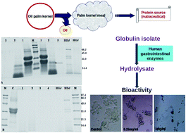
Globulins are the major seed storage proteins (41%) present in oil palm kernel. The study describes the ex vivo digestibility of globulin isolate prepared from oil palm kernel meal. The bioactivity and nutritional value of the ex vivo digest of globulin isolate was also investigated. Globulin isolate (85% protein content) was prepared by a salt extraction method and its digestibility was studied using human gastroduodenal juices (HGDJ). By SDS-PAGE analysis it was evident that the globulin isolate was completely digested by HGDJ. Easy digestibility of a dietary protein is essential from the nutritional point of view. In the present ex vivo study, the digestion profile and degree of hydrolysis of globulin isolate were observed to be comparable with that of casein. RADVFNPR and KLPLVERIP were the two peptides identified by MS/MS analysis in the final hydrolysate. By pepsinolysis, it was evident that the novel globulin protein preparation was devoid of pepsin resistant proteins that are otherwise considered to have allergenic potential. The ex vivo hydrolysate exhibited potent ACE-inhibitory activity (IC50 50 μg ml−1) and anticancer activity against human colon epithelial cancer HT-29 cells and hepatocarcinoma HepG2 cells. Easy digestibility, bioactivity, the presence of nutritionally important free essential amino acids and non-protein amino acids (γ-aminobutyric acid and citrulline) in the gastrointestinal digest suggests that the globulin isolate prepared from palm kernel meal can be used as a nutraceutical protein for food applications.

1. Introduction

Oil palm (Elaeis guineensis), one of the highest oil-yielding crops in the world, is a species of particular economic importance as it provides one of the most important sources of edible oil, the most cost-effective feedstock for biodiesel and also animal feed.1 Palm kernel meal (PKM) is the by-product obtained after extraction of oil from the oil palm kernels and is widely used as a protein source in animal diets.2 The proteins of palm kernels contain all the essential amino acids except lysine and tryptophan. The amino acid profiles of kernel proteins are dominated by glutamic acid, followed by arginine and aspartic acid.1 With an increasing demand for new alternative plant protein source for food application, PKM with 14–21% protein can be used as a new valuable dietary protein source.2,3 Information about extraction and production of palm kernel meal protein isolate is very limited. There are only few reports on extraction of protein from palm kernel meal by methods like alkali extraction methods/enzyme assisted methods.4,5 Though PKM contains a good amount of protein, there are not many reports on the potential of its usage as a protein source for human consumption.

Salt soluble seed storage proteins are commonly known as globulins. In non-cereal grains more nutritiously balanced salt soluble globulins are usually predominant storage proteins.6 The seed storage globulins from various sources make a significant contribution to the human diet. Most of the oil seeds like soybean, sunflower, sesame and groundnut contain globulin as the major seed storage protein. Protein rich oil seed meal was being utilized to recover globulin to be marketed as food ingredients in developed countries.6 During gastrointestinal digestion, a protein resistant to proteolytic digestion has increased probability of stimulating immune reactions.7 Many seed storage proteins like 2S albumins (Ara h2, Ara h6, Ses i1, Sin a1), 11S globulins (sesame, cashew nut, peanut) and 7S globulins (sesame, soybean, peanut) have been identified as allergens and most of these proteins are resistant to proteolytic digestion.7 Seed storage proteins have often been associated with specific reactivity towards IgE from patient serum and it is generally believed that digestive resistant protein can retain sufficient structural integrity to have an increased probability of stimulating immune response.7 Therefore, digestibility is a crucial predictive tool for a novel protein allergenicity assessment. In the present study, ex vivo digestion using human gastro-duodenal enzymes and in vitro digestion by commercial enzymes (pepsin and pancreatin) has been performed to study the digestibility of palm kernel globulin.

Dietary proteins can be precursors for many different bioactive peptides, which are usually released during gastrointestinal enzymatic digestion. These peptides otherwise are buried within the sequence of parent proteins in an inactive form. Food-derived bioactive peptides with health-enhancing property can be released during gastrointestinal digestion. There have been relatively few studies examining bioactive potentials of protein hydrolysate obtained after digestion using human digestive enzymes. These bioactive peptides generated must be resistant to the gastrointestinal environment until absorbed and transported to the target organs to impart bioactive properties. Therefore, demonstration of bioactivity is of particular importance with respect to human intestinal physiology as these bioactive molecules may be rendered inactive during the human digestive processing.8

Sreedhara & Kurup9 have reported that the globulin fraction from palm kernel prepared by salt extraction method exhibited hypocholesterolemic and antiatherogenic action in rats. The globulin isolate prepared from the oil palm kernel is a novel protein preparation and has not been used before for human consumption purpose. The human digestive juice is a better choice to study the digestibility of novel proteins from oil palm kernel. Usage of human gastric and duodenal juice for protein digestibility studies may provide a better knowledge about digestion of seed storage proteins which is essential with respect to novel seed proteins in human nutrition. The current study aims at the preparation of globulin isolate from oil palm kernel meal and studying its human gastrointestinal enzyme digestibility and bioactivity of the hydrolysate. Ex vivo digestibility studies may provide new insights into the utilization of palm kernel globulin isolate for human consumption. Therefore, the present study would give new value addition to the palm kernel meal (an underutilized by-product of palm oil milling industry) as a nutraceutical protein source.

2. Materials and methodology

2.1 Materials

Oil palm seeds (Elaeis guineensis var. tenera) were purchased from Ruchi Soya Industries Limited, Kabini Colony, Mysore, Karnataka, India. The palm kernel meal was prepared by defatting the kernel meal obtained after the oil extraction. Angiotensin-converting enzyme (ACE) from rabbit lung and ACE peptide inhibitor were procured from Sigma-Aldrich Chemicals Co. (St Louis, Mo, USA). Pepsin and pancreatin were purchased from HiMedia Laboratories PVT. Ltd (Mumbai, India). All other chemicals were of analytical grade. All the solutions were freshly prepared. The human colon epithelial cancer HT29 cells were obtained from the European Cell Culture Collection via the Public Health Laboratory Service (Porton Down, Wiltshire, UK). Hepatocarcinoma cells (HepG2) were a kind gift from Dr Milind Vaidya, ACTREC, Navi Mumbai, India. The aspiration of human contents (gastric and duodenal juices) were approved by the Norwegian Ethics Committee, and all volunteers signed an agreement before participating in the study.

2.2 Fractionation of oil palm kernel protein

Extraction and fractionation of defatted palm kernel meal were conducted according to the method described by Horax, Hettiarachchy, Over, Chen & Gbur.10 Distilled water, 1 M NaCl, 70% ethanol and 0.5 M NaOH were used consecutively to extract the protein. The defatted palm kernel meal was sequentially extracted by stirring, using the meal to solvent ratio of 1[thin space (1/6-em)]:[thin space (1/6-em)]10 (w/v), for 1 h. The insoluble residue was removed by centrifugation at 8000 rpm for 30 min. Extraction with each solvent was repeated three times, and all supernatants from each extract were pooled and the nitrogen content of the kernel meal was determined by the micro-Kjeldahl method.11

2.3 Preparation of globulin isolate (GI)

Oil palm kernel globulin isolate was prepared at pilot scale. All preparation procedures were performed at room temperature. Defatted oil palm kernel meal was dispersed in 10% (w/v) NaCl in 1[thin space (1/6-em)]:[thin space (1/6-em)]10 ratio and the slurry was stirred for 1 h. The slurry was passed through cheese cloth and then centrifuged (8000 rpm) to remove the insoluble material. The pH of the supernatant was adjusted to 4.5 with 2 M HCl, and the insoluble protein fraction was collected by centrifugation (8000 rpm, 30 min). The precipitate collected by centrifugation was dispersed in distilled water and neutralized to pH 7 with 2 M NaOH before freeze drying (Lyodryer-LT5S, lyophilization Systems Inc., USA). The total protein content in the globulin isolate was measured by the micro-Kjeldahl method.11

2.4 SDS-PAGE

Sodium dodecyl sulphate polyacrylamide gel electrophoresis (SDS-PAGE) was carried out for the visualization of protein bands of digested samples. The assay was performed on gels (5% stacking and 12% separating gels) according to the protocol as described by Laemmli.12 Electrophoresis was performed and the protein bands were stained with Coomassie Brilliant Blue R-250 to visualize the separated proteins and released fragments.

2.5 Digestibility of GI

Ex vivo digestibility of GI using human gastroduodenal juice was performed according to the method of Almaas et al.13 with slight modifications. Human gastric and duodenal juices were aspirated from healthy volunteers and the aspiration of human contents was approved by the Norwegian Ethics Committee. 5 ml protein solution (3%, w/w) was mixed with 2.5 ml of human gastric juice; the pH was adjusted to 2 and incubated at 37 °C. After 1 h incubation, the pH was adjusted to 7.5 and 2.5 ml of human duodenal juice was added, the pH was again checked for 7.5 and incubated for 2 h at 37 °C. Aliquots at different time intervals were collected to determine the degree of hydrolysis. Visualization of digestibility profile was done by SDS-PAGE. The final hydrolysate was used for free amino acid analysis and resistant peptide sequence identification.

The in vivo digestion of GI using commercial enzymes was performed according to the method described by Zhu, Chen, Tang & Xiong.14 Sequential digestion of GI was performed by pepsin (4% w/w) for 1 h followed by pancreatin (4% w/w) for 2 h at 37 °C. Aliquots were collected at the different time interval for the visualization of hydrolysis.

Pepsinolysis was performed according to the methodology prescribed by Thomas et al.15 A ratio of 10 U of pepsin activity per μg test protein was selected for all tests and the digestions were performed at pH 1.2 and 2. The digestibility profile was assessed by SDS-PAGE visualization.

2.6 Degree of hydrolysis

The degree of hydrolysis (DH) was determined according to the method described by De Castro & Sato16 with slight modification. 1 ml aliquot of the hydrolysate withdrawn at different time intervals was added to 1 ml 12% trichloroacetic acid (TCA). The mixture was centrifuged at 8000 rpm for 15 min. The protein content in TCA-soluble fraction and the hydrolysate (without the addition of TCA) were determined by Lowry's method17 and the DH value, expressed as a percentage, was calculated as the ratio of TCA-soluble protein to total protein in the hydrolysate.

2.7 Free amino acid estimation

Estimation of free amino acid in the hydrolysate was performed by the method described by Qureshi, Vegarud, Abrahamsen & Skeie.18 5 g of sample (hydrolysate) was mixed with 15 ml 0.1 M HCl containing 0.4 μmol ml−1 L-norvalin and 0.4 μmol ml−1 piperidine-4-carboxylic acid as internal standards. After mixing for 15 min, centrifugation (40 min, 3400 rpm, 4 °C) was carried out and 0.5 ml of the supernatant was added to 0.5 ml 4% TCA. After mixing on vortex, the sample was placed on ice for 30 min and centrifuged (5 min, 13[thin space (1/6-em)]000 rpm, 4 °C). The sample was filtered with 0.2 μm MFS-13 CA filter and stored in the freezer until use. The separation of the free amino acids was performed using RP-HPLC.

2.8 Peptide identification by mass spectrometry

Peptide identification was performed according to the method described by Almaas et al.13 In brief, freeze-dried hydrolysates were dissolved in 0.1% (v/v) formic acid (FA). The samples were concentrated and desalted by self-made columns consisting of C18 column material (3 M Empore C18 extraction discs; 3 M Bioanalytical Technologies, St Paul, MN, USA) inserted into micropipette tips. The peptides were eluted using 2 μl 70% acetonitrile −0.1% formic acid (v/v) and eluted samples were diluted in 1% formic acid before loading onto nano LC-MS and subjected to data-dependent tandem MS analysis. The processed data were searched against National Center for Biotechnology Information (NCBI) non-redundant protein sequence databases using the Mascot protein identification tool (http://www.matrixscience.com) for identification of proteins. The fragment mass tolerances were 0.1 Da.

2.9 ACE-inhibition activity

The ACE inhibitory activity was assayed according to the modified method described by Rao, Sun, Liu, Zeng, Su & Yang.19 100 μl sample was incubated with 100 μl (1 unit per ml) of ACE solution (3.3 units per mg) and 100 μl of 5 mM HHL (hippuryl-L-histidyl-L-leucine) in 50 mM sodium borate buffer at pH 8.2 containing 0.3 M NaCl. After incubation at 37 °C for 60 min, the reaction was stopped by adding 300 μl of 1 N HCl. The liberated hippuric acid was extracted with 1 ml of ethyl acetate, dried and re-extracted in 1 ml distilled water. The absorbance at 228 nm was measured to evaluate ACE inhibitory activity. The percent inhibition was measured by the formula: [100 − (absorbance of the sample × 100)/(absorbance of the control)].

2.10 Anticancer activity

The anticancer activity was measured by MTT assay. HT29 and HepG2 cells were cultured in Dulbecco's modified Eagle's medium (DMEM) supplemented with 10% FBS, 100 units per ml penicillin, 100 μg ml−1 streptomycin at 37 °C in 5% CO2. Sub confluent cells HT-29 and HepG2 were seeded 1 × 10⁴ cells per ml in 96 well plates in DMEM with 10% FBS (complete medium) for 48 h. After 48 h of incubation, the growth medium was replaced with DMEM containing 0.5% FBS with various concentrations of hydrolysate (0.25 to 4 mg ml−1) and then cells were incubated for 72 h at 37 °C. At the end of 72 h incubation, the anti-cancer activity was quantified by determining the cell viability, based on the ability of metabolically active cells to reduce the yellow dye MTT to an insoluble blue formazan product. The absorbance was measured using Tecan i-200 microplate reader at 595 nm.

2.11 Statistical analysis

For all the measurements, a minimum of triplicate values were taken for data analysis. All the values were plotted using the Origin 6.1 software. Data were expressed as mean ± standard deviations. One way analysis of variance (ANOVA) was employed to identify significant differences (p < 0.05) between data sets using Origin 6.1.

3. Results and discussion

3.1 Distribution of protein fractions

The oil palm kernel and palm kernel meal after defatting contains 8% and 16% protein respectively. The palm seed storage proteins were mostly soluble proteins and the sum of the protein in the soluble protein fractions amounted to 89% (w/w) of the total protein content. As shown in Fig. 1A the globulins are the major predominant protein fraction in palm kernel accounting to 41% of the total protein, followed by glutelin (19%), albumin (17%) and prolamin (11%). Kwan, Park & Rhee,20 opines that fractionation of plant proteins on the basis of solubility is only an approximation of the actual protein composition and therefore, differences in protein extraction conditions and determination may lead to different values. Previous studies report that globulin and glutelin are the predominant protein fractions in the oil palm kernel.21,22 Oil palm and coconut both being commercially valuable trees belong to same Arecaceae family. Like oil palm, coconut also contains globulin as the major seed storage protein accounting to 40–60% of the total protein.20,23
image file: c5ra25232f-f1.tif
Fig. 1 (A) Distribution (%) of protein fractions in oil palm kernel meal. The protein fractions of defatted kernel meal are given according to their solubility. Effect of pH (B) and NaCl concentration (C) on protein extractability (%) from oil palm kernel meal.

3.2 Globulin isolate (GI)

The protein extractability in water (1[thin space (1/6-em)]:[thin space (1/6-em)]10 meal to water ratio) at different pH (2–10) suggests that the overall protein extractability is less in water irrespective of the pH conditions used (Fig. 1B). The extractability of globulin fraction from PKM depends on the salt concentration used. Maximum extractability was observed at 10% NaCl (1[thin space (1/6-em)]:[thin space (1/6-em)]10 meal to salt ratio) concentration, beyond which there was a decreasing trend in extractability (Fig. 1C). This decreasing trend is due to the abundance of the salt ions at higher salt concentration, which reduces the solubility of protein.4 Globulins being the predominant protein in palm kernel meal, globulin isolate can be prepared by salt extraction method. The GI prepared by this method contains 85% protein content. Our findings were in line with the previous study by Arifin et al.4 who reported that salt extraction method was most suitable for PKM protein extraction. Sodium hydroxide treatment denatures the protein, and it is only suitable for low NaOH concentration (low pH).4 Extraction of palm kernel protein by high alkali and enzyme assisted methods are reported by previous studies.5,24 Recently, Zheng, Li, Zhang, Zhang, Zhang & Zhao,25 reported on using NaCl in dilute alkaline solution (pH 9) for extraction of palm kernel protein.

3.3 Digestibility of GI

The present work mimics the in vivo situation focused on the in vitro digestibility of globulin isolate by human enzymes. Human gastric and duodenal juices from healthy volunteers were taken as a source of proteolytic enzyme for this study. The degradation profile of palm kernel globulin after sequential treatment with human gastric juice (HGJ) and duodenal juice (HDJ) was observed by SDS-PAGE. From the Fig. 2A, it was evident that the digestion with HGJ showed major degradation of globulin proteins except the 21 kDa protein band indicated with an arrow. After this first degradation step, digestion of globulin seemed to produce lower molecular weight products (Fig. 2A). During the second enzymatic step, 21 kDa protein band and the lower molecular weight fragments from HGJ digest were further digested by HDJ and all the bands were vanished, suggesting complete degradation of globulin. The digestibility profile of casein digestion by the human gastrointestinal enzyme is as given in Fig. 2B. The digestibility of GI is comparable with easily digestible protein, casein. The degree of hydrolysis of globulin isolate and casein was almost same with no significant difference reaching 59% and 53% respectively at the end of HGJ digestion step and reached more than 90% at the end of HDJ digestion (Fig. 3). The sequential digestion of palm globulins by HGJ followed by HDJ resulted in complete hydrolysis. Orruno & Morgan,7 opines that the complete digestion is attributed to enhanced proteolytic susceptibility of the protein due to conformational changes and increased access to enzymes during digestion.
image file: c5ra25232f-f2.tif
Fig. 2 (A) SDS-PAGE pattern of globulin isolate (GI) sequentially digested with human gastric and duodenal enzymes. Lane M-marker, Lane HDJ-human duodenal juice, Lane HGJ-human gastric juice, Lane 1-unhydrolysed GI, Lane 2-GI hydrolyzed with HGJ, Lane 3-GI hydrolyzed with HGJ followed by HDJ. The arrow indicates the pepsin resistant protein band. (B) SDS-PAGE of casein sequentially digested with human gastric and duodenal enzymes. Lane M-Marker, Lane HDJ-human duodenal juice, Lane HGJ-human gastric juice, Lane C-unhydrolysed casein, Lane 1 & 2-casein hydrolyzed with HGJ, Lane 3 & 4 – casein hydrolyzed with HGJ followed by HDJ.

image file: c5ra25232f-f3.tif
Fig. 3 The degree of hydrolysis of globulin isolate and casein after in vitro sequential digestion by human gastrointestinal enzymes (HGJ-human gastric juice, HDJ-human duodenal juice). Values were expressed as mean values ± SD and error bars were the standard deviations of the values. ** Denotes that the differences are not statistically significant.

The sequential hydrolysis of globulin isolate by commercial enzymes, pepsin (first step) at pH 2 followed by pancreatin (second step) at pH 7.5 results in complete hydrolysis of globulin isolate as evident by SDS-PAGE (Fig. 4A). The pattern observed in the hydrolysis of globulin isolate by using HGJ–HDJ is same as that observed in in vitro hydrolysis performed by using commercial enzymes (pepsin–pancreatin). Similarly, Kopper, Odum, Sen, Helm, Stanley & Burks,26 reported that the in vitro digestion with pepsin and porcine gastric fluid of peanut protein allergen (Ara h1) resulted in similar hydrolysis patterns. The low molecular weight products observed after hydrolysis with human gastric enzymes were not found in hydrolysates obtained after commercial pepsin digestion. According to Eriksen et al.27 commercial porcine enzymes (pepsin or pancreatic enzymes) appear to digest whey proteins more efficiently compared to human digestive juices and this could lead to conflicting results after comparing commercial pepsin–pancreatic enzymatic digestion with human in vivo protein digestion when used at similar enzyme activities.


image file: c5ra25232f-f4.tif
Fig. 4 (A) SDS-PAGE pattern of sequential in vitro digestibility profile of globulin isolate (GI) by commercial enzymes pepsin and pancreatin. Lane 1-unhydrolyzed GI, Lane 2-GI hydrolyzed with pepsin (0th min), Lane 3-GI hydrolyzed with pepsin (30th min), Lane 4-GI hydrolyzed with pepsin (1 h), Lane 5-GI hydrolyzed with pepsin (1 h) followed by pancreatin (1 h), Lane 6-GI hydrolyzed with pepsin (1 h) followed by pancreatin (2 h). The arrow indicates the pepsin resistant protein band. SDS-PAGE pattern of the pepsinolysis of GI with pepsin at pH 1.2 (B) & pH 2 (C) respectively. Lane 1-unhydrolysed GI, Lane 2-GI hydrolyzed with pepsin (0th min), Lane 3-GI hydrolyzed with pepsin (1 h), Lane 4-pepsin. Arrow indicates the pepsin bands.

Earlier, human gastrointestinal enzymes were used to study the in vitro digestibility of milk proteins like caprine whey protein,13,27 lactoferrin,28 casein and whey protein in equine, bovine, caprine and human milk.29 The human digestive juice is a better choice to study the digestibility of novel proteins. The digestive system contains all the essential components for the digestion of the food components; thus the use of human digestive juices might be preferred for digestibility studies. Although this is the first study on digestibility of oil palm seed storage protein using human gastrointestinal enzymes, further research is essential in this regard with respect to other seed storage proteins.

According to the Codex Alimentarius Commission-2003 a “weight of evidence” approach is recommended for the assessment of potential allergenicity of novel proteins and this approach includes an in vitro pepsin digestion assay.15 The digestibility of novel proteins in simulated gastric fluid is considered to be an indicator of reduced risk of allergenic potential in food.30 There are many reports, on food allergens which are resistant to pepsin digestion, on non-allergenic proteins which are pepsin resistant and as well as on allergenic proteins sensitive to pepsin digestion.15 Pepsin resistant 21 kDa protein band was observed in both human gastrointestinal enzymatic digestion and commercial pepsin–pancreatic digestion methods. Most of the allergenic proteins remain resistant to pepsin digestion even if the pepsin to protein concentration is increased up to 1[thin space (1/6-em)]:[thin space (1/6-em)]5 ratio.7 The palm globulin isolate was subjected to pepsinolysis to check for the presence of any pepsin resistant proteins according to the methodology prescribed by Thomas et al.15 Hydrolysis of GI was performed at both the pH 1.2 (Fig. 4B) and pH 2 (Fig. 4C) as recommended for in vitro pepsin digestion assay. From the Fig. 4B and C it is evident that the globulin isolate is not resistant to pepsin and the 21 kDa protein band was not observed after pepsinolysis.

3.4 Free amino acid and peptide analysis

The free amino acid composition of the hydrolysate obtained after the human gastrointestinal enzymatic digestion is summarized in Table 1. Arginine (28%) is the most abundant free amino acid detected followed by phenylalanine (13.59%) leucine (13.3%) and valine (12.69%). The presence of essential amino acid content suggests the good nutritive quality of the globulin isolate. The percentage ratio of essential to total amino acids (E/T%) is 59%, which is necessary for an ideal protein to meet the amino acid requirements. Non-polar aliphatic (34.7%) and positively charged (33.9%) amino acids are the most abundant present in free form followed by non-polar aromatic (20.5%), polar amino acids (10.1%) and negatively charged (0.6%) amino acids. Our findings were in line with the previous study by Zheng et al.21 who have reported globulin to be rich in arginine. Arginine and leucine were the most abundant amino acids found in both tenera and clonal palm kernel proteins.1 Along with the free amino acids, 0.015 μmol g−1 of citrulline and GABA (γ-aminobutyric acid) were detected. Citrulline is an amino acid that is not involved in protein synthesis but is tightly linked to arginine metabolism and it also stimulates muscle protein synthesis (MPS) in a post-prandial and post-absorptive state.31,32 GABA (γ-aminobutyric acid, a inhibitory neurotransmitter) is a non-protein amino acid with antidiabetic, diuretic, hypotensive, tranquilizing and antiproliferative effect used considerably in pharmaceuticals and in foods with health beneficial effect.33
Table 1 The free amino acid composition of the palm globulin and casein hydrolysate prepared by digesting with human gastrointestinal enzymes
Free amino acid μmol g−1 hydrolysate (palm) % of total amino acid μmol g−1 hydrolysate (casein) % of total amino acid
a Non-protein amino acids.
L-Aspartic 0.09 0.18 0.07 0.11
L-Glutamic 0.22 0.42 0.18 0.29
L-Asparagine 0.50 0.74 0.19 0.30
L-Serine 0.67 0.93 0.44 0.68
L-Glutamine 3.05 4.89 4.83 7.39
L-Threonine 0.78 1.29 1.06 1.62
L-Methionine 1.32 2.33 1.62 2.49
L-Histidine 0.55 0.92 0.37 0.57
L-Arginine 14.83 28.06 7.27 11.11
L-Lysine 2.52 4.95 10.64 16.27
L-Glycine 0.35 0.47 0.26 0.40
L-Alanine 2.62 4.62 2.62 1.82
L-Valine 6.44 12.69 8.52 13.03
L-Isoleucine 2.03 3.69 1.16 1.78
L-Leucine 6.86 13.31 9.91 15.15
L-Tyrosine 3.17 6.09 5.41 8.28
L-Phenylalanine 6.72 13.59 10.17 15.55
L-Tryptophan 0.44 0.84 2.07 3.17
L-Citrullinea 0.015   0.048  
L-Ornithinea   0.079  
γ-Aminobutyric acida 0.015    


The sequences of the resistant peptides present in the GI and casein hydrolysate obtained after the human gastrointestinal enzymatic digestion were identified by MS/MS analysis (Table 2). Only two resistant peptides RADVFNPR and KLPLVERIP were identified in the GI hydrolysate (Fig. 5). Both the peptides were analyzed by BIOPEP database34 to get information regarding the presence of any allergenic potential. The allergenic epitopes were absent in these two peptides. It can be opined that the novel globulin isolate prepared from the oil palm kernel can be considered as safe to be used for the human consumption purpose as there are no digestion resistant peptides released with the allergenic epitope.

Table 2 Peptide sequences identified by MS/MS analysis in palm globulin and casein hydrolysate prepared by digesting with human gastrointestinal enzymesa
Peptide sequences from palm globulin hydrolysate
a The peptide sequences were searched against NCBI database for oil palm and UNIPROT database for casein.
Protein name: glutelin (Elaeis guineensis)
RADVFNPR
[thin space (1/6-em)]
Protein name: histone-lysine N-methyltransferase EZ3
KLPLVERIP

Peptide sequences from commercial casein hydrolysate
Protein name: CASA2_BOVIN
ITVDDKHY, ITVDDKHYQ, KITVDDKHYQ, KTVDMESTEV, PIVLNPWDQ, QGPIVLNPWDQ, QGPIVLNPWDQV, YQGPIVLNPWDQ, YQGPIVLNPWDQV
[thin space (1/6-em)]
Protein name: G5E5H7_BOVIN, LACB_BOVIN
TPEVDDEALEK, TPEVDDEALEKFD, VEELKPTPEGDL, VEELKPTPEGDLE, VEELKPTPEGDLEI, VYVEELKPTPEGDLE, YVEELKPTPEGD, YVEELKPTPEGDL, YVEELKPTPEGDLE, YVEELKPTPEGDLEI
[thin space (1/6-em)]
Protein name: CASA1_BOVIN
FFVAPFPEV, FFVAPFPEVFG, FVAPFPEV, FVAPFPEVF, FVAPFPEVFG, FVAPFPEVFGK, GIHAQQKEPM, HIQKEDVPSE, HQGLPQEV, HQGLPQEVL, KEGIHAQQKEPM, NENLLRFFVAPFPEVFG, SDIPNPIGSENSE, SDIPNPIGSENSEK, VPQLEIVPN, YKVPQLEIV
[thin space (1/6-em)]
Protein name: tr|E1BBK7|E1BBK7_BOVIN-DECOY
LHPVLPLP, LLHPVLPLP
[thin space (1/6-em)]
Protein name: ALBU_BOVIN
FYAPELLYYANK, KVPQVSTPTLVEVSR, LVNELTEFAK, RHPEYAVSVLLR, YNGVFQECCQAEDK
[thin space (1/6-em)]
Protein name: sp|P81265|PIGR_BOVIN
KSPIFGPEEV, KSPIFGPEEVT
[thin space (1/6-em)]
Protein name: CASB_BOVIN
DVENLHLPLP, DVENLHLPLPL, DVENLHLPLPLL, DVENLHLPLPLLQ, DVENLHLPLPLLQS, GPVRGPFPI, HKEMPFPK, HQPHQPLPPT, HQPHQPLPPTV, HQPHQPLPPTVMFPP, HQPHQPLPPTVMFPPQ, HQPHQPLPPTVMFPPQS, HQPHQPLPPTVMFPPQSV, LEELNVPGEIVE, LGPVRGPFPI, LTDVENLH, LTDVENLHLPLP, LTDVENLHLPLPL, LTLTDVENLH, LTLTDVENLHLP, LTLTDVENLHLPLP, LTLTDVENLHLPLPL, LVYPFPGPI, LVYPFPGPIPN, LYQEPVLGPV, LYQEPVLGPVRGPFPI, MHQPHQPLPPT, MHQPHQPLPPTV, MHQPHQPLPPTVM, MHQPHQPLPPTVMFPP, MHQPHQPLPPTVMFPPQ, MHQPHQPLPPTVMFPPQS, MHQPHQPLPPTVMFPPQSV, NIPPLTQTPVVVPPFLQPE, NIPPLTQTPVVVPPFLQPEV, PPLTQTPVVVPPFLQPEVMGVSKVKE, PQNIPPLTQT, PQNIPPLTQTPVVVPPFLQPE, PQNIPPLTQTPVVVPPFLQPEV, PQNIPPLTQTPVVVPPFLQPEVMG, PVEPFTESQS, PVVVPPFL, PVVVPPFLQ, PVVVPPFLQP, PVVVPPFLQPE, PVVVPPFLQPEV, PVVVPPFLQPEVM, PVVVPPFLQPEVMG, PVVVPPFLQPEVMGV, PVVVPPFLQPEVMGVS, QEPVLGPVRGPFPI, QEPVLGPVRGPFPII, QEPVLGPVRGPFPIIV, QPHQPLPPTVMFPPQS, QPLPPTVMFPPQ, QPLPPTVMFPPQS, QPLPPTVMFPPQSV, QSLVYPFPGPI, QSLVYPFPGPIPN, QTEDELQDKIHP, QTEDELQDKIHPF, RELEELNVPGEI, TDVENLHLPLPL, TDVENLHLPLPLLQ, TEDELQDKI, TEDELQDKIHP, TEDELQDKIHPF, TLTDVENLH, TLTDVENLHLPLP, TLTDVENLHLPLPL, TPVVVPPFLQPE, TPVVVPPFLQPEV, TPVVVPPFLQPEVMG, TQSLVYPFPGPI, TQSLVYPFPGPIPN, VENLHLPLP, VENLHLPLPL, VYPFPGPI, YQEPVLGPV, YQEPVLGPVRGPF, YQEPVLGPVRGPFP, YQEPVLGPVRGPFPI, YQEPVLGPVRGPFPIIV, ALINNQFLPYP, KNQDKTEIPT, KNQDKTEIPTIN, KNQDKTEIPTINT, KNQDKTEIPTINT, RELEELNVPGEIVE, RELEELNVPGEIVESL, RELEELNVPGEIVESLS, SLPQNIPPLTQTPVVVPPFLQPE, SLTLTDVENLHLPLP, SLTLTDVENLHLPLPL, SLVYPFPGPI, SLVYPFPGPIP, SLVYPFPGPIPN, SLVYPFPGPIPNS, SLVYPFPGPIPNSLPQ, TDVENLHLPLP


image file: c5ra25232f-f5.tif
Fig. 5 ESI-MS/MS spectrum of human gastrointestinal enzyme resistant peptides (A) RADVFNPR and (B) KLPLVERIP identified in globulin hydrolysate.

Dietary proteins are digested into their constituent free amino acids/oligopeptides before absorption in gastrointestinal tract. Intestinal absorption of biologically active peptides as well as proteins has been reported before and is considered to be a normal physiological process in humans and animals.35–39 Both the resistant peptides were further analyzed for bioactivity in BIOPEP database.40 Both the peptides are shown to exhibit ACE-inhibitory activity and dipeptidyl inhibitory activity. ACE-inhibitors are used to treat hypertension and dipeptidyl peptidase-4 inhibitors are used as oral antihyperglycemic agents to treat diabetes mellitus type-2. Presence of these probable bioactive peptides in the hydrolysate with antihypertensive and hypoglycemic activity suggests its potent nutraceutical applicability.

3.5 Bioactivity of GI hydrolysate

The bioactivities like ACE inhibition and anticancer activity of dietary protein hydrolysates has been well-documented in the literature, but relatively few studies have been conducted to evaluate their activity after digesting with human gastrointestinal enzymes. An effective protein exerting bioactivity like ACE-inhibition and anticancer activity after getting digested in our gastrointestinal tract is greatly desirable, as it can be used directly as dietary protein/food additive against hypertension and to reduce the risk of developing cancer. In the present study, the GI hydrolysate obtained after human gastrointestinal enzymatic digestion was checked for its bioactivities like anticancer activity and ACE-inhibitory activity.
3.5.1 Anticancer activity. The goal of the present study was to investigate the effect of GI hydrolysate generated as a result of human physiological digestion on the cancer cells. Food derived proteins/peptides are known to aid in cancer prevention and treatment. The focus is towards elucidating anti-colon cancer and anti-liver cancer properties of hydrolysate produced during digestion, which can exhibit its activity in the gastrointestinal tract as well after absorption and thus may aid to reduce the risk of developing cancer. In this study, human colon (HT-29) and hepatic (HepG2) cancer cell lines were chosen to determine the anticancer activity of the GI hydrolysate. The colon and hepatic cancer cells were treated with different concentration of GI hydrolysate and the viability were assessed. Fig. 6A shows MTT-based cell viability assay results which confirm inhibitory actions of the hydrolysate on colon and liver cancer cells, respectively. The anticancer ability of hydrolysate against cancer cells increased in a dose dependent manner. Exposure of hydrolysate inhibited HT-29 (IC50 0.65 mg) and HepG2 (IC50 1.25 mg) cell proliferations by 87% and 83% respectively when the cells were exposed to 4 mg ml−1 (protein basis) hydrolysate. From Fig. 6B, it is clearly evident that the hydrolysate is a potent cancer cell growth inhibitor. Numerous studies on plant and animal protein hydrolysates have been reported to exhibit anticancer activity. Resistant peptides present in gastrointestinal enzyme digest can have actual biological activity in terms of bioavailability and delivery.41 The observed activity could be related to the anticancer activity of peptides present in hydrolysate exerting a cytotoxic effect on cancer cells, as previously reported for a haemoglobin derived peptide42 or for an anchovy hydrophobic peptide.43 These evidences on anticancer activity of the GI hydrolysate suggests that the palm kernel globulin isolate has the potential as a human antitumor bioactive agent and may be considered as a promising bioactive food component.
image file: c5ra25232f-f6.tif
Fig. 6 (A) Anticancer activity of human gastrointestinal enzyme digested globulin hydrolysate at different concentrations against human colon (HT-29) and hepatic (HepG2) cancer cells. (B) The picture depicts the untreated (control) HT-29 cells and anticancer effect on HT-29 cells at 72 h treated with 0.25 mg ml−1 & 3 mg ml−1 hydrolysate. (C) ACE inhibitory activity of human gastrointestinal enzyme digested globulin hydrolysate at different concentrations.
3.5.2 ACE inhibitory activity. To verify whether ACE-inhibitory peptides are generated by gastrointestinal digestion, the hydrolysate was assayed for potent ACE-inhibitory activity. The oil palm kernel hydrolysate exhibited significant ACE-inhibitory activity (85%) at the concentration of 0.2 mg ml−1 (Fig. 6C). The IC50 value of hydrolysate and standard inhibitor (ACE peptide inhibitor) was 50 μg ml−1 and 2 μg ml−1 respectively. Presence of ACE inhibition in hydrolysate is consistent with the presence of hydrophobic (aromatic or branched amino acids), positively charged amino acids and proline at the C-terminal ends of the peptides identified in the hydrolysate, which are necessary for binding ACE to exert inhibition.44 Both the identified gastroduodenal enzyme resistant peptides RADVFNPR and KLPLVERIP contain the above-mentioned amino acids at the C-terminal end. To exert antihypertensive effect, the gastrointestinal digestive resistant peptides have to be absorbed into the bloodstream.19 It is likely that these peptides generated during human gastrointestinal enzymatic digestion of palm kernel globulin can be absorbed to exhibit the antihypertensive effect. Further research in this regards using animal studies has to be carried out with the synthesized peptides.

4. Conclusions

Oil palm kernel contains globulin as the major seed storage protein. The globulin isolate (GI) can be prepared by salt extraction method. The globulin protein isolate is easily digestible and not resistant to digestion by human gastrointestinal enzymes and commercial digestive enzymes. The present study on ex vivo digestion by human enzymes will provide better knowledge about the degradation of palm kernel globulin isolate in the digestive system that would be relevant for the development of easily digestible products for human food applications. The promising bioactivity of globulin hydrolysate obtained by digesting with human gastrointestinal enzymes suggests its potential use as a novel nutraceutical ingredient. From the results obtained it can be concluded that palm kernel globulin could be used as a health-beneficial dietary protein. Further research on purification and identification of oil palm kernel globulins is needed for better understanding of palm seed storage protein. As a future plan of work, the peptides identified would be synthesised and checked for bioavailability.

Acknowledgements

The authors greatly acknowledge financial support in the form of Indo-Norwegian DST-RCN jointly funded project awarded by Department of Science and Technology, Government of India. The authors thank Dr Milind Vaidya, ACTREC, Navi Mumbai, India for providing hepatocarcinoma cells (HepG2) to carry out the work. We thank the Director, CSIR-CFTRI, Mysore, India for providing the facilities to carry out the work in the Institute.

References

  1. S. Kok, O. M. Abdullah, G. E. Chenglian and P. Namasivayam, Comparison of nutrient composition in kernel of tenera and clonal materials of oil palm (Elaeis guineensis Jacq.), Food Chem., 2011, 129, 1343–1347 CrossRef CAS.
  2. A. R. Alimon, The nutritive value of palm kernel cake for animal feeds, Palm Oil Dev., 2004, 40, 12–14 Search PubMed.
  3. M. Boateng, D. B. Okai, J. Baah and A. Donkoh, Palm kernel cake extraction and utilisation in pig and poultry diets in Ghana, Livest. Res. Rural Dev., 2008, 20(7), 1–8 Search PubMed.
  4. B. Arifin, A. Bono, Y. Y. Farm, A. L. L. Ling and S. Y. Fui, Protein extraction from palm kernel meal, J. Appl. Sci., 2009, 9(17), 2996–3004 CrossRef CAS.
  5. K. I. Chee, H. K. Ling and M. K. Ayob, Optimization of trypsin-assisted extraction, physico-chemical characterization, nutritional qualities and functionalities of palm kernel cake protein, LWT--Food Sci. Technol., 2012, 46(2), 419–427 CrossRef CAS.
  6. M. F. Marcone, Y. Kakuda and Y. R. Yada, Salt soluble seed globulins of various dicotyledonous and monocotyledonous plants-I. Isolation/purification and characterization, Food Chem., 1998, 62(1), 27–47 CrossRef CAS.
  7. E. Orruno and M. R. A. Morgan, Resistance of purified seed storage proteins from sesame (Sesamum indicum L.) to proteolytic digestive enzymes, Food Chem., 2011, 128, 923–929 CrossRef CAS.
  8. B. Nandhini and M. Palaniswamy, Anticancer effect of goat milk fermented by Lactobacillus plantarum and Lactobacillus paracasei, Int. J. Pharm. Pharm. Sci., 2013, 5(3), 898–901 Search PubMed.
  9. N. Sreedhara and P. A. Kurup, Studies on the effect of globulin fraction isolated from palm kernel on the metabolism of glycosaminoglycans in rats, Nutr. Res., 1992, 12(1), S129–S135 CrossRef CAS.
  10. R. Horax, N. Hettiarachchy, K. Over, P. Chen and E. Gbur, Extraction, fractionation and characterization of bitter melon seed proteins, J. Agric. Food Chem., 2010, 58, 1892–1897 CrossRef CAS PubMed.
  11. Association of Official Analytical Chemists, Official methods of analysis, Gaithersberg, 18th edn, 2005 Search PubMed.
  12. U. K. Laemmli, Cleavage of structural proteins during the assembly of the head of bacteriophage T4, Nature, 1970, 227, 680–685 CrossRef CAS PubMed.
  13. H. Almaas, E. Eriksen, C. Sekse, I. Comi, R. Flengsrud, H. Holm, E. Jensen, M. Jacobsen, T. Langsrud and G. E. Vegarud, Antibacterial peptides derived from caprine whey proteins, by digestion with human gastrointestinal juice, Br. J. Nutr., 2011, 106, 896–905 CrossRef CAS PubMed.
  14. L. Zhu, J. Chen, X. Tang and Y. L. Xiong, Reducing, radical scavenging and chelation properties of in vitro digests of alcalase-treated zein hydrolysate, J. Agric. Food Chem., 2008, 56(8), 2714–2721 CrossRef CAS PubMed.
  15. K. Thomas, M. Aalbers, G. A. Bannon, M. Bartels, R. J. Dearman, D. J. Esdaile, T. J. Fu, C. M. Glatt, N. Hadfield, C. Hatzos, S. L. Hefle, J. R. Heylings, R. E. Goodman, B. Henry, C. Herouet, M. Holsapple, G. S. Ladics, T. D. Landry, S. C. MacIntosh, E. A. Rice, L. S. Privalle, H. Y. Steiner, R. Teshima, R. Van Ree, M. Woolhiser and J. Zawodny, A multi-laboratory evaluation of a common in vitro pepsin digestion assay protocol used in assessing the safety of novel proteins, Regul. Toxicol. Pharmacol., 2004, 39, 87–98 CrossRef CAS PubMed.
  16. R. J. S. De Castro and H. H. Sato, Antioxidant activities and functional properties of soy protein isolate hydrolysates obtained using microbial proteases, Int. J. Food Sci. Technol., 2014, 49, 317–328 CrossRef CAS.
  17. O. H. Lowry, N. J. Rosebrough, A. L. Farr and R. J. Randall, Protein measurement with the folin phenol reagent, J. Biol. Chem., 1951, 193, 265–275 CAS.
  18. T. M. Qureshi, G. E. Vegarud, R. K. Abrahamsen and S. Skeie, Characterization of the Norwegian autochthonous cheese gamalost and its angiotensin I-converting enzyme (ACE) inhibitory activity during ripening, Dairy Sci. Technol., 2012, 92(613), 625 Search PubMed.
  19. S. Rao, J. Sun, Y. Liu, H. Zeng, Y. Su and Y. Yang, ACE inhibitory peptides and antioxidant peptides derived from in vitro digestion hydrolysate of hen egg white lysozyme, Food Chem., 2012, 135, 1245–1252 CrossRef CAS PubMed.
  20. K. Kwan, K. H. Park and K. C. Rhee, Fractionation and Characterization of Proteins from Coconut (Cocos nucifera L.), J. Agric. Food Chem., 1996, 44, 1741–1745 CrossRef.
  21. Y. Zheng, Y. Li, Y. Zhang, R. Zhang, Q. Zhang, Y. Zhang and S. Zhao, Fractionation, physicochemical properties, nutritional value, antioxidant activity and ACE inhibition of palm kernel expeller protein, RSC Adv., 2015, 5, 12613–12623 RSC.
  22. S. K. Chang, A. Ismail, T. Yanagita, M. N. Esa and M. T. H. Baharuldin, Biochemical characterisation of the soluble proteins, protein isolates and hydrolysates from oil palm (Elaeis guineensis) kernel, Food Biosci., 2014, 7, 1–10 CrossRef CAS.
  23. S. J. A. S. Samson, R. N. Khaund, C. M. Cater and K. F. Mattil, Extractability of coconut proteins, J. Food Sci., 1971, 36, 725–728 CrossRef.
  24. O. O. Balogun, Extraction of nitrogen from palm kernel meal and evaluation of digestibility of protein isolate from the meal by in vitro method, J. Agric. Food Chem., 1982, 30(6), 1163–1166 CrossRef CAS.
  25. Y. Zheng, Y. Li, Y. Zhang, R. Zhang, Y. Zhang and S. Zhao, Effects of limited enzymatic hydrolysis, pH, ionic strength and temperature on physicochemical and functional properties of palm (Elaeis guineensis Jacq.) kernel expeller protein, J. Food Sci. Technol., 2015, 52(11), 6940–6952 CrossRef CAS.
  26. R. A. Kopper, J. N. Odum, M. Sen, R. M. Helm, J. S. Stanley and A. W. Burks, Peanut protein allergens: gastric digestion is carried out exclusively by pepsin, J. Allergy Clin. Immunol., 2004, 114(3), 614–618 CrossRef CAS PubMed.
  27. E. K. Eriksen, H. Holm, E. Jensen, R. Aaboe, T. G. Devold, M. Jacobsen and G. E. Vegarud, Different digestion of caprine whey proteins by human and porcine gastrointestinal enzymes, Br. J. Nutr., 2010, 104, 374–381 CrossRef CAS PubMed.
  28. C. B. Furlund, E. K. Ulleberg, T. G. Devold, R. Flengsrud, M. Jacobsen, C. Sekse, H. Holm and G. E. Vegarud, Identification of lactoferrin peptides generated by digestion with human gastrointestinal enzymes, J. Dairy Sci., 2013, 96(1), 75–88 CrossRef CAS PubMed.
  29. R. A. Inglingstad, T. G. Devold, E. K. Eriksen, H. Holm, M. Jacobsen, K. H. Liland, E. O. Rukke and G. E. Vegarud, Comparison of the digestion of caseins and whey proteins in equine, bovine, caprine and human milks by human gastrointestinal enzymes, Dairy Sci. Technol., 2010, 90, 549–563 CrossRef CAS.
  30. R. A. Herman, V. A. Korjagin and B. W. Schafer, Quantitative measurement of protein digestion in simulated gastric fluid, Regul. Toxicol. Pharmacol., 2005, 41, 175–184 CrossRef CAS PubMed.
  31. G. Ventura, P. Noirez, D. Breuillé, J. P. Godin, S. Pinaud, M. Cleroux, C. Choisy, S. Le Plénier, V. Bastic, N. Neveux, L. Cynober and C. Moinard, Effect of citrulline on muscle functions during moderate dietary restriction in healthy adult rats, Amino Acids, 2013, 45, 1123–1131 CrossRef CAS PubMed.
  32. C. Moinard and L. Cynober, Citrulline: a new player in the control of nitrogen homeostasis, J. Nutr., 2007, 137(6), 1621S–1625S CAS.
  33. R. Dhakal, V. K. Bajpai and K. H. Baek, Production of GABA (γ-aminobutyric acid) by microorganisms: a review, Braz. J. Microbiol., 2012, 43, 1230–1241 CrossRef CAS PubMed.
  34. M. Dziuba, P. Minkiewicz and M. Dabek, Peptides, specific proteolysis products as molecular markers of allergenic proteins – in silico studies, Acta Sci. Pol., Technol. Aliment., 2013, 12(1), 101–112 CAS.
  35. E. Ziv and M. Bendayan, Intestinal absorption of peptides through the enterocytes, Microsc. Res. Tech., 2000, 49, 346–352 CrossRef CAS PubMed.
  36. W. Rubas and G. M. Grass, Gastrointestinal lymphatic absorption of peptides and proteins, Adv. Drug Delivery Rev., 1991, 7(1), 15–69 CrossRef CAS.
  37. M. L. G. Gardner, Absorption of intact peptides: studies on transport of protein digests and dipeptides across rat small intestine in vitro, Q. J. Exp. Physiol., 1982, 67, 629–637 CrossRef CAS PubMed.
  38. M. L. G. Gardner, Amino acid and peptide absorption from partial digests of proteins in isolated rat small intestine, J. Physiol., 1978, 284, 83–104 CrossRef CAS.
  39. M. L. G. Gardner, Absorption of amino acids and peptides from a complex mixture in the isolated small intestine of the rat, J. Physiol., 1975, 253, 233–256 CrossRef CAS.
  40. P. Minkiewicz, J. Dziuba, A. Iwaniak, M. Dziuba and M. Darewicz, BIOPEP database and other programs for processing bioactive peptide sequences, J. AOAC Int., 2008, 91, 965–980 CAS.
  41. A. Kannan, N. Hettiarachchy, M. G. Johnson and R. Nannapaneni, Human Colon and Liver Cancer Cell Proliferation Inhibition by Peptide Hydrolysates Derived from Heat-Stabilized Defatted Rice Bran, J. Agric. Food Chem., 2008, 56, 11643–11647 CrossRef CAS PubMed.
  42. E. Y. Blishchenko, O. V. Sazonova, O. A. Kalinina, E. V. Moiseeva, A. A. Vass, A. A. Karelin and V. T. Ivanov, Antitumor effect of valorphin in vitro and in vivo: combined action with cytostatic drugs, Cancer Biol. Ther., 2005, 4(1), 118–124 CAS.
  43. Y. G. Lee, J. Y. Kim, K. W. Lee, K. H. Kim and H. J Lee, Peptides from anchovy sauce induce apoptosis in a human lymphoma cell (U937) through the increase of caspase-3 and -8 activities, Ann. N. Y. Acad. Sci., 2003, 1010, 399–404 CrossRef CAS PubMed.
  44. E. Haque and R. Chand, Antihypertensive and antimicrobial bioactive peptides from milk proteins, Eur. Food Res. Technol., 2008, 227, 7–15 CrossRef CAS.

This journal is © The Royal Society of Chemistry 2016
Click here to see how this site uses Cookies. View our privacy policy here.