Shilin Liu*,
Ya Zhu,
Wei Li,
Yan Li and
Bin Li
College of Food Science & Technology, Huazhong Agricultural University, Wuhan, Hubei 430070, China. E-mail: slliu2013@mail.hzau.edu.cn
First published on 14th January 2016
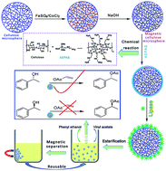
Surface modification of the magnetic cellulose particles has been conducted by using AEAPS, the modified magnetic cellulose particles were then used for the immobilization of lipase for catalysis reaction. The highest specific activity for AEAPS modified magnetic cellulose scaffold was increased to about 30 fold that of the free lipase without support. Furthermore, the reusability of the immobilized lipase on AEAPS modified magnetic cellulose particles was also improved, the relative activity of the ARCM-2 and AMRCM1-2 after being reused for 5 times still remained 72.8% and 71.2%, respectively. Moreover, the immobilized lipase on AEAPS modified magnetic cellulose particles was easily recovered without significant decrease in the specific activity. The immobilization of lipase on AEAPS modified magnetic cellulose particles led to the improvement in stability of activity and reusability for non-aqueous transesterification.
Cellulose is the most abundant natural material with significant properties of biodegradability, and biocompatibility, it is also a very promising raw material in chemical and biological industries.21,22 However, the potential material has not been fully exploited for its difficulty in dissolution by using conventional solvents, which was correlated to the strong inter- and intra-molecular hydrogen bonding in the macromolecules.23–25 We have put an intensive research on cellulose dissolving and construction of functional cellulose materials from the developed solvents. In our previous work, aqueous solvents containing alkaline and urea have been developed for cellulose dissolving.26 The regenerated cellulose materials prepared from LiOH/urea or NaOH/urea aqueous solution had porous structure, which could be used as scaffolds for the in situ synthesis of inorganic nanoparticles,27 or incorporation of curable prepolymers for the construction of functional cellulose materials,28 or in situ polymerization of conductive polymer monomer in the scaffolds.29 It indicated that the porous structured microstructure of the cellulose scaffolds could provide a favorable reaction environment, allowing for the effective diffusion of the reactants. Therefore, cellulose scaffold was expected to serve as a versatile support material and an effective microstructured reactor site for various catalysts, including enzymes. Inspired by our previous research results, in this work, porous structured cellulose microspheres were prepared from aqueous LiOH/urea solution, and then magnetic CoFe2O4 nanoparticles were synthesized in situ in the cellulose microspheres for the preparation of magnetic responsive cellulose materials. The obtained magnetic cellulose microspheres were used as supports for the immobilization of lipase. The highest specific activity for the lipase immobilized on magnetic cellulose microspheres was increased about 7 folds. While for the magnetic cellulose microspheres after being modified with AEAPS, the highest specific activity was increased to about 30 folds than that of the lipase without supports. Furthermore, the reusability of the immobilized lipase on AEAPS modified magnetic cellulose microspheres was also improved, the relative activity of lipase immobilized on RCMs and MRCMs was only 10.4% and 29.7% after being reused for 5 times, respectively, while for the ARCM-2 and AMRCM1-2, the relative activity still remained 72.8% and 71.2% after being reused for 5 times, respectively. Moreover, the immobilized lipase on AEAPS modified magnetic cellulose microspheres was easily recovered without significant decrease in a specific activity. Thus, the immobilization of lipase on AEAPS modified magnetic cellulose microspheres led to the improvement of activity and reusability for non-aqueous transesterification, without notable loss of selectivity.
AI (mg) = 200 − (AE1 + AE2) |
Specific activity (U g−1) = V/m |
![]() | ||
Scheme 1 Schematic illustration of synthesis strategy for ARCMs, the enzyme immobilization onto AMRCMs, their catalysis and reusability. |
The loading of inorganic magnetic components in the cellulose microspheres had an obvious influence on the microstructure, as it was shown in Fig. 2. The shape of the magnetic cellulose microspheres was regular sphere and the mean diameter was about 300 μm, and there was a slightly decrease in the particle size when compared with that of pristine cellulose microspheres, it would be ascribed to the synthesized inorganic particles in the cellulose matrix, which increased the interaction of cellulose matrix and inorganic components. Therefore, the magnetic microspheres exhibited relatively denser structure than that of pristine cellulose microspheres. The cross-section images of the magnetic cellulose microspheres were shown Fig. 3, the magnetic cellulose microspheres constituted porous structure in the inner parts, and the specific surface area of the composite microspheres ranged from 97 to 66 m2 g−1 with the increasing the concentration of CoFe2O4 precursor solution from 0.02 to 0.08 mol L−1, as it was shown in Table 1, and the obtained magnetic cellulose microspheres with high surface area had great potential application in engineering field.
![]() | ||
Fig. 2 SEM images of the surface morphology of the magnetic regenerated cellulose microspheres, (a–d) were for MRCM-1, MRCM-2, MRCM-3 and MRCM-4, respectively. |
![]() | ||
Fig. 3 SEM images of the cross-section morphology of the magnetic regenerated cellulose microspheres, (a–d) were for MRCM-1, MRCM-2, MRCM-3 and MRCM-4, respectively. |
Sample | CoFe2O4 content (wt%) | Porosity (%) | SBET (m2 g−1) | Average diameter (μm) | Lipase amount mg | U (μmol product per min) | Specific activity (U per mg protein) |
---|---|---|---|---|---|---|---|
a The mass of the free lipase used. | |||||||
Free lipase | — | — | — | — | 17.0a | 0.23 | 0.013 |
RCM | — | 97 ± 5 | 109 | 281 ± 2.3 | 0.195 | 0.028 | 0.143 |
MRCM-1 | 4.63 | 96 ± 4 | 97 | 278 ± 1.8 | 0.330 | 0.031 | 0.094 |
MRCM-2 | 7.79 | 96 ± 8 | 79 | 273 ± 1.7 | 0.327 | 0.028 | 0.086 |
MRCM-3 | 11.32 | 96 ± 5 | 75 | 275 ± 1.8 | 0.563 | 0.027 | 0.048 |
MRCM-4 | 15.28 | 95 ± 8 | 66 | 273 ± 2.4 | 0.313 | 0.016 | 0.051 |
ARCM-1 | — | 70 ± 5 | 48 | 277 ± 7.2 | 1.580 | 0.63 | 0.399 |
ARCM-2 | — | 67 ± 4 | 46 | 235 ± 5.4 | 1.742 | 0.79 | 0.453 |
ARCM-3 | — | 65 ± 5 | 58 | 287 ± 8.6 | 1.387 | 0.57 | 0.411 |
AMRCM1-1 | 4.63 | 67 ± 3 | 53 | 288 ± 5.8 | 1.292 | 0.50 | 0.387 |
AMRCM1-2 | 4.63 | 64 ± 4 | 50 | 301 ± 4.8 | 1.512 | 0.63 | 0.417 |
AMRCM1-3 | 4.63 | 63 ± 3 | 50 | 312 ± 3.6 | 1.330 | 0.51 | 0.383 |
In order to further clarify the inorganic components in the magnetic cellulose microspheres, TEM test was carried out by ultrathin sectioning of resin-embedded magnetic cellulose microspheres, and followed by removing of the resin on the copper grid. Fig. 4 shows the images of the magnetic cellulose microspheres. A large amount of inorganic nanoparticles were dispersed in the cellulose matrix. The particle size of the CoFe2O4 nanoparticles was less than 10 nm and it increased slightly with the increasing the precursor contention. As discussed above, the incorporated Co2+ and Fe3+ ions were readily bound to cellulose nanofibrils via electrostatic interactions between the electron-rich oxygen atoms in the cellulose macromolecules and the electropositive metal cations, the Co2+ and Fe3+ ions were anchored tightly to the cellulose nanofibrils. When it was treated with NaOH solution, CoFe2O4 nanoparticles were synthesized in situ in the cellulose scaffolds. The cellulose scaffolds enhanced the formation of CoFe2O4 nuclei and preventing the particles from overgrowth, leading to the homogeneously distribution of CoFe2O4 nanoparticles in the cellulose matrix. The content of the inorganic nanoparticles increased from 4.63 to 15.28 wt% with the increasing the concentration of precursor solution from 0.02 to 0.08 mol L−1. Our previous works indicated that the porous structured cellulose scaffolds was a promising template for the in situ synthesis of inorganic nanoparticles with particle size in 20 nm, and the concentration of the precursor had an influence on the content but little on the particle size of the resulted inorganic nanoparticles.27,28
![]() | ||
Fig. 4 TEM images of magnetic nanoparticles, (a–d) were MRCM-1, MRCM-2, MRCM-3 and MRCM-4, respectively. |
The room-temperature magnetization hysteresis loops of the magnetic cellulose microspheres were shown in Fig. 5. The forward and backward magnetization curves of the magnetic cellulose microspheres were similar. All of the curves went through zero magnetization point at H = 0. The absence of hysteresis and coercivity was characteristic of superparamagnetic behavior.33,34 It was well known that magnetic particles with particle size lower than the critical particle size could be called single domains. As the particle size continued to decrease below the single domain value, the particles exhibited superparamagnetic properties. This result indicated that the particle size of the prepared CoFe2O4 nanoparticles in cellulose matrix was small. The magnetization of the magnetic cellulose microspheres did not exhibit saturation even at magnetic field strength as high as 20 kOe, it would be ascribed to the rotation of the magnetic moment depending on the magnetic field. Moreover, the magnetic cellulose microspheres had novel magnetic responsibility. They could move toward to the magnet direction quickly when put a magnet near to them, demonstrating that the magnetic cellulose microspheres possessed novel magnetic properties and had potential application in magnetic field-assisted manipulation.
The magnetic cellulose microspheres were used as supports for lipases immobilization as a model system to demonstrate the applications in the field of biocatalysis. The protein content loaded on the microspheres and specific activities of the free and immobilized lipases were summarized in Table 1. The amount of immobilized lipases on pure cellulose microspheres was about 0.195 mg g−1, while for the magnetic cellulose microspheres, the amount of immobilized lipases increased from 0.33 mg g−1 for MRCM-1 to 0.563 mg g−1 for MRCM-3, and then deceased to 0.313 mg g−1 for MRCM-4, as it was shown in Fig. 6. Correspondingly, the specific activity for the RCM was 0.143, and it decreased from 0.094 for MRCM-1 to 0.048 for MRCM-3, and then increased to 0.051 for MRCM-4, while for the free lipase without supporting matrix had a low specific activity (0.013). It has been reported that free lipase was easily aggregated in non-polar solvent, leading to a poor specific activity. The immobilized lipase on the biosupport had an obviously increase in the specific activity, and it was increased to about 10 times than that of the lipase without supports. While for the magnetic cellulose supports, the specific activity of the lipase decreased when compared it with that of pristine cellulose microsphere supports, while the content of the encapsulated lipase in the magnetic cellulose supports was higher than that in the cellulose microsphere supports. It would be ascribed to the specific surface properties of the magnetic cellulose microspheres, with the incorporation of magnetic components in the cellulose matrix, the interaction between the lipase and the supports increased, and the immobilized lipase could penetrate into the inner structure and increased the bounded content of lipase in the magnetic cellulose microspheres, therefore, it was difficult for the substrates to interact with the lipases that penetrated in the inner section of the magnetic cellulose matrix, which resulted in negative effects on the specific activity of the lipases.
In order to further increase the specific activity of the magnetic cellulose microspheres supported lipases, the surface modification of the magnetic cellulose supports was carried out. Amino group modified magnetic cellulose supports was prepared via silane hydrolysis in ethanol–water medium in the presence of magnetic cellulose microspheres. After modification of the surface of magnetic cellulose microspheres, the amino functionalized supports could increase the interaction of the supports and the lipases. Fig. 7 shows the FT-IR spectra of RCMs, ARCM-2, MRCM-1, and AMRCM1-2. The peaks at 3700–3200 cm−1 were for the O–H flex vibrations of cellulose, 1060 cm−1 was for the C–O bending vibrations of the ether bond in cellulose molecules and 1157 cm−1 was correlated to the C–O antisymmetric bridge stretching.35,36 According to the spectra of ARCM-2 and AMRCM1-2, the peak at 1575 cm−1 attributed to the –NH2 bending vibrations, which verified the successful silylation modification by AEAPS. The peak for secondary amine groups in AEAPS and the H–O–H bending of the absorbed water was overlapping at 1650 cm−1. The broad absorption peak around 1150 cm−1 was assigned to Si–O–C bond. The N1s spectra of the cellulose microspheres and magnetic cellulose microspheres after being modified with AEAPS were shown in Fig. 8. The weak peak at 399.3 eV was assigned to –NH2.37 Combined with the FT-IR and XPS data, it indicated that the microspheres were modified with AEAPS successfully. After being calcined at 800 °C in air atmospheres by using TGA, the residue content of ARCM-1, ARCM-2, and ARCM-3 was 3.65, 5.55 and 6.28 wt%, respectively. And the residue content of AMRCM-1, AMRCM-2 and AMRCM-3 was 6.22, 7.58 and 8.74 wt%, which was further verified the successful modification by AEAPS.
![]() | ||
Fig. 7 FT-IR of the pristine cellulose and magnetic cellulose microspheres with and without modification with AEAPS. |
Fig. 9 shows the surface morphologies of the RCM and MRCM-1 with and without the modification with AEAPS. The surface morphology of the microspheres changed after the modification with AEAPS, and the porous structure of the microspheres was disappeared, the porosity for the RCM decreased from 97 ± 5% to 65 ± 5% for ARCM-3, while it decreased from 96 ± 5% for MRCM-1 to 63 ± 3% for AMRCM1-3. Correspondingly, the SBET of the RCM and MRCM-1 decreased with the modification of the surface by using AEAPS, and the specific area decreased with the increasing the concentration of the reacted AEAPS, as it was shown in the Table 1. Thus, combined with the results from the FT-IR and TGA analyses, it suggested that the surface of the cellulose and magnetic cellulose microspheres had been modified by using AEAPS. The specific activity of lipase on the pristine cellulose microspheres was about 0.143 U, and it increased to 0.399 U for ARCM-1. With the increasing the concentration of the used AEAPS for the modification of RCM, the specific activity for the immobilized lipase increased correspondingly, as it was shown in Fig. 10a. It was worth noting, the specific activity for the immobilized lipase on ARCM-3 was about 30 times than that of the free lipase. While for the AEAPS modified magnetic cellulose microspheres, the specific activity for the immobilized lipase also increased obviously when compared with that of the magnetic cellulose microspheres without modification with AEAPS, and the highest specific activity for AMRCM1-2 was increased to about 30 times than that of the lipase without supports. It would be resulted from the introduced –NH2 group from the AEAPS that contributed to the improvement of lipase activity for non-aqueous transesterification, so that the substrate was more likely to get close to the catalytic site and the specific activity of the lipase was improved by the AEAPS modified supports. It contributed to a large difference of activities between free and immobilized lipase. It has been reported that surface properties of the supports allowed effective interaction surrounding the catalytic site of lipase, and it contributed to the hyperactivation of lipase via interfacial activation, i.e. the formation of a suitable open structure for efficient contact with the substrates. The reusability of the immobilized enzyme was investigated and shown in Fig. 10b, the relative activity of lipase immobilized on RCMs and MRCMs was only 10.4% and 29.7% after reuse for 5 times, respectively, while for the ARCM-2 and AMRCM1-2, the relative activity still remained 72.8% and 71.2% after reuse for 5 times, respectively. These results indicated an acceptable activity and reusability of the immobilized lipase on amino-modified magnetic cellulose microspheres for (R)-1-phenylethanol. It suggested that the amino groups introduced into the magnetic cellulose microsphere were effective for the immobilization of lipases. The difference in the activity would relate to the supports for the immobilized lipase, and it indicated that the surface properties of the supports had an important influence on the stability and activity of the immobilized lipase. The advantages of the AEAPS modified magnetic cellulose microsphere as a support over conventional materials included its excellent practical utility, in that it was lightweight, biocompatible and easy to handle. In particularly, the immobilized lipase on AEAPS modified magnetic cellulose microspheres was easily recovered and reused without significant decrease in a specific activity. Thus, the immobilization of lipase on AEAPS modified magnetic cellulose microspheres led to the improvement of activity and reusability for non-aqueous transesterification, without notable loss of selectivity.
Footnote |
† Electronic supplementary information (ESI) available. See DOI: 10.1039/c5ra24984h |
This journal is © The Royal Society of Chemistry 2016 |