
Covalent modification of graphene oxide by metronidazole for reinforced anti-corrosion properties of epoxy coatings
Zongxue Yu*a,
Liang Lva,
Yu Maa,
Haihui Dia and
Yi He*ab
aSchool of Chemistry and Chemical Engineering, Southwest Petroleum University, Chengdu, 610500, China. E-mail: haiqingy@163.com; heyi@swpu.edu.cn; Fax: +86 028 83037315; Tel: +86 15208397092 Tel: +86 13881958071
bState Key Lab of Oil and Gas Reservoir Geology and Exploitation, Chengdu, 610500, China
First published on 15th January 2016
Abstract
Graphene can be used as an excellent protection material because of its barrier properties. This work reports a promising application of metronidazole (MET) modified graphene oxide (GO) composites (GME) for the corrosion protection of steel. The composites were synthesized using the carboxyl group of GO and hydroxyl group of MET with the help of maleic anhydride (MA), dispersing these sheets into epoxy resin at a low weight fraction of 0.2 wt%. A UV-vis absorption spectrum revealed that MET can be detected in NaCl (3.5 wt%) solution. An electrochemical impedance spectroscopy (EIS) test showed that the corrosion resistance performance is significantly enhanced by the addition of GME hybrids to epoxy compared to GO. A scratch test illustrated that fewer corrosion products formed in the scratch on the steel coated by GME. The strategy of using corrosion inhibitor modified graphene oxide can be extended to develop new graphene-based materials with other excellent materials for the protection of metal components.
1. Introduction
Recently, graphene has attracted tremendous interest for its unusual physico-chemical properties and possible applications in various fields,1,2 such as catalysis,3 biosensors,4 water purification,5,6 etc. However, graphene is a promising candidate for anti-corrosion coating, due to its unique properties (e.g. ref. 7–9). The Nobel Prize Laureate K. S. Novoselov has shown that the inertness and impermeability of graphene can be used as a protective coating through in situ growth.10 Singh et al. proved that a graphene oxide–polymer composite coating is an effective protective shield to the oxidation and corrosion of metal.11 Our previous study demonstrated that the corrosion protection properties of nano-graphene/epoxy composites on a metal surface are capable of enhancing the corrosion protection of pure metals effectively.12,13 Due to the complexity of a corrosion environment, a simple coating cannot provide the required protection. It is necessary to study active protection in addition to time-limited passive protection. One of the most promising approaches to develop a more effective anti-corrosion coating is based on the use of a corrosion inhibitor. Several systems have been reported: microcapsules, porous microparticles or stratified layers made of pH sensitive polyelectrolytes.14–18 Frederico Maia et al. studied a nano-container based on a silicon packaged MBT and the results showed that the release amount of MBT is affected by the different concentrations of pH and Cl−.19 N. Pirhady Tavandashti et al. introduced an inorganic corrosion inhibitor into SiO2/epoxy and added this to boehmite; a test demonstrated that cerium nitrate can be released from the defects of the coating and provide protection for the metal.20A polymer epoxy coating is an effective and simple approach for anti-corrosion. But there are defects in the process of epoxy coating solidification, such as micro-pores, bubbling etc.21 Thus, in order to overcome this disadvantage, our communication focuses on developing a route to prepare a covalent modification of graphene oxide by metronidazole (MET),22 as illustrated in Fig. 1. Via a versatile grafting process, maleic anhydride (MA) is grafted onto a GO surface utilizing hydroxyl and epoxy bond addition reactions to increase the content of carboxyl groups, which come from the GO and MA (GM), respectively. In a subsequent step, the covalent attachment of MET onto the modified GM was accomplished by an esterification mechanism (GME). The composite epoxy coating is fully tested.
 | ||
| Fig. 1 Flow chart for the synthesis of GME hybrid material. | ||
2. Experimental methods
2.1 Materials
Deionized (DI) water was produced by a water purification machine (UPC-III-40L, Ulupure), and natural graphite powder was purchased from Chendong Organic Chemicals. 4-(Dimethyl amino)pyridine (DMAP), dehydrated N,N-dimethylformamide (DMF), maleic anhydride (MA), metronidazole (MET) and dicyclohexylcarbodiimide (DCC) were purchased from Kanto Chemical Co, Inc. Concentrated sulfuric acid (98%), potassium permanganate (KMnO4), sodium nitrate (NaNO3), hydrogen peroxide aqueous solution (30%), anhydrous ethanol (analytical reagent grade), hydrochloric acid (HCl), and sodium hydroxide (NaOH) were obtained from Chengdu Kelong Chemical Reagent Factory.2.2 The preparation of GO
Graphene oxide (GO) was synthesized by the modified Hummers method.23 In brief, at a low temperature, 0.8 g of graphite was mixed with 24 mL concentrated H2SO4 by ultra-sonication for 20 min, and then the mixture was placed in an ice bath for half an hour. 1 g NaNO3 was added into the mixture within 35 min and the temperature of the mixture was maintained to not exceed 5 °C. KMnO4 (4.8 g) was mixed in slowly in small portions to keep the reaction temperature below 5 °C, and this was followed by stirring for 2 h. Then the mixture was heated up to 39 °C and stirred for 2.5 h. This was followed by the addition of 41 mL of DI water, and the reaction temperature was raised to 95 °C for 1.5 h. 120 mL of 10% H2O2 was added to the mixture for one hour. After this, HCl (10%, 100 mL) was added to the solution to remove the metal ions. Finally, the oxidation mixture was separated by centrifuge and washed with aqueous ethanol. The obtained product was exfoliated through ultra-sonication for 4 h, dried at 60 °C under vacuum for 12 h and ground into powder.2.3 Modification and graft of the graphene oxide (GO)
Subsequently, 0.3541 g GO and MA (0.073 mol) were mixed in a 250 mL round-bottomed flask with stirring and maintained at 75 °C for 3 h, and then at 85 °C for 16 h. Some studies24 show that the content of hydroxyl groups is more than that of carboxyl groups, but this research focuses on the carboxyl groups. We designed a method to modify the hydroxyl groups with MA25 to improve the content of carboxyl groups (Fig. 1). The modified product was named GM and it was obtained by drying the residue in a bag at 60 °C.The target material (GME) was synthesized according to a similar reported method,26 as is shown in Fig. 1. A typical synthetic procedure is as follows: to a solution of GM (0.236 g) and MET (5.9 mmol) in DMF (50 mL) were added DCC (2 mmol) and DMAP (0.04 mmol). After the solution was stirred at 80 °C for 48 h under a nitrogen atmosphere, the reaction mixture was cooled to room temperature and filtered. The filtrate was evaporated under vacuum, washed with hexane (50 mL), and then dried at 100 °C under vacuum for 24 h to give GME as a viscose liquid.
2.4 Preparation of composite coatings
To prepare the coating mixtures, epoxy resin was combined with a known amount of nanofiller (0.2 wt% mass fractions of GO, GM and GME). The composite coatings onto a steel surface after ultrasonic oscillation and mechanical stirring in a homemade container. The steel with the coatings and the redundant mixture were torrefied at 120 °C for 60 min, followed by drying at 220 °C for 90 min in an oven.27 The cured epoxy coatings were named pure epoxy coating, GO epoxy coating, GM epoxy coating and GME epoxy coating, respectively.2.5 Structure and electrochemical characterization
The obtained GO, GM and GME samples were characterized by FT-IR, XRD, TGA, XPS, UV-vis, and SEM. Thermogravimetric analysis (TGA) and Fourier transform infrared (FT-IR) spectroscopy spectra were recorded on a DSC1-1100L (METTLER, Switzerland) in the range of 50–800 °C and on a WQF 520 spectrometer in the range of 500–4000 cm−1. X-ray diffraction (XRD) analysis (X’Pert PRO MPD, Holland) was used to identify the crystallinity of the composite with a voltage of 40 kV and a generator current of 20 mA was used. The scanning rate was 1.5° min−1 and the scanning range was about 5–80°. X-ray photoelectron spectra (XPS) were recorded on an XSAM 800 photoelectron spectrometer (KRATOS) with Al Kα (1486.6 eV), the X-ray electric current set at 12 mA and the high voltage at 12 kV, and C1s (284.8 eV) corrected electron binding energy values. The solid-state 13C NMR experiments were carried out on an Agilent DD2-600 MHz NMR spectrometer.The measurement of corrosion inhibitor release from GME was performed at a neutral pH, adjusted by NaCl. In this work, 0.005 g GO, 0.005 g pure MET and 0.005 g GME powders were submerged in 25 mL NaCl (3.5 wt%) solution for 84 h. The solutions were filtrated by a pumping membrane filter. The filtrated solutions and NaCl (3.5 wt%) pure solution were characterized by an UV-vis spectrometer (UV-1800, Shimadzu).
Electrochemical impedance spectroscopy (EIS) measurements were investigated by an electrochemical workstation (EIS, CHI604D, China) at 104 to 10−1 Hz with a sine wave signal amplitude of 100–10 mV.28
3. Results and discussion
3.1 Characterization of GO, GM and GME
The groups on the surface of GO, GM and GME are characterized by FT-IR analysis (Fig. 2). Different functional groups are found in the FT-IR spectrum and this exhibits several characteristic peaks; O–H at 3407 cm−1, COOH groups at 1726 cm−1, C![[double bond, length as m-dash]](https://www.rsc.org/images/entities/char_e001.gif) C at 1623 cm−1, C–O–C groups at 1249 cm−1, and C–O groups at 1059 cm−1. It is indicated that an abundance of oxygen-containing functional groups exists on the surface of the GO nano-sheets. The absorption peaks at 1394 cm−1 may be associated to a COOH stretch in MA. Different functional groups are found in the FT-IR spectrum of GME (Fig. 2c). For MET, bands29,30 are assigned to OH stretching (3233 cm−1), CN stretching (1637 cm−1),22 NO stretching (1388 cm−1), C–NO2 stretching (844 cm−1) and the related C–H (2926, 2845 cm−1). There is not an obvious difference between ester functional groups and COOH at 1716 cm−1.31–34 The peak at 1413 cm−1 is due to the imidazole ring.31 All results show that the preparation of GME was successful.
C at 1623 cm−1, C–O–C groups at 1249 cm−1, and C–O groups at 1059 cm−1. It is indicated that an abundance of oxygen-containing functional groups exists on the surface of the GO nano-sheets. The absorption peaks at 1394 cm−1 may be associated to a COOH stretch in MA. Different functional groups are found in the FT-IR spectrum of GME (Fig. 2c). For MET, bands29,30 are assigned to OH stretching (3233 cm−1), CN stretching (1637 cm−1),22 NO stretching (1388 cm−1), C–NO2 stretching (844 cm−1) and the related C–H (2926, 2845 cm−1). There is not an obvious difference between ester functional groups and COOH at 1716 cm−1.31–34 The peak at 1413 cm−1 is due to the imidazole ring.31 All results show that the preparation of GME was successful.
 | ||
| Fig. 2 The FT-IR spectra of GO (a), GM (b) and GME (c). | ||
The XRD patterns of the GO, GM and GME are shown in Fig. 3. As shown in Fig. 3a, GO has an intense crystalline peak at 10.2°, corresponding to a d-space of 0.886 nm. The interlayer spacing of the GO sheets can be attributed to its oxygenated functional groups introduced by the modified Hummers method. The XRD of GM is shown in Fig. 3b, and the peak of GO is weaker. The peak of GO is at 8.83° and the d-space is 1.001 nm, which is larger than that of GO due to the introduction of MA.35 The results of d-space analysis demonstrates success of the modification. Compared to GM, in the XRD of GME (Fig. 3c) it could be clearly observed that the amorphous peaks of MET exist, but the characteristics of MA disappear. This could confirm that MET is grafted onto the GM instead of a simple physical mixture.
 | ||
| Fig. 3 The XRD spectra of GO (a), GM (b) and GME (c). | ||
The structural differences between GO, GM and GME are evident from the results obtained by thermogravimetric analysis (TG) and differential thermal analysis (DTA) in Fig. 4. The TG curve of GO (Fig. 4A) exhibited three obvious steps of mass loss as reported in the literature,36 the weight loss before 100 °C is caused by the release of trapped water in these materials. A weight loss of GO occurs from 200 °C to 260 °C (corresponding to a weight loss of GO of about 30%), indicating that there are abundant oxidative groups, corresponding to an obvious exothermic DTA peak (Fig. 4B) at 165 °C. In comparison, the TG curve of GM (Fig. 4A) displays more weight loss than GO, which means it has an enhanced number of the groups in the sheet of GO due to MA, and the exothermic peak is at 196 °C (Fig. 4B) (corresponding to a weight loss of GM of about 48%). However, the curve for GME exhibited two steps of mass loss which correspond to two weak exothermic peaks at 184 °C and 315 °C (Fig. 4B). This suggested that this GO has more groups by grafted MET which enhanced the weight loss of the material.
 | ||
| Fig. 4 (A) TG and (B) DTA spectra of GO, GM and GME. | ||
The surface chemical compositions and differences between GO and GME were investigated by XPS spectra. As shown in Fig. 5a, the main components of GO are C and O, with the two typical peaks at ∼286.4 eV and ∼534.8 eV, respectively. In contrast, a new peak at ∼401 eV appears in the XPS spectra of GME (Fig. 5c). The C/O atomic ratio value of GME calculated from the percentages of C1s to O1s shows a slight increase compared to that of GO (from ∼0.986 in GO to 1.555 for GME), which is attributed to the fact that MET chains possess a high C content in their structures (Fig. 1). The C1s level spectrum of GO with peak-fitting curves in Fig. 5b shows four typical chemically shifted components:37 C–C/CC (284.6 eV), C–O (285.7 eV), CO (287.3 eV), and C (O)–O (288.9 eV). After MET modification, the appearance of C–N and CN are observed in the C1s XPS spectra (Fig. 5c and d), implying a reaction between MET and GO. Compared with the GO spectrum, the GME spectrum exhibits a significant increase in the N peak, as shown in Fig. 5c. Moreover, the C1s XPS spectra of GME in Fig. 5d present peaks at ∼285.7 eV and 287 eV, which are assigned to C–N and CN, respectively. These above mentioned XPS results further demonstrate that GME was successfully prepared, which agrees well with the FTIR and XRD results.
 | ||
| Fig. 5 XPS results and the related C1s spectra of GO (a and b) and GME (c and d). | ||
In order to differentiate the structure of the GME nanofibers in the composite, the solid-state 13C NMR spectra of GO and GME were collected and are shown in Fig. 6. Typical resonances of GO are shown in Fig. 6a, which are ascribed to epoxy and hydroxyl groups, and the intense peak at 130 ppm is assigned to the graphitic carbons in the GO.38,39 However, the solid-state 13C NMR spectrum of GME not only has the typical resonances of GO, but also there are other resonances in the 13C NMR spectrum of GME (Fig. 6b). A schematic representation of GME with the labelling of C atoms used is shown in Fig. 6c. In the 13C NMR spectrum of GME, the signals at 172.29, 151.62, 138.23, 130.02, 60.58 and 44.05 ppm are attributed to the C-6, C-1, C-3, C-2, C-5 and C-4 of MET present on the GO, respectively. These results indicate that MET is indeed grafted onto the GO. The analysis results of 13C NMR are consistent with the results of the FT-IR, XRD, TG, and XPS analyses.
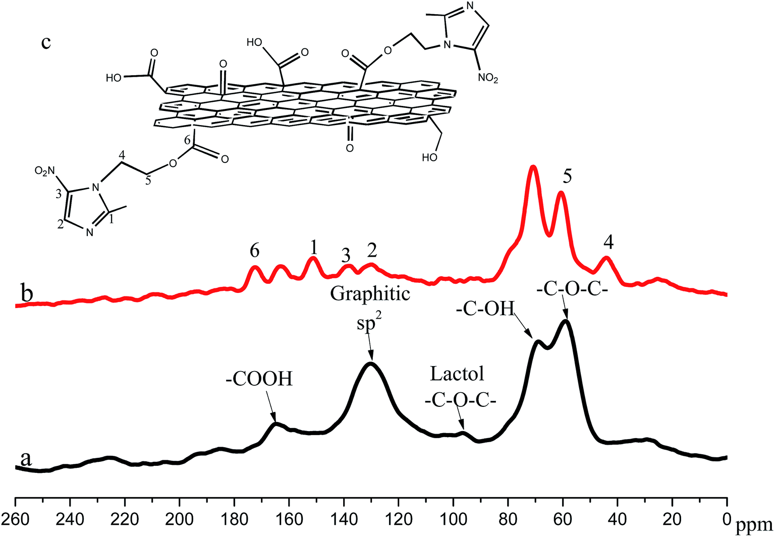 | ||
| Fig. 6 The 13C NMR spectra of GO (a) and GME (b). Schematic representation of GME (c) with the labelling of C atoms used. | ||
3.2 Release properties of GME
Fig. 7 illustrates the release of MET from GME in a NaCl (3.5 wt%) solution after 84 h. As Fig. 7 shows, there is no maximum absorption peak in the GO and NaCl solution spectrum, but a maximum absorption peak appears at 287 nm for the MET solution, indicating the maximum absorption peak of MET is here. As presented in Fig. 7, the absorption spectrum of GME in the solution has a maximum absorption peak at 287 nm, which can be explained as MET being released in NaCl solution from the GME material. As a result, it is expected that the mechanical property of the MET release could be attributed to the fracture of ester bond in the solution from the GO sheet. In addition, the release property of GME can enhance the anti-corrosion performance when the GME/epoxy coating is broken. | ||
| Fig. 7 The release of corrosion inhibitor from GME in NaCl (3.5% wt) solution. | ||
3.3 Thermal stability analysis of the composite coating (TG-DTG)
The thermal weight loss curves of the composite coatings are shown in Fig. 8. The thermal degradation rate of GME/epoxy is higher than the other coatings, and as such the thermal degradation rate of pure epoxy, 0.2 wt% GO/epoxy, 0.2 wt% GM/epoxy and 0.2 wt% GME/epoxy is 67.208%, 69.841%, 76.373% and 77.879%, respectively. According to Table 1, the thermal weight loss of pure epoxy, GO/epoxy, GM/epoxy and GME/epoxy is 56.470%, 59.069%, 64.729% and 62.267%, respectively, at 500 °C. The pure epoxy has the lowest thermal degradation rate, whereas the GM epoxy has the greatest; this can be attributed to there being a lot of oxygen-containing functional groups in GO. The thermal degradation rate shows the same tendency at 600 °C. However, the thermal degradation rate of GME/epoxy is the greatest of all, at 800 °C, this can be attributed to some of the groups disappearing in the reaction process, so the thermal conductivity of GO is enhanced and the thermal degradation rate of GME/epoxy is highest. From the DTG analysis shown in Fig. 8, the decomposition temperature of pure epoxy, GO/epoxy, GM/epoxy and GME/epoxy is 422 °C, 423 °C, 433 °C, and 438 °C, respectively. These results are because GO with fewer groups can improve the thermal conductivity of epoxy, this is consistent with the results of the TG analysis. The results show that the thermal stability of composite coating is improved in the GME/epoxy coating. | ||
| Fig. 8 The TG and DTG curves of the composite coatings. | ||
| Weight loss/% | |||||
|---|---|---|---|---|---|
| Coatings | |||||
| Temperature/°C | |||||
| 350 °C | 450 °C | 500 °C | 600 °C | 800 °C | |
| MET-GO/epoxy | 2.874% | 44.368% | 62.267% | 70.141% | 77.879% |
| GM/epoxy | 3.438% | 53.712% | 64.729% | 71.366% | 76.373% |
| GO/epoxy | 3.118% | 49.615% | 59.069% | 65.726% | 69.841% |
| Pure epoxy | 1.653% | 48.030% | 56.470% | 62.411% | 67.208% |
3.4 EIS studies
EIS is one effective and powerful technique for the investigation of the corrosion resistance of a coating. In this work, Nyquist and Bode plots and phase angle diagrams of 24 h were obtained for the 0.2 wt% GO/epoxy coating and 0.2 wt% GME/epoxy coating during 60 h immersion in 3.5 wt% NaCl aqueous solution (Fig. 9). For quantitative estimation of the anti-corrosion resistance of the composite coating, experimental impedance spectra are fitted using equivalent circuits. For the equivalent circuits depicted in Fig. 9(a and b), Rs accounts for the solution resistance; Rc, Ro and Rct can be assigned respectively to the pore resistance of the coating, resistance of the intermediate oxide layer and charge transfer resistance. CPEc, CPEo and CPEd are the constant phase elements of the coating, oxide and double layer, respectively. | ||
| Fig. 9 Equivalent circuit models used for numerical simulation of the EIS measurements of the coatings at different soaking stages (a and b); (a) 0.2 wt% GO/epoxy; (b) 0.2 wt% GME/epoxy; (a1 and b1) Nyquist and (a2 and b2) Bode diagrams of different times (Z′: the real part of the impedance, Z′′: imaginary impedance, |Z|: impedance module value), (a3 and b3) phase angle diagrams of 24 h. | ||
The barrier effect is one of the important features associated with corrosion protection by organic coatings. Therefore, the barrier properties of the coating system can not be compromised by the modification of the coatings of GO loaded with MET. The Nyquist plots of the GO/epoxy coating show that the impedance of the GO/epoxy coating decreases with longer immersion time (Fig. 9a1). The corresponding Bode plots are illustrated in Fig. 9a2 for the GO/epoxy coating. These show that the impedance of the low frequency range is also decreasing. The two time constants (high frequencies: 101 to 105 Hz) are a characteristic for some systems in the high frequency range (Fig. 9a3).
However, the protection properties of GME/epoxy coating are slowly degraded with time, then the upgrade of the impedance of low frequency of the GME/epoxy coating with time is due to the existence of MET (Fig. 9b1 and b2). The existence of the epoxy coating defects can improve the path for some molecules (H2O, O2 and Cl−) to penetrate the coating and the steel substrate will be corroded. Usually, the impedance moduli at low frequencies also reflect the protective properties of coatings. The phase of the GO/epoxy coatings show the normal properties of polymer coatings, but the GME/epoxy coating contrasts with the GO/epoxy coating at low frequencies. At low frequencies, there is a peak of the GME/epoxy coating in Fig. 9b3, indicating that this coating exhibited a good barrier property against corrosive species. This time constant is the response of the metal and reflects the corrosion behavior of the metal substrate. This result demonstrates that the anti-corrosion performance of the GME/epoxy coating is successful and outstanding.
3.5 Self-healing properties of the scratched specimens
The anti-corrosion performances of the multiplex epoxy coatings loaded with different addition materials were tested in a NaCl (3.5 wt%) solution test chamber for 84 h as shown in Fig. 10. As shown in Fig. 10, it can be clearly seen that corrosion products formed near the scratches of all the coatings. The accumulation of corrosion products near the scratches was more pronounced on the coating without any additional materials. However, there were some corrosion products in the GO epoxy coating, which showed a better anti-corrosion performance than the pure epoxy coating. The epoxy coating of GME showed the best performance of anticorrosion. As Fig. 10 shows, the formation of corrosion products in the scratches of the GME epoxy coating were rare. As a result, it is clear from the figure that the addition of GME to the coating caused a decrease in the amount of corrosion products produced near the scratches. It was also determined that if the ester bond that MET grafted to a GO sheet in NaCl (3.5 wt%) solution was broken, then MET could be released from GME and bring into play an anti-corrosion role to protect the steel, which has been illustrated by EIS in section 3.3. | ||
| Fig. 10 Visual performances of the coatings exposed to an NaCl (3.5% wt) solution test for 84 h ((A): before, (B): after). | ||
A schematic diagram of the anti-corrosion process is shown in Fig. 11. There are some oxygen-containing functional groups, such as hydroxy, epoxy and carboxyl, which have been proven by FTIR (Fig. 2) in GME, so GME can perfectly disperse in the epoxy coating as depicted in Fig. 11a. Though the addition of GO increases the molecular diffusion resistance in the coating matrix, some molecules (H2O, O2 and Cl−) can penetrate through the GO matrix over a long time scale (Fig. 11). A part of molecules, such as H2O, Cl− and O2, etc., via coating and get the substrate, the steel substrate will be corroded. Some studies40 show that GO and steel exposed to corrosive electrolyte, leads to the galvanic corrosion of the steel. GO connects with the substrate either directly or indirectly, which greatly increases the cathode-to-anode area ratio (Fig. 11a and b). The GME/epoxy coating can prolong the useable lifetime of steel, due to the MET cover on the surface of GO and by reducing the conduction of GO. For GME, as shown in Fig. 11c, when a corrosion reaction, such as electrochemical oxidation reaction and oxygen reduction reaction, occurs at the surface of steel, these are greatly inhibited. This is due to the mass transfer which is greatly limited by the coating, leading to an excellent anticorrosion performance of the GME/epoxy coating. The exposure of GME can release MET and change the adsorption film on the substrate to protect the steel. The anticorrosion mechanism of the GME/epoxy coating is the same as other studies.41–43
 | ||
Fig. 11 Schematic diagram of the anti-corrosion process in an NaCl (3.5% wt) solution. (a) Coating defects are exaggerated to advantageously show mass transfer in the coating matrix. The polymer-free GO is in black and the process of molecule penetration is in red. (b) The corrosion of steel generates a large number of electrons in the anode area (the anode sites are noted as  ). Some electrons transmit through the metal substrate and reach the anodic region on the surface of steel to participate in an oxygen reduction reaction. Other electrons will transmit through the GO and participate in an oxygen reduction reaction occurring in the GO cathode area (the cathode sites are noted as ). Some electrons transmit through the metal substrate and reach the anodic region on the surface of steel to participate in an oxygen reduction reaction. Other electrons will transmit through the GO and participate in an oxygen reduction reaction occurring in the GO cathode area (the cathode sites are noted as  and the cathode areas are highlighted in pink). (c) Electron transfer of the GME coating. The MET is marked in yellow. The reveal of GME in epoxy coating dissolved in the solution. The MET can be released from GME and form a film that is marked in yellow to protect the steel. and the cathode areas are highlighted in pink). (c) Electron transfer of the GME coating. The MET is marked in yellow. The reveal of GME in epoxy coating dissolved in the solution. The MET can be released from GME and form a film that is marked in yellow to protect the steel. | ||
4. Conclusions
We have synthesized fine metronidazole grafted graphene oxide nanocomposites via a simple two-step modification and graft without using any extra templates or surfactants. The characteristic functional groups, such as COOH, CN, NO, and C–NO2, of the modification and graft of GO had been proved by FTIR, XPS and 13C NMR. The weight loss as found by TGA of GO, GM and GME were 30%, 48% and 53%, respectively. The thermal weight loss curve of the composite coatings confirmed that the GME/epoxy modified the thermal stability of the composite coating. The comparison with GO reported in this work indicated that GME hybrids displayed an obvious advantage in the enhancement of corrosion resistance and enhanced the epoxy coatings’ corrosion resistance at a low content (0.2 wt%). There are several reasons for this superiority of the hybrids, which include the release of MET from GME, the sheet structure, and the excellent exfoliation in epoxy resin. These results were consistent with the original design ideas. The scratched specimens could be self-healing where the coating was damaged to prolong the life time of the metal. Herein, the GME hybrids presented a “smart” application in the field of nanofillers for anticorrosive epoxy coatings.
Acknowledgements
The authors gratefully acknowledge the financial support from the major cultivated project of the sci-tech achievements transition (15CZ0005) from the education department in Sichuan Province.References
- N. Levi-Polyachenko, D. Carroll and J. I. V. Stewart, Applications of Carbon-Based Nanomaterials for Drug Delivery in Oncology, ed. F. Cataldo and T. Da Ros, in Medicinal Chemistry and Pharmacological Potential of Fullerenes and Carbon Nanotubes, Springer, Netherlands, 2008, pp. 223–266 Search PubMed.
- Z. Zhu, L. Garcia-Gancedo, A. J. Flewitt, H. Xie, F. Moussy and W. I. Milne, A Critical Review of Glucose Biosensors Based on Carbon Nanomaterials: Carbon Nanotubes and Graphene, Sensors, 2012, 12, 5996–6022 CrossRef PubMed.
- R. Leary and A. Westwood, Carbonaceous nanomaterials for the enhancement of TiO2 photocatalysis, Carbon, 2011, 49, 741–772 CrossRef CAS.
- W. Yang, K. R. Ratinac, S. P. Ringer, P. Thordarson, J. J. Gooding and F. Braet, Carbon Nanomaterials in Biosensors: Should You Use Nanotubes or Graphene?, Angew. Chem., Int. Ed., 2010, 49, 2114–2138 CrossRef CAS PubMed.
- J. Xu, L. Wang and Y. Zhu, Decontamination of Bisphenol A from Aqueous Solution by Graphene Adsorption, Langmuir, 2012, 28, 8418–8425 CrossRef CAS PubMed.
- J. Xu and Y.-F. Zhu, Elimination of Bisphenol A from Water via Graphene Oxide Adsorption, Acta Phys.-Chim. Sin., 2013, 29, 829–836 CAS.
- J. C. T. Dhiraj Prasai, R. R. Harl, G. Kane Jennings and K. I. Bolotin, Graphene Corrosion-Inhibiting coating, ACS Nano, 2012, 6, 1102–1108 CrossRef PubMed.
- K.-C. Chang, M.-H. Hsu, H.-I. Lu, M.-C. Lai, P.-J. Liu, C.-H. Hsu, W.-F. Ji, T.-L. Chuang, Y. Wei, J.-M. Yeh and W.-R. Liu, Room-temperature cured hydrophobic epoxy/graphene composites as corrosion inhibitor for cold-rolled steel, Carbon, 2014, 66, 144–153 CrossRef CAS.
- M. L. Zheludkevich, J. Tedim and M. G. S. Ferreira, “Smart” coatings for active corrosion protection based on multi-functional micro and nanocontainers, Electrochim. Acta, 2012, 82, 314–323 CrossRef CAS.
- K. S. Novoselov, V. I. Fal, L. Colombo, P. R. Gellert, M. G. Schwab and K. Kim, A roadmap for graphene, Nature, 2012, 490, 192–200 CrossRef CAS PubMed.
- B. P. Singh, B. K. Jena, S. Bhattacharjee and L. Besra, Development of oxidation and corrosion resistance hydrophobic graphene oxide–polymer composite coating on copper, Surf. Coat. Technol., 2013, 232, 475–481 CrossRef CAS.
- Z. Yu, et al., Preparation of graphene oxide modified by titanium dioxide to enhance the anti-corrosion performance of epoxy coatings, Surf. Coat. Technol., 2015, 276, 471–478 CrossRef CAS.
- Z. Yu, et al., Fabrication of graphene oxide–alumina hybrids to reinforce the anti-corrosion performance of composite epoxy coatings, Appl. Surf. Sci., 2015, 351, 986–996 CrossRef CAS.
- L. Fedrizzi, W. Furbeth and F. Montemor, Self-healing properties of new surface treatments, European Federation of Corrosion Series, Maney Publishing, UK, 2011, vol. 58, p. 58, ISBN: 978 1906540364 Search PubMed.
- E. B. Murphy and F. Wudl, The world of smart healable materials, Prog. Polym. Sci., 2010, 35(1–2), 223–251 CrossRef CAS.
- D. Snihirova, et al., Hydroxyapatite Microparticles as Feedback-Active Reservoirs of Corrosion Inhibitors, ACS Appl. Mater. Interfaces, 2010, 2(11), 3011–3022 CAS.
- R. G. Buchheit, et al., Active corrosion protection and corrosion sensing in chromate-free organic coatings, Prog. Org. Coat., 2003, 47(3–4), 174–182 CrossRef CAS.
- M. L. Zheludkevich, et al., Active protection coatings with layered double hydroxide nanocontainers of corrosion inhibitor, Corros. Sci., 2010, 52(2), 602–611 CrossRef CAS.
- F. Maia, J. Tedim, A. D. Lisenkov, A. N. Salak, M. L. Zheludkevich and M. G. S. Ferreira, Silica nanocontainers for active corrosion protection, Nanoscale, 2012, 4, 1287 RSC.
- N. Pirhady Tavandashti and S. Sanjabi, Corrosion study of hybrid sol–gel coatings containing boehmite nanoparticles loaded with cerium nitrate corrosion inhibitor, Prog. Org. Coat., 2010, 69, 384–391 CrossRef.
- Y. He, et al., Synthesis and properties of iron oxide coated carbon nanotubes hybrid materials and their use in epoxy coatings, Polym. Adv. Technol., 2015, 26(4), 414–421 CrossRef CAS.
- S. M. Megalai, P. Manjula, K. N. Manonmani, N. Kavitha and N. Baby, Metronidazole: A Corrosion Inhibitor for Mild Steel in Aqueous Environment, Port. Electrochim. Acta, 2012, 30, 395–403 CrossRef CAS.
- S. William, J. Hummers and E. Richard, Preparation of graphitic oxide, J. Am. Chem. Soc., 1958, 6, 1399 Search PubMed.
- C. Botas, P. Álvarez, P. Blanco, M. Granda, C. Blanco, R. Santamaría, L. J. Romasanta, R. Verdejo, M. A. López-Manchado and R. Menéndez, Graphene materials with different structures prepared from the same graphite by the Hummers and Brodie methods, Carbon, 2013, 65, 156–164 CrossRef CAS.
- J. Xu, C. Xiao and X. He, Controllable synthesis of a novel poly(vinyl alcohol)-based hydrogel containing lactate and PEG moieties, Polym. Eng. Sci., 2014, 54, 1366–1371 CAS.
- T. Nakamura, X. Chu, T. Shimasaki and M. Shibata, Organogelation behavior and thermal properties of supramolecular polymer network composed of carboxy- and pyridyl-terminated 4-arm star-shaped epsilon-caprolactone oligomers, J. Colloid Interface Sci., 2013, 404, 8–15 CrossRef CAS PubMed.
- H. Yi, C. Chen, F. Zhong and Z. Xu, Preparation of aluminum oxide-coated carbon nanotubes and the properties of composite epoxy coatings research, High Perform. Polym., 2013, 26, 255–264 CrossRef.
- J. Vogelsang and W. Strunz, New interpretation of electrochemical data obtained from organic barrier coatings, Electrochim. Acta, 2001, 46, 3817–3826 CrossRef CAS.
- V. K. Singh, K. Pal, D. K. Pradhan and K. Pramanik, Castor oil and sorbitan monopalmitate based organogel as a probable matrix for controlled drug delivery, J. Appl. Polym. Sci., 2013, 130, 1503–1515 CrossRef CAS.
- J. Yuan and M. H. Liu, Chiral molecular assemblies from a novel achiral amphiphilic 2-(heptadecyl)naphtha 2,3 imidazole through interfacial coordination, J. Am. Chem. Soc., 2003, 125, 5051–5056 CrossRef CAS PubMed.
- H. I. Petersen, P. Rosenberg and H. P. Nytoft, Oxygen groups in coals and alginite-rich kerogen revisited, Int. J. Coal Geol., 2008, 74(2), 93–113 CrossRef CAS.
- M. Deng, et al., Synthesis and Characterization of Biodegradable Poly(ester amide)s with Pendant Amine Functional Groups and In Vitro Cellular Response, Biomacromolecules, 2009, 10(11), 3037–3047 CrossRef CAS PubMed.
- N. Wang, et al., Adsorption of environmental pollutants using magnetic hybrid nanoparticles modified with β-cyclodextrin, Appl. Surf. Sci., 2014, 305, 267–273 CrossRef CAS.
- J. Yuan and M. H. Liu, Chiral molecular assemblies from a novel achiral amphiphilic 2-(heptadecyl)naphtha 2,3 imidazole through interfacial coordination, J. Am. Chem. Soc., 2003, 125, 5051–5056 CrossRef CAS PubMed.
- Q. Shao, J. Tang, Y. Lin, J. Li, F. Qin, J. Yuan and L.-C. Qin, Carbon nanotube spaced graphene aerogels with enhanced capacitance in aqueous and ionic liquid electrolytes, J. Power Sources, 2015, 278, 751–759 CrossRef CAS.
- J. Chen, et al., An improved Hummers method for eco-friendly synthesis of graphene oxide, Carbon, 2013, 64, 225–229 CrossRef CAS.
- J. Chen, B. Yao, C. Li and G. Shi, An improved Hummers method for eco-friendly synthesis of graphene oxide, Carbon, 2013, 64, 225–229 CrossRef CAS.
- W. Gao, et al., New insights into the structure and reduction of graphite oxide, Nat. Chem., 2009, 1(5), 403–408 CrossRef CAS PubMed.
- K.-W. Park and J. H. Jung, Spectroscopic and electrochemical characteristics of a carboxylated graphene–ZnO composites, J. Power Sources, 2012, 199, 379–385 CrossRef CAS.
- W. Sun, et al., Inhibiting the Corrosion-Promotion Activity of Graphene, Chem. Mater., 2015, 27(7), 2367–2373 CrossRef CAS.
- S. Sathiyanarayanan, S. S. Azim and G. Venkatachari, Corrosion protection coating containing polyaniline glass flake composite for steel, Electrochim. Acta, 2008, 53(5), 2087–2094 CrossRef CAS.
- N. Jadhav, C. A. Vetter and V. J. Gelling, The effect of polymer morphology on the performance of a corrosion inhibiting polypyrrole/aluminum flake composite pigment, Electrochim. Acta, 2013, 102, 28–43 CrossRef CAS.
- M. Nematollahi, M. Heidarian, M. Peikari, S. M. Kassiriha, N. Arianpouya and M. Esmaeilpour, Comparison between the effect of nanoglass flake and montmorillonite organoclay on corrosion performance of epoxy coating, Corros. Sci., 2010, 52(5), 1809–1817 CrossRef CAS.
| This journal is © The Royal Society of Chemistry 2016 |