Activated dopamine derivatives as primers for adhesive-patch fixation of bone fractures

K. Olofsson, V. Granskog, Y. Cai, A. Hult and M. Malkoch*
KTH Royal Institute of Technology, Fibre and Polymer Technology, Teknikringen 56-58, Stockholm, SE-10044, Sweden. E-mail: malkoch@kth.se

Received 4th November 2015 , Accepted 3rd March 2016

First published on 4th March 2016


Abstract

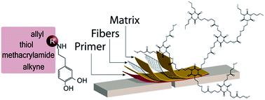
For the stabilization of complex bone fractures, tissue adhesives are an attractive alternative to conventional implants, often consisting of metal plates and screws whose fixation may impose additional trauma on the already fractured bone. This study reports on the synthesis and evaluation of activated dopamine derivatives as primers for fiber-reinforced-adhesive patches in bone-fracture stabilization strategies. The performance of synthesized dopamine derivatives are evaluated with regard to the adhesive shear strength of formed bone patches, as well as cell viability and surface properties. Dopamine-derived primers with methacrylamide, allyl, and thiol functional groups were found to significantly increase the adhesive shear strength of adhesive patches. Furthermore, deprotonation of the primer solution was determined to be essential in order to achieve good adhesion. In conclusion, the primer solutions that were found to give the best adhesion were the once where dopa-thiol was used in combination with either dopa-methacrylamide or dopa-allyl, resulting in shear bond strengths of 0.29 MPa.


Introduction

Conventional methods of stabilizing complex bone fractures rely on implants comprising metal-based plates that are applied by open surgery and fixated with pins and screws. While conventional methods are effective, limitations concerning accessibility, stress shielding and patient discomfort have to be considered. In the development of new minimally-invasive fracture-stabilization techniques, cross-linked bone adhesives are promising candidates.1 Since adhesives do not require drilling, fracture fixation can be achieved via minimally invasive surgery, enabling treatment of fractures where conventional implants are unsuitable. With adhesives, it is possible to obtain a more homogeneous weight-bearing load between bone fragments than when pins and screws are utilized.1 Many materials evaluated as potential bone adhesives have been originally developed for other areas, such as dentistry and soft tissue adhesives, and were thus originally not designed as adhesives for bone. Cyanoacrylates, urethanes, and alkylene bis(oligolactoyl)-methacrylates, as well as a number of dental cements and fibrin-based adhesives that are already in use as sealants or adhesives for soft tissues or in dentistry, are promising candidates that are still unsuitable as bone adhesives due to problems with e.g. toxicity, biodegradability, insufficient mechanical strength, or unsatisfactory stability in wet environment.2–6 The foreseen advantages of using adhesives continue to motivate research in this area. For more load bearing applications, the adhesive can be improved by the formation of a fiber reinforced adhesive patch (FRAP), comprising structural fibers embedded in a cross-linked adhesive matrix applied outside of a fracture.7,8 FRAP fixation makes it possible to tune the properties of the patch according to the loading conditions for each specific fracture by manipulating the number of fiber layers or increasing the adhesion area to allow for adjustments of the final strength of the patch.9 The FRAP-fixation approach, with patching outside of a fracture, is furthermore believed to interfere less with the natural bone healing process, than if adhesive is applied directly in the crevice of a fracture. To facilitate the surgical procedure, curing upon external stimuli such as e.g. UV or high-energy-visible (HEV) light is desirable. In the pursuit of a suitable matrix for FRAP, the thiol–ene coupling (TEC) reaction between thiols and unsaturated bonds is envisioned as a promising cross-linking strategy. Aspects promoting the use of the TEC cross-linking in surgical procedures are: insensitivity to oxygen inhibition, selectivity, and possibilities of cross-linking using minimally invasive optical fibers.10

We have previously shown that the triazine-based-building blocks; tris[2-(3-mercaptopropionyloxy)ethyl]isocyanurate (TAT) and 1,3,5-triallyl-1,3,5-triazinane-2,4,6-trione (TAA), with thiol and allyl functional groups respectively, can be cross linked into a promising matrix for adhesion to bone substrates.11 To further improve the adhesion of the cross linked triazine-based matrix (TA-matrix), primers utilizing the remarkable adhesive abilities of marine mussels were envisioned. Marine mussels secrete byssal threads built up of so called mussel foot proteins (mfps) that are able to bind strongly to various substrates both in aqueous and dry environments.12 The adhesive properties of mfps is associated with the sequences expressing the amino acid 3,4-dihydroxyphenyl-L-alanine (DOPA).13–16 The ability to form strong hydrogen bonds and the reported affinity toward hydroxyapatite, an abundant compound in bone, has in combination with the unique cohesive strength of DOPA in aqueous environments resulted in several reports with focus on DOPA-based compounds in the development of adhesives.14,17–24 Most of these reports have focused on DOPA-based adhesives for soft tissues and other substrates, but little work has so far been done on evaluating the materials as bone adhesives.22,24 Herein, we present an alternative approach for the fabrication of E-glass-fiber-reinforced adhesive thiol–ene patches for the fixation of bone fractures, exploring the use of derivatives of dopamine as adhesive primers.

Results and discussion

In previously published results we showed that the triazine-based-building blocks; tris[2-(3-mercaptopropionyloxy)ethyl]isocyanurate (TAT) and 1,3,5-triallyl-1,3,5-triazinane-2,4,6-trione (TAA), with thiol and allyl functional groups respectively, could successfully be cured into a matrix with promising adhesion to bone substrates.11 In light of these results, activated dopamine derivatives were envisioned as promising primers to improve the adhesion between the triazine-based matrix (TA-matrix) and bone. A library of dopamine derivatives was carefully synthesized to include two important features; (1) unsaturated or thiol functionalities that enable the formation of covalent bonds between primer and matrix and (2) DOPA groups to enhance the adhesion to the bone through e.g. hydrogen bonding. This was achieved by activating the primary amine of dopamine via amidation reactions, enabling the anchorage of allyl, methacrylamide, thiol, and alkyne-functional groups, according to Scheme 1. The synthesis of primers was straight forward with acceptable yields after purification and the structure was confirmed using NMR spectroscopy. Evaluation of the performance of the derivatives as primers was conducted via formation of FRAPs for bone-fracture stabilization. The primers were furthermore evaluated with respect to cell viability and surface properties.
image file: c5ra23142f-s1.tif
Scheme 1 Synthetic pathways to dopamine derivatives: dopamine 1, dopa-allyl 2, dopa-methacrylamide 3, dopa-thiol 4, dopa-alkyne 5, and dopa-sp-allyl 6.

Cytotoxicity of dopamine derivatives

In developing materials intended for us in the medical field, it is important to investigate the viability of cells in contact with the material. Primer toxicity toward human dermal fibroblasts (hDF) was thus evaluated using alamarBlue® Assay, where primer solutions with concentrations of 0.1–1000 μg ml−1 were added to hDF-cell culture. Cell viability, indicated by the metabolic activity from the alarmarBlue® Assay, decreased with increasing concentration of dopa-primer and dose limitations were calculated according to ISO 10993-5 standards, in which materials causing cell-viability reduction by less than 30% are considered to be non-cytotoxic.25 Dose limitations of DOPA-primers were 2.5 μg ml−1 (10.6 μM) for dopa-allyl, 30 μg ml−1 (136 μM) for dopa-methacrylamide, 45 μg ml−1 (237 μM) for dopamine, 90 μg ml−1 (309 μM) for dopa-alkyne, 150 μg ml−1 (587 μM) for dopa-thiol, and 250 μg ml−1 (423 μM) for dopa-sp-allyl, and thus affected by small differences in the structures of the primers. These dose limitations are in the same range as values reported by Ben-Shachar et al. in 2004, who reported that extracellular dopamine could cause cell death at concentrations above 200 μM.26 The in vitro elution test for the TA-matrix showed no signs of toxicity, with cell-viability values of 111% ± 9.

Surface properties

The idea of a primer is to improve the interactions between a substrate and an adhesive through the application of a thin intermediate layer of primer. The low-molecular-weight primers synthesized herein had slightly different molecular structures and different functional end groups believed to be able to interact covalently with the TA-matrix. For this to occur good wetting is essential and an evaluation of the surface properties of both primers and TA-matrix was therefore foreseen as a means to predict the performance of the primers. To evaluate the surface properties of primer surfaces, circular glass substrates (15 mm in diameter) were spin-coated with primer solution (5 μL, 84.5 mM) and analyzed using static contact-angle (CA) measurements. CAs were measured for water, ethylene glycol, and diiodomethane to enable calculations of surface free energy according to the van Oss-Chaudhury-Good (OCG) theory.27 CAs showed that all primers were significantly more hydrophilic than the cross-linked TA-matrix (Table 1). Whereas the TA-matrix had a water CA of 86°, all primers, apart from dopa-methacrylamide and dopamine, displayed a water CA around 20°. Dopa-methacrylamide, however, displayed a water CA of 55°, much more similar to that of the TA-matrix. Dopa-methacrylamide and the TA-matrix also had significantly more similar CAs for ethylene glycol than any of the other substrates.
Table 1 Contact angles of the cured TA-matrix and FRAP-primer surfaces measured for water, ethylene glycol, and diiodomethane
Surface CA (water) [°] CA (ethylene glycol) [°] CA (diiodomethane) [°]
TA-matrix 86.1 ± 1.6 53.5 ± 0.6 28.8 ± 1.3
Dopamine 5.4 ± 1.1 12.5 ± 0.4 44.0 ± 0.8
Dopa-sp-allyl 20.3 ± 1.0 27.8 ± 2.1 43.0 ± 0.7
Dopa-alkyne 23.7 ± 0.8 13.9 ± 1.7 36.5 ± 0.4
Dopa-allyl 17.8 ± 0.3 13.0 ± 0.5 39.4 ± 0.6
Dopa-methacrylamide 55.4 ± 2.1 43.5 ± 1.5 37.5 ± 3.1
Dopa-thiol 19.1 ± 2.2 12.1 ± 1.6 37.0 ± 2.0


Additionally, surface free energy was determined for the TA-matrix and for all primer systems according to the van Oss-Chaudhury-Good theory27,28 (ESI). It was found that the surface free energy of dopa-methacrylamide was more similar to the TA-matrix than the other primers. Combined with a lower hydrophilicity of the dopa-methacrylamide surface compared to the other primers, as measured by CA, it was expected that dopa-methacrylamide would have a stronger interaction with the TA-matrix than the other primers and thereby result in stronger adhesion.

Fiber-reinforced-adhesive-patch (FRAP) fixation of generic bone fractures

A FRAP with a thiol–ene coupled matrix in combination with the adhesive properties provided by a dopa-functional primer and the load-bearing advantages of fibers, was foreseen as a robust strategy for bone-fracture fixation. Other studies on dopa-functional materials as bone adhesives have shown that these materials have potential,22,24 why dopa-functional compounds were foreseen as promising primers for the FRAP-fixation approach used herein. Differences in formulations, substrates, test setup and other conditions between the studies, however make comparisons of actual values from the different studies inconclusive. The commercial dental primer thiol–ene was therefore used herein for comparison.

Due to large variations between bone samples from e.g. different animal species, anatomical sites, and individuals, it is difficult to get a representative model substrate in vitro. To minimize influence of external factors and focus this investigation on the adhesion between the bone and the patch, bone pieces with sizes of 50 × 15 × 3 mm were cut from bovine marrow pipe and simple generic fractures were created using a bow saw by dividing the bone pieces in two. The bone substrates were furthermore sanded to minimize influence of differences in surface roughness and patches spanning across the fracture were formed in layers on a 10 × 15 mm area on each side of the fracture (Fig. 2a). 4 μL aqueous primer solution (84.5 mM) was applied to moist bone, followed by TA-matrix alternated with five layers of E-glass fibers. The matrix layers were cured using HEV light with a total dose of 142 kJ cm−2. Patches were prepared on both sides of each bone sample to generate a double lap-shear mode at testing and stored in PBS buffer for 24 h before tensile tests were performed (Fig. 2b).


image file: c5ra23142f-f1.tif
Fig. 1 Dose limitations of dopamine derivatives (defined as the concentration where the cell viability of human dermal fibroblasts is 70%).

image file: c5ra23142f-f2.tif
Fig. 2 Patch fabrication on bone specimen: (a) schematic simplified patch build up (only two fiber layers are shown even though five layers of fibers were used in the study), (b) a schematic representation of a stabilized bone specimen, and (c) a photograph of a FRAP-stabilized bone specimen ready for testing.
Influence of dopa primers on adhesive shear strength of FRAPs. The adhesive shear strength of the patches was evaluated using lap-shear tests. Additionally, NaOH was added as an additive to the primer solution to deprotonate the acidic phenols and thereby increase the adhesive properties of the patches through hydrogen bonding. The shear bond strengths calculated from the lap-shear tests (Fig. 3), confirmed that activated derivatives of dopamine could successfully be used as primers to improve the shear bond strength of FRAPs. The shear-bond strengths for the individual primer solutions with NaOH added (Fig. 3 grey, no pattern) show that the primers performed differently depending on their structure. Primers with alkyne, allyl, methacrylamide, and thiol, all enhanced the adhesive strength of formed FRAPs, compared to FRAPs prepared without primer. Dopa-thiol and dopa-methacrylamide (0.18 and 0.17 MPa, respectively), gave the strongest patches, closely followed by dopa-allyl (0.14 MPa). Compared to patches without primer, patches with dopa-thiol and dopa-methacrylamide displayed approximately two times as high bond strengths. The differences between dopa-allyl and dopa-methacrylamide could possibly be explained by differences in reactivity towards TAT in the TA-matrix. Although dopa-allyl should have a higher reactivity during TEC, dopa-methacrylamide should be more prone to homo-polymerization,29 which might explain why FRAPs with dopa-methacrylamide showed stronger shear bond strengths than FRAPs with dopa-allyl. Polarity differences between the amide bonds might also have an effect, as the polarity would influence the secondary forces between the primer molecules and/or the bone tissue.
image file: c5ra23142f-f3.tif
Fig. 3 Shear bond strength of FRAPs determined by tensile tests (dental-adhesive CLEARFIL™ SE BOND used as a comparison).
Influence of addition of NaOH. In order to investigate the impact of NaOH addition, the patches displaying the highest adhesive strength (i.e. the patches with dopa-methacrylamide and dopa-thiol) were also prepared without addition of NaOH. The effect of NaOH on primer structure was investigated using NMR analysis of a solution of dopa-methacrylamide prepared in 50% D2O in MeOH with a 1[thin space (1/6-em)]:[thin space (1/6-em)]1 molar ratio addition of NaOH to dopa-groups. The 13C-NMR spectra clearly indicated a change in the structure of the catechol, as the peaks for the carbon atoms attached to the hydroxyl groups shifted from 143.8 and 145.3 ppm to 151.2 and 153.6 ppm (ESI, Fig. S3). The other aromatic carbons also shifted slightly, while the rest of the peaks were unaffected by the NaOH addition. The 13C-NMR spectra did not imply oxidation from catechol to quinone, as that would have resulted in peaks around 180 ppm30 and it was concluded that the catechol group was deprotonated by the addition of NaOH. Interestingly, the addition of NaOH to primer solutions did indeed increase the adhesive shear strength of the patches 6.3 and 5.1 times, using primer solutions of dopa-methacrylamide and dopa-thiol, respectively (Fig. 3).

Dopa-sp-allyl 6 was initially synthesized to evaluate the impact of adding a spacer between the dopa-group and the allyl group. Dopa-sp-allyl was envisioned to have better adhesion than the shorter dopa-allyl primer, due to the increased mobility brought by the tri(ethylene oxide) spacer. Additionally, dopa-alkyne 5 was envisioned to improve adhesion due to the possibilities of binding to more than one TAT molecule in the matrix via thiol–yne chemistry (TYC). Unfortunately, these two primers did not perform as good as anticipated (Fig. 3). In fact, dopa-sp-allyl showed the lowest adhesion of all tested dopamine derivatives, prompting further investigation. NMR spectra of the dopa-alkyne primer before and after addition of NaOH (ESI, Fig. S4), revealed that the increase in pH caused hydrolytic cleavage of the ester bonds in the structure. The same sensitivity towards hydrolysis is expected for dopa-sp-allyl due to the presence of similar ester bonds. Degradation of the primers in this way after addition of NaOH would explain why dopa-sp-allyl and dopa-alkyne did not increase the adhesion of FRAPs as expected, as the primers could no longer interact with both bone and matrix as envisioned. The significant differences in the adhesion of patches where the degraded primers were used compared to patches where primers with higher hydrolytic stability were used, furthermore support the theory that the added functional groups promoted interactions towards the matrix.

Influence of allyl-to-thiol ratio. Earlier published results, showed that off-stoichiometry between allyl and thiol groups in the TA-matrix can influence the properties of the cured matrix.31 The efficiency of the HEV-induced crosslinking of an equimolar mixture of TAA and TAT was monitored by FT-Raman analysis. The disappearance of the thiol and allyl peaks, at 2574 and 1645 cm−1 respectively, showed that the matrix could be efficiently cured using HEV-light irradiation (ESI, Fig. S5). Adding a layer of dopa-thiol underneath the matrix, however, resulted in reduced curing efficiency of the matrix (ESI, Fig. S6), where small peaks corresponding to both allyl and thiol groups were visible even after curing. This could thus influence the adhesive bond strength of the patches and an ene-to-thiol ratio of 1.1 should thus be preferred. The best results were in fact achieved when dopa-thiol was used in combination with either dopa-allyl or dopa-methacrylamide, both combinations showing shear bond strengths of 0.29 MPa, more than 1.6 times higher than for the individual primer solutions (Fig. 3). With ene and thiol combined, both compounds in the primer solution had a dopa group able to interact with the bone and a functional group able to interact with the matrix, while maintaining an ene-to-thiol ratio of 1[thin space (1/6-em)]:[thin space (1/6-em)]1 in the entire primer-matrix system.

The combination of dopa-thiol with either dopa-methacrylamide or dopa-allyl interestingly gave adhesive strengths similar to that of the commercially available dental primer: CLEARFIL™ SE BOND. Statistical evaluation (ESI, Table S3) even showed that there was no significant difference between the dopa-thiol/dopa-allyl combination and CLEARFIL™ SE BOND. It should however be noted that, when CLEARFIL™ SE BOND is used in dentistry, another sample-preparation procedure is used, which may influence the results.

Experimental

Materials

All chemicals were purchased from Sigma Aldrich and used as received, unless otherwise specified. E-glass fabric (25 g m−2, finish FE 800, plain) was purchased from R&G Faserverbundwerkstoffe GmbH and used as received. CLEARFIL™ SE Bond, a commercial dental primer was purchased from Kuraray America, Inc. All cell-culture reagents were purchased from Thermo Scientific™ HyClone™.

Instrumentation

1H and 13C-NMR analysis were performed on a Bruker Avance NMR instrument. 1H-NMR spectra were acquired at 400 MHz with a spectral window of 20 ppm, an acquisition time of 4 s, and a relaxation delay of 1 s. 13C-NMR spectra were acquired at 101 MHz with a spectral window of 240 ppm, an acquisition time of 0.7 s, and a relaxation delay of 2 s.

Static contact-angle measurements were performed on a CAM200 contact-angle meter (KSV Instruments, Helsinki, Finland), using the sessile-drop technique and an automatic dispenser. Contact angles were measured for water, ethylene glycol, and methyl diiodate at 23 °C and at a relative humidity of 50%, using a drop volume of 5 μl for water and ethylene glycol, and a drop volume of 3 μl for methyl diiodate (the drop volume had to be decreased to 3 μl because of the low surface tension of the liquid). OneAttention software, using Laplace fitting, was used for calculating the contact angles at three different positions for each substrate. Surfaces for contact-angle measurements were prepared via spin coating of the primer solutions on glass substrates.

Curing was performed using a lamp intended for light-cured dental materials, Bluephase® 20i, predominately radiating HEV light with wavelengths of 410 nm and 470 nm. Two different irradiation programs were used: the TURBO program (2000 mW cm−2) and the HIGH POWER program (1200 mW cm−2).

All mechanical tests were performed on an Instron 5566 static-testing machine with a 10 kN load cell and a cross-head speed of 5 mm min−1, using a preload of 1 N and a pre-load speed of 2 mm min−1. All measurements were conducted at 23 °C and at a relative humidity of 50%. Samples were acclimatized to the testing temperature prior to measurements and kept in PBS buffer as long as possible before being attached to the machine. Data was collected using Bluehill software.

Synthesis of dopamine derivatives

The synthesis paths for all primers used in this study are displayed in Scheme 1.
Synthesis of N-(3,4-dihydroxyphenethyl)pent-4-enamide – dopa-allyl (2). Dopamine hydrochloride 1 (8.00 g, 42.1 mmol) was dissolved in a solution of dimethyl sulfoxide (DMSO) (40.0 ml) and triethylamine (TEA) (7.07 ml, 50.7 mmol). 4-Pentenoic anhydride (6.94 ml, 38.0 mmol) was slowly added, after which the reaction was allowed to proceed under agitation overnight. Diethyl ether (1000 ml) was added and the mixture was extracted four times with 100 NaHSO4(aq) (10 w%). The organic phase was dried over MgSO4 and subsequently filtered and the solvent removed under reduced pressure. Further purification was carried out by flash chromatography using ethyl acetate and heptane to give 2 as a white solid (4.84 g, 54% yield).

1H NMR (400 MHz, CD3OD) δ 6.67 (d, 1H, Ar), 6.62 (d, 1H, Ar), 6.51 (dd, 1H, Ar), 5.76 (m, 1H, –CH[double bond, length as m-dash]), 4.98 (dd, 2H, [double bond, length as m-dash]CH2), 3.31 (t, 2H, –CH2–), 2.61 (t, 2H, Ar-CH2–), 2.29 (m, 2H, –NHCO–CH2–), 2.21 (m, 2H, –CH2–CH[double bond, length as m-dash]); 13C NMR (101 MHz, CD3OD) δ 175.3, 146.2, 144.7, 138.2, 132.0, 121.0, 116.8, 116.3, 115.8, 42.2, 36.4, 35.9, 31.0 (M = 235.28 g mol−1).

Synthesis of N-(3,4-dihydroxyphenylethyl)-methacrylamide – dopa-methacrylamide (3). Dopamine hydrochloride 1 (16.6 g, 87.3 mmol), and TEA (14.8 ml, 106 mmol) were dissolved in DMSO (80.0 ml), after which methacrylic anhydride (11.8 ml, 79.4 mmol) was slowly added (Scheme 1). The reaction mixture was thereafter left under agitation for 90 min until all anhydride had reacted, as analyzed with 13C-NMR. Diethyl ether (1750 ml) was added and the mixture was extracted four times with 200 ml NaHSO4(aq) (10 w%). The organic phase was dried over MgSO4 and subsequently filtered after which a small spoon of methyl hydroquinone was added as inhibitor before evaporation of the solvent. Further purification of the organic phase was carried out by flash chromatography using ethyl acetate and heptane. In addition, product from the water phase was allowed to crystallize to give a total of 9.8 g of 3 as a white solid (56% yield).

1H NMR (CD3OD, 400 MHz) δ 6.68 (d, 1H, Ar), 6.65 (d, 1H, Ar), 6.52 (dd, 1H, Ar), 5.64 (s, H, [double bond, length as m-dash]CHH), 5.34 (s, H, [double bond, length as m-dash]CHH), 3.38 (t, 2H, –CH2–NH–), 2.64 (t, 2H, Ar-CH2–), 1.91 (s, 3H, –CH3); 13C NMR (CD3OD, 101 MHz) δ 172.1, 147.1, 145.6, 142.3, 133.0, 121.9, 121.1, 117.8, 117.2, 43.5, 36.8, 19.6 (MW = 221.26 g mol−1).

Synthesis of N-(3,4-dihydroxyphenetyl)-4-mercapto-butanamide – dopa-thiol (4). Dopamine hydrochloride 1 (25.0 g, 132 mmol), NaHCO3 (23.3 g, 277 mmol), and γ-thiobutyrolactone (12.5 ml, 144 mmol) were dissolved in 250 ml deionized water. The reaction mixture was heated to 95 °C and was allowed to reflux for 2 h before being cooled down to room temperature. 250 ml brine was added and the product was extracted two times with 625 ml THF. The organic phase was dried over MgSO4, filtered, and the solvent evaporated under reduced pressure. Further purification was carried out by flash chromatography using ethyl acetate and heptane, which gave 28.3 g of the product as a slightly yellow solid (84% yield).

1H NMR (400 MHz, CD3OD) δ 6.66 (d, 1H, Ar), 6.61 (d, 1H, Ar), 6.50 (dd, 1H, Ar), 2.61 (t, 2H, Ar-CH2–), 2.44 (t, 2H, –CH2–SH), 2.25 (t, 2H, –NHCO–CH2–), 1.82 (p, 2H, –CH2–CH2–SH); 13C NMR (101 MHz, CD3OD) δ 175.44, 146.40, 144.92, 132.11, 121.18, 116.98, 116.46, 42.33, 36.05, 35.69, 31.49, 24.59 (MW = 255.33 g mol−1).

Synthesis of prop-2-yn-1-yl-4-((3,4-dihydroxyphenethyl)amino)-4-oxobutanoate – dopa-alkyne (5). Dopa-alkyne 5 was synthesized according to a previously published procedure32 as follows: dopamine hydrochloride 1 (5.00 g, 26.4 mmol) was dissolved in a solution of DMSO (25.0 ml) and TEA (4.41 ml, 31.6 mmol), after which 4-oxo-4-(prop-2-yn-1-yloxy)butanoic anhydride (synthesized according to a previously published procedure33) (6.98 g, 23.7 mmol) was slowly added (Scheme 1). After 1 h, diethyl ether (1000 ml) was added and the mixture was extracted four times with 100 ml NaHSO4(aq) (10 w%). The organic phase was dried over MgSO4 and subsequently filtered and the solvent evaporated under reduced pressure. Further purification was carried out by flash chromatography with ethyl acetate and heptane to give 2.61 g of 4 as a white solid (38% yield).

1H NMR (400 MHz, CD3OD/D2O) δ 6.83 (d, 1H, Ar), 6.77 (d, 1H, Ar), 6.66–6.63 (dd, 1H, Ar), 4.71 (d, 2H, –CH2–C[triple bond, length as m-dash]), 3.38 (t, 2H, –CH2–NHCO–), 2.97 (t, 1H, [triple bond, length as m-dash]CH), 2.67 (m, 4H, –NHCO–CH2–CH2–COO– overlap), 2.51 (t, 2H, Ar-CH2); 13C NMR (101 MHz, CD3OD) δ 174.1, 173.5, 146.4, 144.9, 132.2, 121.2, 117.0, 116.5, 78.9, 76.3, 42.5, 36.1, 31.4, 30.3 (MW = 291.30 g mol−1).

Synthesis of (1-(13-oxo-3,6,9-trioxa-12-azaheptadec-16-en-1-yl)-1H-1,2,3-triazol-4-yl)methyl-4-((3,4-dihydroxylphenethyl)amino)-4-oxobutanoate – dopa-sp-allyl (6). Dopa-sp-allyl 6 was synthesized via copper(I)-catalyzed azide–alkyne cycloaddition (CuAAC). Dopa-alkyne 5 (0.537 g, 1.84 mmol) and N-(2-(2-(2-(2-azidoethoxy)ethoxy)ethoxy)ethyl)-pent-4-enamide (0.504 g, 1.68 mmol) were dissolved in 5.00 ml THF after which sodium ascorbate (66.0 mg, 0.335 mmol) and CuIISO4 (42.0 mg, 0.168 mmol) dissolved in 2.00 ml of DI water were added. The reaction was heated to 40 °C and stirred overnight. An additional amount of dopa-alkyne (98.0 mg, 0.336 mmol) was added to ensure full conversion of azide groups and the reaction was followed using 13C NMR. After complete conversion had been achieved, the solvent was removed under reduced pressure, and 100 ml DCM was added. The mixture was extracted four times with 10 ml H2O, after which the combined water phase was extracted with an additional 50.0 ml DCM. The DCM phases were then combined and dried over MgSO4, filtered, whereafter the solvent was removed under reduced pressure. Further purification was carried out using flash chromatography in ethyl acetate and methanol to yield 0.222 g of the pure product (22.4% yield).

1H NMR (400 MHz, CDCl3) δ 7.82 (s, 1H, triazole), 6.79 (d, 1H, Ar), 6.70 (d, 1H, Ar), 6.53 (dd, 1H, Ar), 5.78 (m, 1H, –CH [double bond, length as m-dash]), 5.17 (s, 2H, –COO–CH2–triazole–), 5.05–4.96 (dd, 2H, [double bond, length as m-dash]CH2), 4.50 (t, 2H, –triazole–CH2–CH2–O–), 3.84 (t, 2H, –triazole–CH2–CH2–O–), 3.60 (s, 8H, –O–CH2–CH2–O–), 3.53 (t, 2H, –O–CH2–CH2–NHCO–), 3.41 (m, 4H, Ar-CH2–CH2–NHCO– and –O–CH2–CH2–NHCO–), 2.63 (t, 4H, Ar-CH2– and –NHCO–CH2–CH2–COO–), 2.40 (t, 2H, –NHCO–CH2–CH2–COO–), 2.35 (t, 2H, –CH2–CH2–CH[double bond, length as m-dash]), 2.25 (m, 2H, –CH2–CH2–CH[double bond, length as m-dash]); 13C NMR (101 MHz, CDCl3) δ 173.2, 172.8, 171.9, 144.6, 143.4, 142.5, 137.2, 131.0, 125.5, 120.6, 116.0, 115.8, 115.6, 70.6, 70.5, 70.3, 70.0, 69.4, 57.7, 50.5, 41.1, 35.9, 34.8, 31.0, 29.8, 29.7 (MW = 591.66 g mol−1).

Synthesis of tris[2-(3-mercaptopropionyl-oxy)ethyl]isocyanurate (TAT). TAT was synthesized by mixing 1,3,5-tris(2-hydroxyethyl)isocyanurate (25.0 g, 95.7 mmol), 3-mercaptopropionic acid (91.0 g, 0.857 mol) and p-toluene sulfonic acid monohydrate (11.0 g, 57.8 mmol) in 800 ml toluene. The mixture was heated to 125 °C and the reaction was run for 2 h. The reaction was monitored by NMR. The reaction solution was cooled down to room temperature and washed with H2O, followed by washing with NaHCO3(aq) (10 w%) 3 times. The organic phase was collected and toluene was removed under reduced pressure. Remaining material was dissolved in DCM and washed with NaHCO3(aq) (10 w%) 4 times. The organic phase was collected and dried over MgSO4, followed by evaporation of solvents. 44 g of the product was obtained as a clear oil (88% yield).

1H NMR (400 MHz, CDCl3) δ 4.37–4.34 (t, 6H, –CH2–CH2–O–), 4.20–4.17 (t, 6H, –CH2–CH2–N–), 2.76–2.71 (t, 6H, –CH2–CH2–SH), 2.63–2.60 (t, 6H, –CH2–CH2–COO–), 1.66–1.62 (t, 3H, –SH). 13C NMR (101 MHz, CDCl3) δ 171.62, 149.05, 61.36, 42.16, 38.39, 19.62 (MW = 527.64 g mol−1).

Cytotoxicity study

A cytotoxicity study was performed on solutions of all dopamine-derived primers as well as on eluents from the cured adhesive-matrix. Human dermal fibroblasts (hDF) were cultured in complete growth medium (CGM), containing Dulbecco's Modified Eagle's Medium (DME/F12) comprising 10% fetal bovine serum (FBS), penicillin (100 U ml−1) and streptomycin (100 μg ml−1). Cells were incubated at 37 °C in a humidified atmosphere with 5% CO2. CGM renewal was carried out once every three days. Cells were detached at 80% confluence with trypsin/EDTA and seeded in 48-well tissue-culture plates with 50[thin space (1/6-em)]000 hDFs and 500 μl CGM in each well.

Solutions of either dopamine, dopa-allyl, dopa-methacrylamide, dopa-thiol, dopa-alkyne, or dopa-sp-allyl were added to each well to achieve a concentration series of 0.1–1000 μg ml−1 for each primer. An elution test for the TA-matrix was performed according to ISO 10993-5. A cured sample of the TA-matrix was rinsed in 70% ethanol for sterilization and then washed three times with sterile PBS. The TA-matrix was then submerged in CGM in a tissue-culture plate for 24 h at 37 °C and 5% CO2, with a weight to volume ratio of 0.2 g ml−1. 500 μl of extract medium was then added to each well in a 48-well tissue-culture plate and 50[thin space (1/6-em)]000 hDFs were seeded to each well with extract medium.

All cells were cultured for 24 h and the viability was determined using alamarBlue® Assay (Life Technology) according to the instruction from the manufacturer. The metabolic activity of cells, indicated by the production of resorufin from non-fluorescent resazurin, was measured with a plate reader (Tecan Infinite® M200 Pro) with excitation wavelength at 560 nm and emission wavelength at 590 nm. hDFs cultured in CGM were used as negative control to define the viability of 1 and CGM without addition of cells served as blank control to define a viability of 0. Each data point was made in four replicas.

Procedure for the preparation of fiber reinforced adhesive patches (FRAPs)

Sample preparation. Bone pieces with sizes of 50 × 15 × 1 mm were cut from bovine bone of marrow pipe using an electric tile saw and a bow saw. The pieces were polished using a detail sander to get flat pieces. 3 mm holes were drilled at both distal ends of the bone pieces to facilitate attachment to the Instron 5566 during mechanical testing. A fracture was simulated by sawing the bone pieces straight off into two halves, after which the pieces were polished with P80 sandpaper followed by P320 sandpaper. All bone pieces were kept wet in PBS buffer (pH 7.4, phosphate buffer 0.01 M, NaCl 0.154 M) until FRAPs were prepared.
Primer-solution and matrix-resin preparation. 3.38 × 10⁻⁵ mol primer was dissolved in 400 μl of deionized water to a concentration of 84.5 mM. In primer solutions where NaOH was used, NaOH (2.7 mg, 6.76 × 10⁻⁵ mol) was added to the primer solution.

The matrix resin was prepared with equimolar amounts of TAT and 1,3,5-triallyl-1,3,5-triazinane-2,4,6-trione (TAA). 0.1 w% of the photo initiator camphor quinone was additionally added to the resin to enable HEV-curing.

FRAP-formation and testing. An area of 10 × 15 mm was defined on all bone pieces by covering the other parts of the bone with sealing tape. Wet bone pieces were wiped with surgical pads before 4 μl of primer solution were applied on each moist bone piece and then allowed to dry until the bone surface regained its moist state. Matrix resin and reinforcing biaxial E-glass fiber sheets were then consecutively applied to build up a patch, consisting of five fiber sheets imbedded in the matrix. The first matrix layer was irradiated with HEV-light for 15 s (2 kW cm−2), and subsequent layers were irradiated for 5 s each (2 kW cm−2). After all five layers of fibers and matrix had been applied; the patch was irradiated for an additional 2 × 30 s (1.2 kW cm−2), resulting in an average total dose of 142 kJ cm−2. Patches were prepared on both sides of each bone sample to generate a double lap-shear mode at testing, as can be seen in Fig. 1b.

All specimens were submerged in PBS for 24 h after the FRAPs had been applied, after which lap-shear tests were performed to determine the adhesive shear strength of the patches. Strings were used to attach the specimens to the machine in order to compensate for irregularities of the bone pieces and to enable uniaxial deformation. Triplicates for each patch formulation were used and the maximum bond shear strength of the FRAPs was calculated using eqn (1).

 
τ = F/A (1)
where τ is the shear strength [Pa], F is the load at break [N], and A is the total adhesive area [m2]. The total adhesive area was measured after the mechanical testing using a digital caliper.

Conclusions

Dopamine derivatives were herein synthesized and evaluated as primers for adhesive stabilization of bone fractures to improve the interaction between bone and matrix. Results showed that the use of dopamine-derived primers improved the adhesion between the bone and the patch and also that the addition of NaOH to the primer solution was essential to achieve a good adhesion. It was furthermore concluded that similarities in contact angles and surface free energy could add some understanding into why there was a stronger interaction between dopa-methacrylamide and the matrix compared to the other primers. Results moreover showed that it was beneficial to use a combination of dopa primers with both thiol and allyl or methacrylamide end groups. After combining the results from tensile tests, CA measurements, and cell-viability tests, the mixture with dopa-thiol and dopa-methacrylamide is considered to be the most promising primer solution of the ones tested herein for this application.

In conclusion, the use of primers derived from dopamine, made it possible to enhance the adhesion of FRAPs and resulted in FRAPs with shear strength values up to around 0.3 MPa, comparable with the commercially used dental primer CLEARFIL™ SE BOND.

Acknowledgements

Wilhelm Beckers Jubileumsfond is acknowledged for financial support. Additionally, Professor Mats Johansson at the department of Fiber- and Polymer Technology at KTH Royal Institute of Technology in Stockholm, Sweden, is acknowledged for productive scientific discussions and support.

Notes and references

  1. M. Donkerwolcke, F. Burny and D. Muster, Biomaterials, 1998, 19, 1461–1466 CrossRef CAS PubMed.
  2. L. Grossterlinden, A. Janssen, N. Schmitz, M. Priemel, P. Pogoda, M. Amling, J. M. Rueger and W. Linhart, Biomaterials, 2006, 27, 3379–3386 CrossRef CAS PubMed.
  3. P. Maurer, K. Bekes, C. R. Gernhardt, H. G. Schaller and J. Schubert, Journal of Cranio-Maxillofacial Surgery, 2004, 32, 85–89 CrossRef PubMed.
  4. P. Maurer, K. Bekes, C. R. Gernhardt, H. G. Schaller and J. Schubert, Int. J. Oral Surg., 2004, 33, 377–381 CrossRef CAS PubMed.
  5. A. K. Gosain and P. S. E. F. D. Committee, Plast. Reconstr. Surg., 2002, 109, 2581–2583 CrossRef PubMed.
  6. A. P. Duarte, J. F. Coelho, J. C. Bordado, M. T. Cidade and M. H. Gil, Prog. Polym. Sci., 2012, 37, 1031–1050 CrossRef CAS.
  7. A. Nordberg, H. von Holst, K. Brolin and A. Beckman, Bio-Med. Mater. Eng., 2007, 17, 299–308 CAS.
  8. S. M. R. Tuusa, L. V. J. Lassila, J. P. Matinlinna, M. J. Peltola and P. K. Vallittu, J. Biomed. Mater. Res., Part B, 2005, 75, 334–342 CrossRef CAS PubMed.
  9. A. Nordberg, Doctoral thesis, KTH Royal Institute of Technology, 2009.
  10. K. L. Killops, L. M. Campos and C. J. Hawker, J. Am. Chem. Soc., 2008, 130, 5062–5064 CrossRef CAS PubMed.
  11. A. Nordberg, P. Antoni, M. I. Montanez, A. Hult, H. Von Holst and M. Malkoch, ACS Appl. Mater. Interfaces, 2010, 2, 654–657 CAS.
  12. J. H. Waite, Ann. N. Y. Acad. Sci., 1999, 875, 301–309 CrossRef CAS PubMed.
  13. J. H. Waite and M. L. Tanzer, Science, 1981, 212, 1038–1040 CAS.
  14. J. H. Waite, Int. J. Adhes. Adhes., 1987, 7, 9–14 CrossRef CAS.
  15. V. Vreeland, J. H. Waite and L. Epstein, J. Phycol., 1998, 34, 1–8 CAS.
  16. Q. Lin, D. Gourdon, C. J. Sun, N. Holten-Andersen, T. H. Anderson, J. H. Waite and J. N. Israelachvili, Proc. Natl. Acad. Sci. U. S. A., 2007, 104, 3782–3786 CrossRef CAS PubMed.
  17. W. M. Chirdon, W. J. O'Brien and R. E. Robertson, J. Biomed. Mater. Res., Part B, 2003, 66, 532–538 CrossRef PubMed.
  18. H. Lee, S. M. Dellatore, W. M. Miller and P. B. Messersmith, Science, 2007, 318, 426–430 CrossRef CAS PubMed.
  19. H. Lee, N. F. Scherer and P. B. Messersmith, Proc. Natl. Acad. Sci. U. S. A., 2006, 103, 12999–13003 CrossRef CAS PubMed.
  20. Y. F. Ai, J. Nie, G. Wu and D. Z. Yang, J. Appl. Polym. Sci., 2014, 131, 41102 CrossRef.
  21. J. Sedo, J. Saiz-Poseu, F. Busque and D. Ruiz-Molina, Adv. Mater., 2013, 25, 653–701 CrossRef CAS PubMed.
  22. H. Shao, K. N. Bachus and R. J. Stewart, Macromol. Biosci., 2009, 9, 464–471 CrossRef CAS PubMed.
  23. H. Zhang, L. P. Bre, T. Y. Zhao, Y. Zheng, B. Newland and W. X. Wang, Biomaterials, 2014, 35, 711–719 CrossRef CAS PubMed.
  24. H. Zhang, L. Bre, T. Y. Zhao, B. Newland, M. Da Costa and W. X. Wang, J. Mater. Chem. B, 2014, 2, 4067–4071 RSC.
  25. In ISO 10993-5:2009(E) International standard ISO 10993: Biological evaluation of medical devices, Part 5: Tests for in vitro cytotoxicity, Technical Committee ISO/TC 194, 2009.
  26. D. Ben-Shachar, R. Zuk, H. Gazawi and P. Ljubuncic, Biochem. Pharmacol., 2004, 67, 1965–1974 CrossRef CAS PubMed.
  27. C. J. van Oss, Interfacial Forces in Aqeous Media, Marcel Dekker, Inc., New York, USA, 1994 Search PubMed.
  28. C. D. Volpe and S. Siboni, J. Colloid Interface Sci., 1997, 195, 121–136 CrossRef PubMed.
  29. C. E. Hoyle, T. Y. Lee and T. Roper, J. Polym. Sci., Part A: Polym. Chem., 2004, 42, 5301–5338 CrossRef CAS.
  30. D. G. R. Anderson, Ph.D thesis, University of Iowa, 2011.
  31. S. Mongkhontreerat, K. Oberg, L. Erixon, P. Lowenhielm, A. Hult and M. Malkoch, J. Mater. Chem. A, 2013, 1, 13732–13737 CAS.
  32. K. Oberg, Y. Hed, I. J. Rahmn, J. Kelly, P. Lowenhielm and M. Malkoch, Chem. Commun., 2013, 49, 6938–6940 RSC.
  33. P. Antoni, Y. Hed, A. Nordberg, D. Nystrom, H. von Holst, A. Hult and M. Malkoch, Angew. Chem., Int. Ed., 2009, 48, 2126–2130 CrossRef CAS PubMed.

Footnote

Electronic supplementary information (ESI) available: Calculations of surface free energy and supporting figures and tables. See DOI: 10.1039/c5ra23142f

This journal is © The Royal Society of Chemistry 2016
Click here to see how this site uses Cookies. View our privacy policy here.