DOI:
10.1039/C5RA22452G
(Paper)
RSC Adv., 2016,
6, 6048-6057
Formulation of pea protein for increased satiety and improved foaming properties†
Received
26th October 2015
, Accepted 4th January 2016
First published on 8th January 2016
Abstract
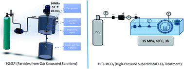
Pea protein has been associated with promoting the satiety effect. One of the issues associated with the incorporation of pea protein in food products is the product homogeneity due to its solubility and dispersibility issues. Within this context, one goal of this study was to exploit the use of supercritical fluid technology to develop Solid Lipid Pea Particle (SLPP) aiming at improving dispersibility in fat-based products. PP was encapsulated by the PGSS® (Particles from Gas Saturated Solutions) technique into glyceryl dipalmitostearate (E471) and olive oil. Different process conditions, namely pressure (7.3–20.7 MPa), temperature (51–75 °C) and equilibrium time (3–37 min) were tested in order to optimize the encapsulation of pea protein via Response Surface Methodology (RSM), following a Central Composite Rotatable Design (CCRD). Results showed that pressure and the interaction between pressure and temperature had a significant impact (p < 0.05) on the protein load and thus on the encapsulation efficiency. The highest encapsulation efficiency (96%) was achieved at 14 MPa, 51 °C and 20 min. Under these conditions, SLPP presented 0.15 mg of protein per mg of particles and 84% of lipase inhibitory activity. When compared with the PP (non-encapsulated), liposoluble pea protein particles contributed to a better product homogenization. The food industry can also take advantage of the ability of pea protein for foam stabilization in aqueous food products. Therefore, PP was treated with high-pressure supercritical CO2 treatment (HPT-scCO2) that has led to improved foaming properties when compared with the non-treated PP.
Introduction
The rise of global obesity prevalence in both adults and children may lead to a decrease in life expectancy.1 Consequently, there is an urgent need to find solutions to help control the rise in obesity. High protein diets are one common strategy to fight obesity, mainly due to some proteins' capacity to induce satiety2 compared to other macronutrients.3,4
Recently, plant protein ingredients have been receiving much attention from industry and consumers, thanks to their good environmental sustainability, health-oriented composition, reliable origin, and attractive price.5 Although, the main plant protein still remains soy protein, there is a growing interest in the use of other proteins, especially those from pulses, such as pea, chickpea, lentil, common bean and lupin, in food formulation.6 Peas (Pisum sativum L.) are used most extensively as a source of commercial protein, fiber and starch over other pulses for several reasons. They are one of the more economically viable pulses to fractionate, they are grown extensively all over the world and the hull is easy to remove.7 The increased acceptance of pea proteins is due to pea manifold qualities, good functional properties in food applications, high nutritional value, availability, and relatively low cost. Additionally, pea beans and their products are a rich source of biologically active components that may exert beneficial health and therapeutic effects.8 Moreover, it is a potential alternative to soybean in Europe, due to their capacity to modulate satiety and, thus, modulate weight loss.
Commercialized pea protein powders are commonly produced by spray drying, after several steps, such as, physical cleaning, grinding, dispersion in water, cyclone separation of starch, fibre decantation and protein flocculation/separation.7 One of the drawbacks of these commercial products is often the poor dispersibility and high sedimentation rates of their powders, due to large protein aggregation during the drying stage.9
To form an acceptable beverage, ingredients used must be dispersible and capable of forming a homogeneous suspension. The settling of solid particles within the suspension is undesirable. Encapsulation technologies could be an alternative to overcome this issue and several methods have been developed so far for protein encapsulation such as, water/oil/water (w/o/w),10 solid/oil/water (s/o/w),11 simple coacervation12 and spray-drying.13 The common point of the above-mentioned methods is the use of toxic volatile solvents, which are harmful for the environment and operators. Furthermore, these methods are not always suitable for the encapsulation of proteins and peptides, once they are labile and do not keep their native structure in the presence of organic solvents and high temperatures.14 In order to avoid the use of solvents, other encapsulation processes have been attempted.15 Supercritical fluid technology, namely Particles from Gas Saturated Solutions (PGSS®) methodology seems to be an alternative to the conventional precipitation processes for the development of liposoluble particles of proteins. Up to date, liposoluble forms of pea protein haven't been reported either using conventional or non-conventional techniques.
PGSS® methodology has been used by several authors to incorporate nutrients and bioactive compounds in lipophilic matrices. In this process, the compounds are melted and mixed with carbon dioxide in supercritical conditions (temperature > 31 °C, pressure > 7.4 MPa) forming a gas-saturated solution which is subsequently expanded to atmospheric conditions through an atomization nozzle. During the expansion, carbon dioxide is suddenly vaporized and intensely cooled down, thus providing the driving force for the solidification of the solute.16 The PGSS® process is especially suited for processing polymers and lipids in which CO2 has a large solubility and a melting depression effect.17 For this application the plasticizing and swelling effect caused by CO2 dissolution are particularly important for the improvement of the active substances incorporation. The high concentration of gas in the liquid phase leads to a considerable reduction in the melting point, viscosity and interfacial tension, helping to render substances sprayable which under classical conditions can hardly be sprayed or can even not be sprayed at all.18 This methodology is totally GRAS without contact with volatile organic solvents and allows operation at mild conditions concerning temperature, thus enhancing the advantages over conventional methods.
Another functional property of pea protein is the ability to contribute for foam formation depending on the surface properties.
Owing to their high interfacial area (and surface free energy), all foams are unstable in the thermodynamic sense. However, some differences can be made, between unstable and metastable foam structures. Protein solutions are metastable foams in which the balance of forces is such that the drainage of liquid stops when a certain film thickness is reached and, in absence of turbulences, these foams would persist almost indefinitely. In addition to foam drainage, the stability of a foam depends on the ability of the liquid films to resist excessive local thinning and rupture which may occur as a result of various disturbances. Gibbs–Marangoni surface elasticity effect is an important stabilizing effect in foams. The increased surface area as a consequence of external disturbance will lead to an increase in the surface tension (Gibbs effect).19
The potential of using pea and other legume proteins as foaming agents exists, depending on the surface properties. To create foam, proteins must migrate and be absorbed at an air/water interface with reduced surface tension. To maintain a stable foam, the protein then needs to provide a viscoelastic film around the air bubble. Unlike emulsification, the ability of proteins to produce stable foams is related to the “exposable hydrophobicity” rather than the surface hydrophobicity.20 It is expected that modifications to the protein that enhance exposure of hydrophobic areas from the interior of globular proteins should improve foaming properties.20 Both scientific and industrial applications are present in this work. The scientific application is the development and optimization of a process using specific materials while the industrial application is the innovation in the Food Industry, trying to solve dispersibility issues. Two main objectives were envisaged in this work: (i) the exploitation of Supercritical Fluid Technology (SFT) to prepare liposoluble forms of pea protein for food incorporation and (ii) the exploitation of SFT for the treatment of pea protein targeting improved foam stability.
In order to prepare liposoluble forms of pea protein, PGSS® (Particles from Gas Saturated Solutions) method was used for the encapsulation of pea protein in a lipophilic carrier for improved dispersibility in fat-based products. Response Surface Methodology (RSM) was used to model the encapsulation of pea protein, into a lipophilic carrier (glyceryl dipalmitostearate). Regarding the pea protein treatment for foam stabilization, high-pressure supercritical CO2 treatment (HPT-scCO2) was conducted. The rationale behind this treatment is that CO2 has affinity to hydrophobic moieties, allowing the improvement of the surface properties of pea protein while stabilizing air/water interfaces.
Experimental
Materials
Vegetable protein isolated from pea (Pisum sativum) (Nutralys® S85F) were kindly supplied by Roquette-frères SA, Lestrem, France.
Reagents used for PGSS® methodology were: carbon dioxide (99.95% purity, Air Liquide, Lisbon, Portugal), Biogapress Vegetal BM297ATO™ (glyceryl dipalmitostearate, HLB = 2.0 Gattefossé, France) and olive oil (purchase from a local market).
Reagents used for phosphate buffer solution (PBS) preparation included sodium phosphate monobasic monohydrate (NaH2PO4·H2O) from Sigma-Aldrich (St Quentin Fallavier, France) and sodium phosphate dibasic dehydrate (Na2HPO4·2H2O) from Riedel-de-Haën (Seelze, Germany).
Reagents used for pancreatic lipase activity included pancreatic lipase (type II), from porcine pancreas and 4-methyllumbelliferyl oleate (4MUO) from Sigma-Aldrich, Steiheim, Germany.
Reagent used for protein quantification was Bovine Serum Albumim (BSA) from Sigma-Aldrich, Steiheim, Germany.
Reagents used for the incorporation assays: propylene glycol from Fagron Iberica SAU, Terrassa, Barcelona.
Reagents used for cytotoxicity and sterilization: all cell culture media and supplements namely, RPMI 1640 medium, Fetal Bovine Serum (FBS), penicillin–streptomycin and trypsin/EDTA were obtained from Invitrogen (Invitrogen Corporation, Paisley, UK). For cytotoxicity assays phosphate buffered saline (PBS) powder, was obtained from Sigma-Aldrich (St. Louis, USA) and CellTiter 96® AQueous One Solution Cell Proliferation Assay was obtained from Promega (Wisconsin, USA). For sterilization experiments Tryptone Soya Broth (TSB) and Tryptone Soya Agar (TSA) were purchased from Oxoid (Hampshire, England).
Methods used for the encapsulation of pea protein
Particles from Gas Saturated Solutions (PGSS®). Lipophilic forms of pea protein were produced using the PGSS® technique. The dispersion was prepared in the thermostated high-pressure stirred vessel (PGSS® equipment) by adding the triacylglycerol (glyceryl dipalmitostearate) used as carrier, the pea protein powder and olive oil with a ratio pea protein
:
carrier of 1
:
4. The schematic representation of the modified PGSS equipment (Separex Supercritical & High Pressure Technology) used to produce the particles is shown in Fig. 1.
 |
| Fig. 1 Experimental setup: (1) CO2 cylinder (2) pneumatic piston pump (3) stirred vessel (electrically thermostated) (4) automated depressurisation valve (5) recovery vessel (6) nozzle. | |
Carbon dioxide was fed by a high-pressure piston pump (29723-71, Haskel International Inc., CA, USA) to a 50 cm3 electrically thermostated high-pressure stirred vessel, containing the dispersion, until the desired working pressure was reached. After an equilibrium time at 150 rpm, the mixture was depressurised by an automated depressurisation valve and atomised through a two-fluid nozzle of 710 μm of diameter with external mixing (Spraying Systems Co., Air atomization 1/4J-SS, Separex, France) to a cyclone, where it was mixed with compressed air (0.7 MPa) for a better drying. Finally the particles were recovered in an 18 L collector vessel at atmospheric pressure.
Experimental design (process optimization). Response Surface Methodology (RSM) was used to model the encapsulation of pea protein. RSM consists of a set of mathematical and statistical methods developed for modelling phenomena and finding combinations of a number of experimental factors (variables) that will lead to optimum responses. With RSM, several variables are tested simultaneously with a minimum number of trials, using special experimental designs that enable to find interactions between the variables which cannot be identified with classical approaches. In the fields of Food Science, Chemistry and Biotechnology, the Central Composite Rotatable Design (CCRD) are the most used design to optimize a process or a formulation. The encapsulation of pea protein through PGSS® was carried out following a CCRD, as a function of three factors: pressure, temperature and equilibrium time. A total of 17 experiments were performed: 8 factorial points (coded levels as (+1) and (−1)); 6 star points (coded as (+α) and (−α)); 3 center points (coded as 0) (Table 1). The pressure varied from 7.3 to 20.7 MPa, the temperature from 51.2 to 74.8 °C and the equilibrium time from 3 to 36.8 min, according to the experimental design (Table 2). The repetitions of the center points are used to determine the experimental error, which is assumed to be constant along the experimental domains.
Table 1 Actual values of the variables for the coded values
Variable, factors, unit |
Levels |
−α |
−1 |
0 |
+1 |
+α |
Pressure, P (MPa) |
7.3 |
10.0 |
14.0 |
18.0 |
20.7 |
Temperature, T (°C) |
51.2 |
56.0 |
63.0 |
70.0 |
74.8 |
Equilib. time, t (min) |
3.0 |
10.0 |
20.0 |
30.0 |
36.8 |
Table 2 Summary of experimental results
Variables |
Responses/results |
Experiment number |
Pressure, P (MPa) |
Temperature, T (°C) |
Equilibrium time, t (min) |
YCP (%) |
PC (mg mL−1) |
PL (mg per mg particles) |
EE (%) |
IPLA (%) |
Pea protein |
|
|
|
— |
3.06 ± 0.046 |
0.153 ± 0.05 |
— |
IC50 = 7.74 mg mL−1 |
1 |
10.0 (−1) |
56.0 (−1) |
10.0 (−1) |
42 |
2.16 ± 0.075 |
0.110 ± 0.004 |
72.15 ± 2.5 |
83 |
2 |
10.0 (−1) |
56.0 (−1) |
30.0 (+1) |
33 |
1.05 ± 0.039 |
0.054 ± 0.006 |
35.12 ± 4.0 |
79 |
3 |
18.0 (+1) |
56.0 (−1) |
10.0 (−1) |
57 |
2.54 ± 0.018 |
0.129 ± 0.001 |
84.59 ± 0.6 |
83 |
4 |
18.0 (+1) |
56.0 (−1) |
30.0 (+1) |
52 |
2.59 ± 0.222 |
0.132 ± 0.011 |
86.34 ± 7.4 |
84 |
5 |
10.0 (−1) |
70.0 (+1) |
10.0 (−1) |
56 |
2.14 ± 0.195 |
0.109 ± 0.010 |
71.47 ± 6.5 |
83 |
6 |
10.0 (−1) |
70.0 (+1) |
30.0 (+1) |
39 |
2.15 ± 0.154 |
0.110 ± 0.008 |
71.76 ± 5.1 |
83 |
7 |
18.0 (+1) |
70.0 (+1) |
10.0 (−1) |
73 |
2.04 ± 0.038 |
0.104 ± 0.002 |
67.95 ± 1.3 |
82 |
8 |
18.0 (+1) |
70.0 (+1) |
30.0 (+1) |
66 |
2.26 ± 0.326 |
0.115 ± 0.011 |
75.37 ± 7.1 |
83 |
9 |
14.0 (0) |
51.2 (−1.68) |
20.0 (0) |
60 |
2.90 ± 0.037 |
0.148 ± 0.002 |
96.63 ± 1.2 |
84 |
10 |
14.0 (0) |
74.8 (+1.68) |
20.0 (0) |
76 |
1.87 ± 0.037 |
0.095 ± 0.002 |
62.36 ± 1.2 |
80 |
11 |
7.3 (−1.68) |
63.0 (0) |
20.0 (0) |
64 |
1.54 ± 0.102 |
0.079 ± 0.005 |
51.52 ± 6.9 |
80 |
12 |
20.7 (+1.68) |
63.0 (0) |
20.0 (0) |
68 |
2.18 ± 0.154 |
0.111 ± 0.008 |
72.73 ± 5.1 |
84 |
13 |
14.0 (0) |
63.0 (0) |
3.2 (−1.68) |
59 |
2.51 ± 0.240 |
0.128 ± 0.008 |
83.74 ± 5.3 |
82 |
14 |
14.0 (0) |
63.0 (0) |
36.8 (+1.68) |
45 |
2.56 ± 0.084 |
0.131 ± 0.004 |
85.44 ± 2.8 |
82 |
15 |
14.0 (0) |
63.0 (0) |
20.0 (0) |
69 |
1.96 ± 0.115 |
0.100 ± 0.006 |
65.23 ± 3.8 |
82 |
16 |
14.0 (0) |
63.0 (0) |
20.0 (0) |
70 |
2.14 ± 0.215 |
0.109 ± 0.011 |
71.47 ± 7.2 |
83 |
17 |
14.0 (0) |
63.0 (0) |
20.0 (0) |
70 |
2.02 ± 0.101 |
0.103 ± 0.005 |
67.50 ± 3.4 |
82 |
Method used for the high-pressure supercritical CO2 treatment
High-pressure supercritical CO2 treatment (HPT-scCO2). 2 g of pea protein (PP) powder was mixed with glass spheres (to improve CO2 diffusivity along the sample) and placed in the high-pressure cell, which was immersed in a water bath at 40 °C, and then pressurized by CO2. The trial was carried out at a pressure of 15 MPa, temperature of 40 °C and contact time of 3 h (supercritical conditions). At the end of the treatment, the pressure was slowly released and the sample was removed from the cell (Fig. 2). The glass spheres were carefully removed from the treated PP powder.
 |
| Fig. 2 Apparatus used for the HPT-scCO2 of pea protein powder. (a) Pea protein and glass spheres; (b) high pressure cell; (c) electrical heating jacket (d) CO2 pump (e) water bath. | |
Characterization of pea protein (PP) and solid lipid pea particles (SLPP)
Scanning electron microscopy (SEM). Morphology of PP and SLPP was observed by scanning electron microscopy (FEG-SEM) (Jeol, JSM-5310 model, Japan) at 20/25 kV, samples were coated with approximately 300 Å of gold in argon atmosphere.
Differential scanning calorimetry (DSC). Differential scanning calorimetry measurements were carried out on a DSC TA instruments Q200 with module MDSC, to check the melting point of the particles and associated enthalpy. The samples were placed in an aluminium pan and sealed; the probes were heated from 20 °C to 80 °C at a rate of 1°C min−1 under nitrogen atmosphere. Measurements were done in triplicate.
Yield of collected SLPP. The yield of collected SLPP produced by PGSS® was determined by the ratio (%) of the amount of the obtained particles in the sample collector and the amount of mass (carrier and protein) initially introduced in the mixing chamber.
Total protein quantification. Solutions containing protein were prepared in PBS 0.01 M, pH 7.4 in a concentration of 20 and 80 mg mL−1 (w/v) for PP and SLPP, respectively. The solutions were left in the ultrasonication bath for 1 h, especially to ensure total destruction of the SLPP and the total release of protein. After, the samples were centrifuged at 14
000 rpm during 10 minutes and the supernatants were filtered through a PVDF membrane (0.45 μm pore size). Protein concentration of samples was determined using Direct Detect™ assay-free sample card (Cat. no. DDAC00010-8P) and the Direct Detect™ quantitation system (Cat. no. DDHW00010-00). All measurements were performed using 2 μL of sample solution per membrane position (PTFE). The standard curve was prepared using BSA. A series of dilutions (in triplicates) spanning the range from 0.125 mg mL−1 to 5 mg mL−1 was used to prepare a robust calibration curve. The protein concentration in the extraction solution (mg mL−1), the encapsulation load (mg protein per mg of particles) and the encapsulation efficiency (%) were calculated and expressed as a mean of triplicates. The encapsulation efficiency (%) was defined as the ratio between the amount of pea protein encapsulated in the triglyceride carrier and the amount of pea protein in the initial mixture (3.06 mg mL−1).
Pancreatic lipase activity. Pancreatic lipase activity was measured according to Sugiyama et al., 2007 using 4-methylumbelliferone oleate (4MUO) as the substrate.21 Solutions containing protein were prepared in PBS 0.01 M, pH 7.4 in a concentration of 20 and 80 mg mL−1 (w/v) for PP and SLPP, respectively. Briefly, 25 μL of the sample solution dissolved in water and 25 μL of the pancreatic lipase solution (1 mg mL−1) were mixed in the well of a microtiter plate. Fifty microliters of 4MUO solution (0.1 mM) dissolved in Dulbecco's phosphate buffered saline was then added to initiate the enzyme reaction. After incubation at 23 °C for 20 min, 100 μL of 0.1 M sodium citrate (pH 4.2) was added to stop the reaction. The amount of 4-methylumbelliferone released by lipase was measured using a fluorescence microplate reader (FLx800 Fluorescence Reader, Biotek, Winooski, US) at an excitation wavelength of 320 nm and an emission wavelength of 450 nm. The inhibitory activity was expressed as a percentage of the control. The 50% inhibition concentration (IC50) of the test sample was calculated from dose–response curves using software GraphPad Prism (GraphPad Software, Inc., La Jolla, CA) fit.
Foam stability. Foam stability (FS) of the protein isolates were determined using a glass foaming tube with a sintered glass grid (porosity 0) at the bottom and an inner diameter of 2 cm. Foam was made in the protein solution by introduction of air (200 cm3 min−1) to 5 mL of 1.0% protein in 0.05 M phosphate buffer (pH 7.4) in a glass tube for 15 s. Volume of the foam was recorded right after air introduction stopped (0 time), and 15 min after air introduction stopped. The foaming stability (FS) was calculated from the following equation: FS = V0 × Δt/ΔV, where ΔV was a decrease in the volume of the foam at the interval time of Δt (15 min), and V0 was the volume of the foam at 0 time. The FS was expressed in percentage.
FTIR-ATR spectroscopy. A Thermo Scientific FTIR Spectrometer (San Jose, USA), Class 1 Laser Product Nicolet 6100, was used. The equipment included an accessory with a diamond ATR crystal. The crystal provided an angle of incidence of 45. The software used for FTIR data collection was Omnic version 7.3 (Thermo Electron Corporation). Before analysis the instrument was purged with nitrogen for 15 min. As reference, the background spectrum of air was collected before the acquisition of the sample spectrum. After each sample, the crystal was rinsed with acetone and then dried with a soft tissue. To record spectra, the solid sample was poured on the ATR crystal. Spectra were recorded with a resolution of 4 cm−1, and 32 scans were averaged for each spectrum (scan 4000–550 cm−1) were used.
Particle sterilization. Sample sterilization was carried out as previously described by Li and co-workers (2013) with some modifications.22 Briefly, PP, SLPP (best conditions) and the processed carrier (glyceryl dipalmitostearate) were put directly in contact with UV irradiation during 1 hour at room temperature in a Biological Safety Cabinet (Nuaire, USA). To further confirm sterility, samples were incubated in TSB at 37 °C. After 24 hours, samples were plated in TSA Petri dishes (during 24 hours at 37 °C) to ensure the absence of bacterial contamination (data not shown).
Cell culture. Human colon carcinoma Caco-2 cells were purchased from Deutsche Sammlung von Mikroorganismen und Zellkulturen (DSMZ, Braunschweig, Germany) and were routinely grown in a standard medium: RPMI 1640 supplemented with 10% (v/v) of inactivated FBS (fetal bovine serum) and 5000 U of penicillin–streptomycin (PS). Stock cells were maintained as monolayers in 80 cm2 culture flasks. Cells were subcultured every week at a split ratio of 1 to 4 by treatment with 0.1% trypsin and 0.02% EDTA and incubated at 37 °C in a 5% CO2 humidified atmosphere. For cytotoxicity experiments Caco-2 were assayed in RPMI 1640 culture media with 0.5% of FBS and without penicillin–streptomycin.
Cytotoxicity assay. Cell toxicity assays were performed using human Caco-2 cells which are a good model of the intestinal barrier.23 Briefly, Caco-2 were seeded in 96-well plates at a density of 2 × 10⁴ cells per well and the medium was changed every 48 hours. After reaching confluence, different concentrations of sterilized samples (0.19–6.25 mg mL−1 of pea protein particles; 0.02–0.875 mg mL−1 of pea protein powder and 0.16–5.37 mg mL−1 of carrier) diluted in RPMI medium with 0.5% FBS were added to Caco-2 cells and incubated at 37 °C and 5% CO2 humidified atmosphere. After 4 hours, samples were removed and cells washed 2 times with sterile PBS. One hundred microliters of CellTiter 96® AQueous One Solution Cell Proliferation Assay reagent (MTS) diluted in RPMI medium 0.5%FBS was added to each well and left to react for 2 hours. MTS is bio-reduced by cells into a coloured formazan product that is soluble in tissue culture medium. The quantity of formazan produced was quantified spectrophotometrically at 490 nm in a microplate reader (EPOCH, Bio-Tek, USA) and is directly proportional to the number of living cells in culture. Results were expressed in terms of percentage of cellular viability relative to control (%). Experiments were performed in triplicate in three independent assays.
Dispersion in oily solutions. Propylene glycol was used as a model to incorporate PP and SLPP, once it is a transparent fluid, enabling the visualization of the behaviour of the samples in solution. 5 mg mL−1 of PP and SLPP were added to a glass container and pictures were taken to access their behaviour throughout time.
Experimental design/statistical analysis. The results of the CCRD, concerning the encapsulation efficiency (EE), yield of collected particles (YCP) and inhibition of pancreatic lipase activity (IPLA) were analyzed using the software Statistica™, version 10, from Statsoft (Tulsa, USA). Both linear and quadratic effects of each factor under study, as well as their interactions were calculated. Their significance was evaluated by analysis of variance. A surface, described by a second-order polynomial equation, was fitted to each set of experimental data points. First- and second-order coefficients of the polynomial equations were generated by regression analysis. The fit of the models was evaluated by the determination coefficients (R2) and adjusted R2 (Radj2).24,25
Results and discussion
Modelling of PP encapsulation through PGSS®
The encapsulation experiments were carried out according to the Central Composite Rotatable Design (CCRD) previously described (Tables 1 and 2). The obtained results of the experiments, i.e. encapsulation efficiency (EE), protein load (PL), protein concentration (PC), yield of the collected particles (YCP) and inhibition of pancreatic lipase activity (IPLA) are shown in Table 3. The results concerning the YCP, EE and IPLA were used to estimate both linear and quadratic effects of the variables and also their linear interactions (see ESI†). The negative significant interaction between T and P shows that as T and P increased, the encapsulation of pea protein decreased. This results can be probably explained by the denaturation of protein at higher T5 and the lack of homogeneity of the dispersion under higher P.26 PL and PC results were not considered in this analysis since they were used to calculate de EE. The response surfaces (Fig. 3) fitted to the EE and YCP can be described by second-order polynomial models as a function of pressure, temperature and equilibrium time (Table 3). For the IPLA, a lack of fit of the polynomial models exhibited by low values of R2 and Radj2 was observed. In these fitted response profiles, the significant effects p < 0.05 and those having confidence range smaller than the value of the effect, or smaller the standard deviation (data not shown), were included in the model equations of these surfaces to avoid missing an important factor.25 The good values for both R2 and Radj2 of these models (Table 3) suggest a close agreement between the experimental data and the theoretical values predicted by the model. About 77% and 78% of the observed results concerning the EE and YCP respectively, are explained by the respective models (see ESI†). Optimum conditions were observed in the response surfaces for the yield of collected particles (17.66 MPa, 70.43 °C, 17.29 min) to achieve 76.74% of YCP. Concerning the EE, only the identification of the region corresponding to the best response could be achieved.
Table 3 Model equations for the response profiles fitted to the values of encapsulation efficiency (EE) and yield of collected particles (YCP), as a function of pressure (P), temperature (T) and equilibrium time (t), and respective R2 and Radj2
Polynomial model equations |
R2 |
Radj2 |
EE = −54.355 − 1.481T + 0.044T2 + 25.327P − 0.251P2 + 0.023t2 − 0.284TP − 0.032Tt + 0.069Pt |
0.77 |
0.54 |
YCP = −282.173 + 7.621T − 0.054T2 + 7.50P − 0.213P2 + 2.872t − 0.083t2 |
0.78 |
0.65 |
 |
| Fig. 3 Fitted response surfaces to the EE and YCP as a function of temperature and pressure, as plotted for two variables with the other fixed at middle settings. | |
Analysing Fig. 3, the P had a positive effect on the encapsulation efficiency. Thus, higher pressures led to an increase in the protein encapsulation. This effect was more pronounced at lower temperatures. The best conditions were achieved at 14 MPa, 51.2 °C and 20 min. At these conditions, the particles presented a protein load of 0.148 ± 0.002 mg mg−1 and EE of 96%. Under optimum conditions, 4% of protein was not encapsulated due to product losses across the process (equipment vessel and fittings) and due to dispersibility issues (i.e. if the T and P conditions don't allow a good protein dispersion, higher losses will occur). Some previous studies were conducted by other authors on the encapsulation of proteins using PGSS® technique. For instance, Tran et al., 2015 have developed a formulation method namely modified-PGSS, for the encapsulation of lysozyme into polymeric nanoparticles in CO2 media. Moreover, isosorbide dimethyl ether, a non-toxic solvent was used in the formulation. An EE of 65% was obtained in this study.14 Another study was conducted to encapsulate bovine serum albumin (BSA) in poly(lactic-co-glycolic acid) (PLGA), polylactic acid (PLA) and Polaxamer 407 (PEO-PPO-PEO) using PGSS®.
The EE ranged between 96.85 and 101.75%. However the process yield varied between 19.7% and 41.74%.15 Caliceti, et al., 2010, have investigated the production of lipid-PEG particles incorporating ribonuclease A by PGSS® to obtain solid micro- and nanoparticles. The product yield was of about 30–35%.27 As shown in Table 2, the yield of collected particles (mass of particles collected/mass of product introduced in the pressure vessel) ranged from 33 to 76%, indicating a loss of product and possibly of fine particles. This product yield is highly correlated with the process parameters (P and T). This variability should not be considered in absolute terms when scaling-up the process. Overall, the work presented in this manuscript enabled the obtention of both higher EE (≈96%) and higher process yield (≈76%) compared to the works mentioned above. The repeatability (coefficient of variation) of the encapsulation process through PGSS® was 4.6%, taking into account three samples of the design (centre points).
Pancreatic lipase activity of PP (pea protein) and SLPP (solid lipid pea particles)
Pancreatic lipase, a key enzyme responsible for triglyceride absorption in the small intestine, is secreted from the pancreas and hydrolyses triglyceride into glycerol and fatty acids. Suppression and delay of triglyceride digestion and absorption through inhibition of lipase is considered to be one of the more effective strategy to control of hyperlipidaemia and obesity.28,29 The inhibition of pancreatic lipase activity was tested for the PP (IC50) and all samples of the design (SLPP).
The IC50 value for pancreatic lipase inhibition of PP was shown to be 7.74 mg mL−1 (see ESI†). Similar results were found for lentils, which have shown IC50 values between 6.26 and 9.26 mg mL−1.29 In a recent study, Lee et al., 2015, have conducted a study to determine the inhibitory potential of seven selected legumes against pancreatic lipase. The IC50 of the tested samples ranged between 5.90 and 8.14 mg mL−1.30 Slanc et al., 2009 studied the pancreatic lipase inhibition of plant extracts, including Pisum sativum.31 However, the inhibitory activity was not determined. Regarding SLPP, the inhibition of pancreatic lipase activity ranged between 79 and 84% for protein concentrations from 1 to 3 mg mL−1 (Table 3). From the results it was possible to visualize that there is a linear correlation between the protein content and the inhibition of pancreatic lipase activity with a correlation factor (R2) of 0.65 (Fig. 4). These results can probably suggest that the encapsulated form (SLPP) can be even more effective in weight management than the PP. Moreover, concerning the colloidal aspects of protein digestion, in a study conducted by Marciani et al., 2009, a comparison between an acid-stable and an acid-unstable emulsion was investigated. The acid-unstable emulsion broke and rapidly layered in the stomach. Gastric emptying of meal volume was slower for the acid stable emulsion. Additionally, the rate of energy delivery of fat from the stomach to the duodenum was not different but the acid stable emulsion induced fullness, decreased hunger and decreased appetite.32 This result may propose that the SLPP can have the same behaviour while in the stomach, once the carrier used in the formulation of pea protein is a sustained release agent.
 |
| Fig. 4 Correlation between protein concentration and inhibition of pancreatic lipase activity. | |
Accordingly, these results suggest that PP and thus SLPP can be a useful dietary adjunct for the management of body weight and obesity.
Particle morphology by scanning electron microscopy
SEM microphotographs of the powders produced by PGSS® process for comparable conditions are shown in Fig. 5. When processing at higher pressures (18 MPa), a greater fragmentation was observed than when processing at lower pressures (10 MPa). Furthermore, as pressure is increased, larger amounts of CO2 are dissolved in the melted carrier and higher pressure drop is produced across the nozzle; therefore, more CO2 gas bubbles are formed increasing the cooling rate which originates porous particles as the gas cannot diffuse out of the particles perforating particle surface. The average size of particles obtained was 40 μm, approximately.
 |
| Fig. 5 SEM pictures at different conditions of pressure and temperature and an equilibrium time of 20 minutes. | |
According to other studies of encapsulation of proteins using PGSS®, the mean particle sizes ranged between 4 and 80 μm.14,15,27
From Fig. 5, it is noticeable that the PP was totally encapsulated in glyceryl dipalmitostearate.
Determination of the melting point for the particles with higher protein content
The melting point was evaluated for the particles with higher protein content. The thermogram is presented in Fig. 6. From the thermogram presented above, the melting temperature of the particles having the highest protein content was 56.06 °C. The DSC measurement was carried out from 20 °C to 80 °C once higher temperatures would led to protein denaturation, forming aggregates through hydrophobic and covalent interactions.33
 |
| Fig. 6 Thermogram of particles with highest betacyanin content. | |
Cytotoxicity evaluation
The incidence of UV irradiation during 1 hour directly into the samples showed to be an effective sterilization method, as confirmed by the absence of microorganism's growth in TSA plates (data not show). This simple procedure allowed the evaluation of cytotoxicity of samples without the interference of microorganism's contamination, which could promote the MTS reduction to formazan crystals contributing to an overestimated absorbance.
The cytotoxicity of the encapsulated and non-encapsulated pea protein as well as the processed carrier (P. carrier) were evaluated on human colon carcinoma Caco-2 cells after incubation of increasing concentrations of samples. The results of toxicity experiments revealed that samples did not show cytotoxicity in the concentrations tested (Fig. 7) relatively to the control (100% of cell viability) after 4 hours of incubation.
 |
| Fig. 7 Cell viability of SLPP, processed carried and PP after 4 hours of incubation in Caco-2 cells. | |
Incorporation in propylene glycol
The PP and the SLPP were added to propylene glycol to check their dispersion and homogeneity in solution. Pictures have been taken each 15 min and after 150 min and the images are shown in Fig. 8. From the images it is possible to see a good homogenization and dispersion of SLPP and a high sedimentation rate of PP throughout time.
 |
| Fig. 8 Incorporation of PP and SLPP in propylene glycol. | |
Impact of high pressure supercritical CO2 treatment (HPT-scCO2) on physical properties of pea protein (PP) powder
To evaluate the impact of Supercritical Fluid Technology (SFT) on foam stabilization, PP was treated using HPT-scCO2. The structural properties of globular proteins are heat- and high pressure sensitive, making their functional properties greatly dependant on food processing conditions.34 PP was put in contact with supercritical CO2, trying to improve foam stability in aqueous food products. The rationale behind this process is that CO2 has affinity to hydrophobic moieties, possibly allowing the improvement of the surface properties of PP while stabilizing air/water interfaces. The ability of proteins to produce stable foams is related to “exposable hydrophobicity”, meaning that the protein needs to be more unfolded.20
In Fig. 9, the results obtained after HPT-scCO2 of PP are presented, concerning morphology (a), structure (b), protein quantification (c) and foam stability (d). In this work, it was possible to obtain a PP through high-pressure supercritical carbon dioxide treatment with improved surface properties concerning hydrophobicity and thus, improved foam stabilization of aqueous solutions compared to non-treated PP without protein denaturation.
 |
| Fig. 9 Results obtained for the HPT-scCO2 of PP regarding: (a) morphology; (b) structure; (c) protein quantification and (d) foam stability. | |
Regarding the morphology (a), it was possible to visualize modifications upon HPT-scCO2. The surface area has become higher due to CO2 compression. Nevertheless, the structure of PP has not been modified during HPT-scCO2, confirmed by FTIR-ATR (b). In one review article,35 the authors referred that changes in pressure can alter the native protein structure and most proteins unfold when exposed to pressures of 0.6–0.7 GPa. The application of high pressures causes a protein to adopt the smallest possible volume, inducing unfolding via a pathway distinct from thermal unfolding.36
Concerning the functional properties, the protein determination of both PP and HPT-PP has led to the conclusion that the contact with CO2 had an impact on protein solubility, once there was a slightly decay on protein concentration of HPT-PP (c). When the HPT-PP was evaluated for foam stability, improvements were observed (d). The effect of dynamic high pressure homogenization on the aggregation state of the soy protein isolate was investigated by Keerati-u-rai & Corredig, 2009.37 The dispersions were subjected to homogenization at two different pressures, 26 and 65 MPa. The results demonstrated that dynamic high pressure homogenization causes changes in the supramolecular structure of the soy proteins. Silva & Weber, 1993 have reported that high pressure treatment modifies protein conformation by affecting hydrogen and hydrophobic interactions, disrupting the tertiary and/or quaternary structure of most globular protein. Depending on the conditions, the changes can be reversible.38 Relkin, 1998, reported that some functional groups of milk proteins initially covered in their core (high ordered folded structures), turn out to be more exposed to the surface upon to heat induced and – high-pressure transitions.34 Moreover, the yielded denatured proteins offered an increased surface hydrophobicity together with solubility and structure changes namely, aggregation and film-forming abilities around oil droplets or gas bubbles.37,39 Further investigations are needed regarding hydrophobicity and protein unfolding to better understand the impact of this process on protein structure and conformation.
Conclusions
In this work, pea protein loaded particles were successfully produced through supercritical fluid technology, namely PGSS®. By using the statistical tool of response surface methodology, it was possible to find the best conditions for protein encapsulation (14 MPa, 51.2 °C and 20 min). Under these conditions, pea protein particles presented 0.148 mg of protein per mg of particles and exhibited an enhanced inhibition of pancreatic lipase activity (relatively to pea protein), allowing a good dispersion after incorporation in an oily model. From the results obtained it can be concluded that PGSS® can be considered as an adequate technology to develop lipophilic forms of a pea protein for the application in food industry. To evaluate the impact of Supercritical Fluid Technology (SFT) on foam stabilization, pea protein was treated using HPT-scCO2 that has led to improved foam stability when compared with the non-treated pea protein powder. To conclude, it would be of interest to develop tailored pea proteins for specific food applications.
Acknowledgements
This work acknowledges Fundação para a Ciência e Tecnologia (FCT) through PEst-OE/EQB/LA0004/2011, the financial support received from FCT through the doctoral (SFRH/BDE/51856/2012) fellowship and iNOVA4Health-UID/Multi/04462/2013, a program financially supported by FCT/Ministério da Educação e Ciência, through national funds and co-funded by FEDER under the PT2020 Partnership Agreement.
References
- P. W. Franks, R. L. Hanson, W. C. Knowler, M. L. Sievers, P. H. Bennett and H. C. Looker, N. Engl. J. Med., 2010, 362, 485–493 CrossRef PubMed.
- A. Jahan-Mihan, B. L. Luhovyy, D. El Khoury and G. H. Anderson, Nutrients, 2011, 3, 574–603 CrossRef PubMed.
- G. H. Anderson and S. E. Moore, J. Nutr., 2004, 134, 974S–979S CAS.
- M. S. Westerterp-Plantenga, Curr. Opin. Clin. Nutr. Metab. Care, 2003, 6, 635–638 CrossRef PubMed.
- E. Sirtori, I. Isak, D. Resta, G. Boschin and A. Arnoldi, Food Chem., 2012, 134, 113–121 CrossRef.
- J. Boye, F. Zare and A. Pletch, Food Res. Int., 2010, 43, 414–431 CrossRef CAS.
- S. Arntfield and H. Maskus, in Handbook of food proteins, Woodhead Publishing Limited Cornwall, UK, 2011, pp. 233–266 Search PubMed.
- F. Roy, J. I. Boye and B. K. Simpson, Food Res. Int., 2010, 43, 432–442 CrossRef CAS.
- D. Sağlam, P. Venema, E. van der Linden and R. de Vries, Curr. Opin. Colloid Interface Sci., 2014, 19, 428–437 CrossRef.
- A. Aubert-Pouëssel, M. C. Venier-Julienne, A. Clavreul, M. Sergent, C. Jollivet, C. N. Montero-Menei, E. Garcion, D. C. Bibby, P. Menei and J. P. Benoit, J. Controlled Release, 2004, 95, 463–475 CrossRef PubMed.
- T. Morita, Y. Sakamura, Y. Horikiri, T. Suzuki and H. Yoshino, J. Controlled Release, 2000, 69, 435–444 CrossRef CAS PubMed.
- C. Thomasin, H. P. Merkle and B. A. Gander, Int. J. Pharm., 1997, 147, 173–186 CrossRef CAS.
- B. Gander, P. Johansen, H. Nam-Trân and H. P. Merkle, Int. J. Pharm., 1996, 129, 51–61 CrossRef CAS.
- M. K. Tran, A. Swed, B. Calvignac, K. N. Dang, L. N. Hassani, T. Cordonnier and F. Boury, J. Mater. Chem. B, 2015, 3, 1573–1582 RSC.
- D. R. Perinelli, G. Bonacucina, M. Cespi, A. Naylor, M. Whitaker, G. F. Palmieri, G. Giorgioni and L. Casettari, Int. J. Pharm., 2014, 468, 250–257 CrossRef CAS PubMed.
- E. de Paz, A. Martin, C. M. Duarte and M. J. Cocero, Powder Technol., 2012, 217, 77–83 CrossRef CAS.
- E. Lack, E. Weidner, Z. Knez, S. Gruner, B. Weinreich and H. Seidlitz, Proceedings of the 1st Vienna International Conference: Micro- and Nano-Technology, Vienna, Austria, 2005 Search PubMed.
- E. Weidner, J. Supercrit. Fluids, 2009, 47, 556–565 CrossRef CAS.
- D. J. Shaw, in Introduction to Colloid and Surface Chemistry, ed. D. J. Shaw, Butterworth-Heinemann, Oxford, 4th edn, 1992, pp. 262–276, DOI:10.1016/B978-0-08-050910-5.50014-1.
- K. D. Schwenke, Nahrung/Food, 2001, 45, 377–381 CrossRef CAS PubMed.
- H. Sugiyama, Y. Akazome, T. Shoji, A. Yamaguchi, M. Yasue, T. Kanda and Y. Ohtake, J. Agric. Food Chem., 2007, 55, 4604–4609 CrossRef CAS PubMed.
- L. Li, K. Mak, J. Shi, C. Leung, C. Wong, C. Leung, C. Mak, K. Chan, N. Chan and E. Wu, Microelectron. Eng., 2013, 111, 310–313 CrossRef CAS.
- Y. Sambuy, I. de Angelis, G. Ranaldi, M. L. Scarino, A. Stammati and F. Zucco, Cell Biol. Toxicol., 2005, 21, 1–26 CrossRef CAS PubMed.
- M. C. Gacula Jr, J. Singh, J. Bi and S. Altan, Statistical methods in food and consumer research, Academic Press, 2008 Search PubMed.
- P. D. Haaland, Experimental design in biotechnology, CRC press, 1989 Search PubMed.
- C. Saldanha do Carmo, A. N. Nunes, A. T. Serra, S. Ferreira-Dias, I. Nogueira and C. M. M. Duarte, Green Chem., 2015, 17, 1510–1518 RSC.
- K. Vezzú, D. Borin, A. Bertucco, S. Bersani, S. Salmaso and P. Caliceti, J. Supercrit. Fluids, 2010, 54, 328–334 CrossRef.
- J. X. Bao, K. H. Li, N. Z. Yi, H. L. Jeong and K. S. Chang, Arch. Pharmacal Res., 2005, 28, 180–185 CrossRef.
- B. Zhang, Z. Deng, D. D. Ramdath, Y. Tang, P. X. Chen, R. Liu, Q. Liu and R. Tsao, Food Chem., 2015, 172, 862–872 CrossRef CAS PubMed.
- S. S. Lee, N. Mohd Esa and S. P. Loh, J. Food Biochem., 2015, 39, 485–490 CrossRef CAS.
- P. Slanc, B. Doljak, S. Kreft, M. Lunder, D. Janeš and B. Štrukelj, Phytother. Res., 2009, 23, 874–877 CrossRef CAS PubMed.
- L. Marciani, R. Faulks, M. S. J. Wickham, D. Bush, B. Pick, J. Wright, E. F. Cox, A. Fillery-Travis, P. A. Gowland and R. C. Spiller, Br. J. Nutr., 2009, 101, 919–928 CrossRef CAS PubMed.
- M. A. de la Fuente, H. Singh and Y. Hemar, Trends Food Sci. Technol., 2002, 13, 262–274 CrossRef CAS.
- P. Relkin, Int. J. Biol. Macromol., 1998, 22, 59–66 CrossRef CAS PubMed.
- J. K. Raynes, J. A. Carver, S. L. Gras and J. A. Gerrard, Trends Food Sci. Technol., 2014, 37, 42–50 CrossRef CAS.
- S. Mangialardo, F. Piccirilli, A. Perucchi, P. Dore and P. Postorino, J. Raman Spectrosc., 2012, 43, 692–700 CrossRef CAS.
- M. Keerati-U-Rai and M. Corredig, J. Agric. Food Chem., 2009, 57, 3556–3562 CrossRef CAS PubMed.
- J. L. Silva and G. Weber, Annu. Rev. Phys. Chem., 1993, 44, 89–113 CrossRef CAS PubMed.
- J. Lefebvre, Surface activity of proteins: Chemical and physicochemical modifications, 1996, p. 181 Search PubMed.
Footnote |
† Electronic supplementary information (ESI) available. See DOI: 10.1039/c5ra22452g |
|
This journal is © The Royal Society of Chemistry 2016 |
Click here to see how this site uses Cookies. View our privacy policy here.