Maryam Shekofteh-Gohari and
Aziz Habibi-Yangjeh*
Department of Chemistry, Faculty of Science, University of Mohaghegh Ardabili, P.O. Box 179, Ardabil, Iran. E-mail: ahabibi@uma.ac.ir; Fax: +98 045 33514701; Tel: +98 045 33514702
First published on 23rd December 2015
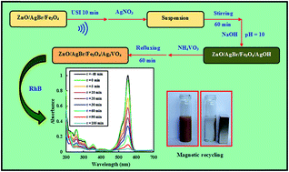
In the present work, novel magnetically separable ZnO/AgBr/Fe3O4/Ag3VO4 nanocomposites with different weight percentages of Ag3VO4 were successfully prepared. The preparation method is a facile large-scale method with a low temperature of 96 °C. Characterization techniques of XRD, EDX, SEM, TEM, UV-vis DRS, FT-IR, PL, and VSM were applied to study the microstructure, purity, morphology, spectroscopic, and magnetic properties of the resultant samples. Photocatalytic activity was evaluated by degradation of rhodamine B under visible-light irradiation. The results indicated that the nanocomposite with 10% Ag3VO4 has the best activity. The photocatalytic activity of this nanocomposite is about 2.8 and 3.5-fold higher than those of the ZnO/AgBr/Fe3O4 and ZnO/Ag3VO4/Fe3O4 nanocomposites, respectively. The highly enhanced activity was attributed to the formation of tandem n–n heterojunctions between ZnO, AgBr, and Ag3VO4 which suppresses recombination of the photogenerated electron–hole pairs. Furthermore, the enhanced activity of the nanocomposite for degradation of two more dye pollutants and a colorless pollutant was confirmed. Finally, the magnetic photocatalyst was recycled using an external magnetic field with reasonable activity after four runs.
On the other hand, after using photocatalysts in the reaction system, they cannot be entirely recycled, leading to generation of secondary pollution. Consequently, a major problem in large-scale utilization of photocatalysts is removal of them from the treated solutions. Magnetic photocatalysts, as new generation of photocatalysts, could tackle the above mentioned problem.29 However, to the best of our knowledge, there is no report about the preparation and photocatalytic activity of the magnetic ZnO/AgBr/Fe3O4/Ag3VO4 nanocomposites with tandem n–n heterojunctions.
Based on the above considerations, we report novel magnetically separable ZnO/AgBr/Fe3O4/Ag3VO4 nanocomposites with different weight percents of Ag3VO4 prepared using a facile refluxing method at a low temperature of 96 °C. The microstructure, purity, morphology, spectroscopic, and magnetic properties of the prepared samples were studied using X-ray diffraction (XRD), energy dispersive analysis of X-rays (EDX), scanning electron microscopy (SEM), transmission electron microscopy (TEM), UV-vis diffuse reflectance spectroscopy (UV-vis DRS), Fourier transform-infrared spectroscopy (FT-IR), photoluminescence spectroscopy (PL), and vibrating sample magnetometry (VSM) techniques. Photocatalytic activity of the nanocomposites was investigated by degradations of rhodamine B (RhB), methylene blue (MB), methyl orange (MO), and phenol under visible-light irradiation. To further elucidate the photocatalytic degradation mechanism, a series of scavengers for the reactive species were employed. It was demonstrated that weight percent of Ag3VO4, refluxing time, and calcination temperature have considerable influence on the degradation reaction. Finally, the nanocomposite was magnetically separated from the treated solution after four successive cycles.
In a typical procedure for preparation of the ZnO/AgBr/Fe3O4/Ag3VO4 (10%) nanocomposite, where 10% is the weight percent of Ag3VO4, 0.450 g of ZnO/AgBr/Fe3O4 nanocomposite was dispersed into 150 mL of water by ultrasonic irradiation for 10 min. Then, silver nitrate (0.058 g) was dissolved in the suspension and mechanically stirred for 60 min. After that, an aqueous solution of NaOH (5 M) was dropwise added into the solution under stirring at room temperature until the pH of the solution reached to 10. Then, an aqueous solution of ammonium metavanadate (0.013 g in 50 mL of water) was slowly added to the formed suspension. Finally the suspension was refluxed at 96 °C for 60 min. The suspension was centrifuged to get the precipitate out and washed twice with water and ethanol and dried in an oven at 60 °C for 24 h. The schematic diagram for preparation of the nanocomposites can be illustrated in Scheme 1.
![]() | ||
Fig. 1 XRD patterns for the Fe3O4, ZnO/Fe3O4, ZnO/AgBr/Fe3O4, ZnO/Ag3VO4/Fe3O4, and ZnO/AgBr/Fe3O4/Ag3VO4 nanocomposites with different weight percents of Ag3VO4. |
Purity of the samples was verified by EDX spectra and the results for the ZnO/Fe3O4, ZnO/AgBr/Fe3O4, ZnO/Ag3VO4/Fe3O4, and ZnO/AgBr/Fe3O4/Ag3VO4 (10%) samples are shown in Fig. 2a. It is evident that the samples have reasonable purity and all of the peaks are simply ascribed to elements of the resultant samples. Other peaks in these spectra are related to the elements applied for sputter coating of the samples on the EDX stage. Weight percents of Zn, O, Ag, Br, Fe, and V elements in the ZnO/AgBr/Fe3O4/Ag3VO4 (10%) nanocomposite are 37.5, 23.8, 17.0, 3.8, 11.4, and 6.4, respectively. Furthermore, EDX mapping was employed to display the element distributions in the ZnO/AgBr/Fe3O4/Ag3VO4 (10%) nanocomposite and the results are shown in Fig. 2b–h. It is evident that there is homogeneous distribution for the elements. Hence, it was concluded that the counterparts of the nanocomposite have uniformly combined to each other.
![]() | ||
Fig. 2 (a) EDX spectra for the ZnO/Fe3O4, ZnO/AgBr/Fe3O4, ZnO/Ag3VO4/Fe3O4, and ZnO/AgBr/Fe3O4/Ag3VO4 (10%) samples. (b)–(h) EDX mapping of the ZnO/AgBr/Fe3O4/Ag3VO4 (10%) nanocomposite. |
Morphology and junction between constituents of the ZnO/AgBr/Fe3O4/Ag3VO4 (10%) nanocomposite was investigated by SEM and TEM techniques and the results are shown in Fig. 3. For this nanocomposite, particles of Fe3O4, AgBr, and Ag3VO4 are clearly seen on ZnO sheets (Fig. 3a). Moreover, the TEM images of the nanocomposite confirm distribution of Fe3O4, AgBr, and Ag3VO4 particles on the ZnO plates (Fig. 3b and c).
![]() | ||
Fig. 3 (a) SEM image of the ZnO/AgBr/Fe3O4/Ag3VO4 (10%) nanocomposite. (b) and (c) TEM images of the ZnO/AgBr/Fe3O4/Ag3VO4 (10%) nanocomposite with two magnifications. |
It is believed that photocatalytic activity is closely related to extent of generation, separation, and transfer of the photogenerated charge carriers.34 UV-vis spectra of photocatalysts give useful information about extent of electron–hole pairs generated under the light irradiation. Hence, UV-vis DRS spectra of the resultant samples were provided and the results are shown in Fig. 4a. As can be seen, the ZnO/AgBr/Fe3O4, ZnO/Ag3VO4/Fe3O4, and ZnO/AgBr/Fe3O4/Ag3VO4 nanocomposites have intense absorption in the visible-light range. Consequently, under visible-light irradiation, more electron–hole pairs are generated on the nanocomposites, resulting in enhanced photocatalytic activity.
To characterize structural properties of the resultant samples, FT-IR spectra of the Fe3O4, ZnO/AgBr/Fe3O4, ZnO/Ag3VO4/Fe3O4, and ZnO/AgBr/Fe3O4/Ag3VO4 (10%) samples were provided and the spectra are shown in Fig. 4b. For the all samples, the broad absorption bands around 3200–3400 cm−1 are related to the O–H stretching vibration of adsorbed water. For the Fe3O4 nanoparticles, two characteristic peaks at 435 and 610 cm−1 are related to the vibrations of Fe–O bond.29 For the samples with ZnO, the peak at 560 cm−1 is ascribed to the stretching vibration of the Zn–O bond.28 In the case of the samples containing Ag3VO4, the peaks for Ag–V and V–O bonds were appeared about at 770, 880, and 920 cm−1.24 For the ZnO/AgBr/Fe3O4/Ag3VO4 (10%) nanocomposite, the peaks for Fe–O, Zn–O, Ag–V, and V–O bonds are clearly seen in the wavenumbers similar to its counterparts.
It is well known that magnetic properties of photocatalysts affect on magnetic separability of them. Hence, magnetization of the pure Fe3O4 nanoparticles and ZnO/AgBr/Fe3O4/Ag3VO4 (10%) nanocomposite were investigated and the results are shown in Fig. 5. As can be seen, saturated magnetization for the Fe3O4 nanoparticles and the nanocomposite at 8500 Oe are 55.5 and 6.26 emu g−1, respectively. Decrease of the magnetization of the nanocomposite is simply related to the presence of the nonmagnetic ZnO, AgBr, and Ag3VO4 particles along with the magnetic Fe3O4 nanoparticles. As can be seen in inset of Fig. 5, saturated magnetization of the nanocomposite is high enough to separate it from the solution after decontamination processes.
![]() | ||
Fig. 5 Magnetization curves for the Fe3O4 nanoparticles and ZnO/AgBr/Fe3O4/Ag3VO4 (10%) nanocomposite. |
To investigate visible-light photocatalytic activity of the prepared samples, RhB, a widely used dye, was selected as a target pollutant. The degradation reaction was performed over the Fe3O4, ZnO/Fe3O4, ZnO/AgBr/Fe3O4, ZnO/Ag3VO4/Fe3O4, and ZnO/AgBr/Fe3O4/Ag3VO4 nanocomposites with different weight percents of Ag3VO4 and the results are displayed in Fig. 6a. Furthermore, photolysis of RhB under the visible-light irradiation (in absence of any photocatalyst) was carried out and the results are shown in this figure. It is evident that RhB has reasonable stability under the light irradiation for 360 min. It is observed that photocatalytic activity of the ZnO/AgBr/Fe3O4/Ag3VO4 nanocomposites with different weight percents of Ag3VO4 is higher than those of the Fe3O4, ZnO/Fe3O4, ZnO/AgBr/Fe3O4, and ZnO/Ag3VO4/Fe3O4 samples. Among the prepared samples, the ZnO/AgBr/Fe3O4/Ag3VO4 (10%) nanocomposite has the superior activity in the degradation reaction. After the light irradiation for 105 min, about 21, 16, 69, 65, and 98% of RhB were degraded over the Fe3O4, ZnO/Fe3O4, ZnO/AgBr/Fe3O4, ZnO/Ag3VO4/Fe3O4, and ZnO/AgBr/Fe3O4/Ag3VO4 (10%) samples, respectively. Fig. 6b–e shows plots of absorbance versus wavelength for degradation of RhB over the ZnO/Fe3O4, ZnO/AgBr/Fe3O4, ZnO/Ag3VO4/Fe3O4, and ZnO/AgBr/Fe3O4/Ag3VO4 (10%) samples at various irradiation times. It is evident that intensity of the absorption peaks continuously decreases and there are not any blue-shifts for RhB absorption peaks centered at 554 nm. Therefore, it was concluded that degradations of RhB over the nanocomposite takes place by aromatic ring-opening mechanism.35,36
In order to describe quantitatively photocatalytic activity of the resultant samples, observed first-order rate constants (kobs) of the degradation reaction over the samples was calculated using the slope of equation ln[RhB] = ln[RhB]o − kobst,11 in which [RhB] and [RhB]o are concentrations of RhB in time of t and zero, respectively and the results are shown in Fig. 7a. As can be seen, the degradation rate constant increases with weight percent of Ag3VO4 up to 10 and then decreases. The degradation rate constants of RhB over the Fe3O4, ZnO/Fe3O4, ZnO/AgBr/Fe3O4, ZnO/Ag3VO4/Fe3O4, and ZnO/AgBr/Fe3O4/Ag3VO4 (10%) samples 4.92 × 10⁻⁴, 4.74 × 10⁻⁴, 102 × 10−4, 82.6 × 10⁻⁴, and 290 × 10−4 min−1, respectively. Hence, activity of the nanocomposite is about 59, 61, 2.8, and 3.5-fold larger than those of the Fe3O4, ZnO/Fe3O4, ZnO/AgBr/Fe3O4, and ZnO/Ag3VO4/Fe3O4 samples, respectively. The emission intensity in a PL spectrum is closely related to recombination of photogenerated electron–hole pairs. Consequently, PL spectra have useful information about separation of photogenerated charge carriers. To disclose the enhanced activity of the ZnO/AgBr/Fe3O4/Ag3VO4 (10%) nanocomposite relative to the ZnO/Fe3O4, ZnO/AgBr/Fe3O4, ZnO/Ag3VO4/Fe3O4, ZnO/AgBr/Fe3O4/Ag3VO4 (5%), and ZnO/AgBr/Fe3O4/Ag3VO4 (20%) samples, their PL spectra were provided and the results are shown in Fig. 7b. As can be seen, compared with the other samples, intensity of the PL spectrum for the ZnO/AgBr/Fe3O4/Ag3VO4 (10%) nanocomposite shows significant decrease, suggesting efficiently separation of the charge carriers in this nanocomposite relative to the other samples. This result clearly indicates that hybridization of the ZnO/Fe3O4 with AgBr and Ag3VO4 particles promotes the separation of electron–hole pairs in the ZnO/AgBr/Fe3O4/Ag3VO4 nanocomposites. However, similar to many multi-component photocatalysts,37–39 further increase in weight percent of Ag3VO4 leads to a decrease in the degradation rate constant. This may be due to agglomeration of the excess Ag3VO4 particles on the nanocomposites, which reduces the heterojunctions between Ag3VO4 and ZnO/AgBr/Fe3O4 counterparts, thereby separation of the charge carriers decreases. Hence, the amount of Ag3VO4 in the nanocomposites needs an optimum value.
To describe the enhanced activity of the nanocomposite, a hypothesized mechanism for separation of the charge carriers is presented (Fig. 8). The conduction band (CB) and valence band (CB) energies for ZnO, AgBr, and Ag3VO4 at the point of zero charge were calculated using Butler and Ginley model40 and the results are shown in Table 1. Being as n-type semiconductors, the Fermi levels for ZnO, AgBr, and Ag3VO4 are close to their CB levels. The Fermi level of ZnO is more negative than those of AgBr and Ag3VO4. Hence, after contacting of ZnO with AgBr, electrons simply flow from ZnO to AgBr until their Fermi levels coincide.41,42 As a result, negative and positive charges are accumulated on AgBr and ZnO counterparts of the nanocomposite, respectively. Hence, an n–n heterojunction with the same Fermi level is formed (Fig. 8). Separation of the charge carriers by n–n heterojunction produces an internal electrostatic field directed from the ZnO to AgBr. Moreover, similar n–n heterojunction is formed after contacting ZnO with Ag3VO4. In this region, electrons transfer from ZnO to Ag3VO4. After reaching to equilibrium state, an electric field is formed at the interface area, directed from ZnO to Ag3VO4. As a result, two n–n heterojunctions are formed in the contacting region between ZnO with AgBr in one side and ZnO with Ag3VO4 in the other side. Hence, tandem n–n heterojunctions between these n-type semiconductors are formed.28 Due to their narrow band gaps, under the visible-light irradiation, electron–hole pairs are generated on AgBr and Ag3VO4 counterparts of the ZnO/AgBr/Fe3O4/Ag3VO4 nanocomposites. The photogenerated electrons on the CB levels of AgBr and Ag3VO4 simply transfer to the CB of ZnO with the help of the internal electrostatic field produced by tandem n–n heterojunctions. On the other hand, the VB energies of AgBr and Ag3VO4 are less positive than that of the ZnO. Consequently, the holes cannot transfer from the VB levels of AgBr and Ag3VO4 to that of the ZnO. Finally, after transfer of the electrons, holes and electrons are accumulated on the narrow band gap semiconductors and ZnO, respectively. As a result, due to formation of tandem n–n heterojunctions between ZnO with AgBr and ZnO with Ag3VO4, the charge carriers are spatially separated from each other, resulting in an increased lifetime for the electron–hole pairs. Afterwards, the separated charge carriers in the ZnO/AgBr/Fe3O4/Ag3VO4 nanocomposites effectively migrate to surface of the photocatalyst and participate in the degradation reactions, leading to enhanced activity relative to their counterparts. It is noteworthy that separation of the photogenerated charge carriers in AgBr and Ag3VO4 could not take place without formation of tandem n–n heterojunctions; because, the CB levels of AgBr and Ag3VO4 are less negative than the CB of ZnO. Hence, the photogenerated electrons cannot transfer from AgBr and Ag3VO4 to ZnO, resulting in more recombination of the photogenerated electron–hole pairs and high intensity of the PL spectrum. However, separation of the charge carriers in the nanocomposite was demonstrated by the PL spectra. Hence, formation of the tandem n–n heterojunctions between the three n-type semiconductors is confirmed.
![]() | ||
Fig. 8 Hypothesized mechanism for separation of the charge carriers in the ZnO/AgBr/Fe3O4/Ag3VO4 nanocomposites via tandem n–n heterojunctions. |
Semiconductor | χ (eV) | VB energy (eV) | CB energy (eV) | Eg (eV) |
---|---|---|---|---|
ZnO | 5.76 | +2.86 | −0.34 | 3.20 |
AgBr | 5.80 | +2.60 | 0.00 | 2.60 |
Ag3VO4 | 5.64 | +2.24 | +0.04 | 2.20 |
In order to explore the role of reactive species responsible for degradation of RhB over the photocatalyst, the degradation reaction was conducted in the presence of a series of scavengers and the results are shown in Fig. 9. In the present work, benzoquinone, ammonium oxalate, and 2-PrOH were used as scavengers of ˙O2−, h+, and ˙OH, respectively.43 It is evident that decrease of the rate constant in the presence of scavengers is as benzoquinone > ammonium oxalate > 2-PrOH. Hence, it is concluded that the effect of superoxide ions and holes on the degradation reaction is higher than that of hydroxyl radicals.
![]() | ||
Fig. 9 The degradation rate constant of RhB over the ZnO/AgBr/Fe3O4/Ag3VO4 (10%) nanocomposite in presence of various scavengers. |
It is well known that preparation time of photocatalysts can considerably affect morphology, crystallinity, aggregation, and size of particles. Hence, the preparation time could remarkably affect photocatalytic activity. To find optimum preparation time, the ZnO/AgBr/Fe3O4/Ag3VO4 (10%) nanocomposite prepared by refluxing for 15, 30, 60, 120, and 240 minutes and the results are shown in Fig. 10a. It can be seen that the degradation rate constant increases with increasing the preparation time up to 60 min and then decreases. Decrease of the photocatalytic activity for the samples prepared with higher refluxing time could be related to more aggregation of the particles, resulting in decreased surface area of the photocatalyst.
To investigate the effect of calcination temperature on the photocatalytic activity, the ZnO/AgBr/Fe3O4/Ag3VO4 (10%) nanocomposite was calcined at 200, 300, 400, 500, and 600 °C for 2 h and the degradation reactions were carried out over them and the rate constant are shown in Fig. 10b. It can be seen that the degradation rate constant enhances with increasing calcination temperature up to 500 °C and then promptly decreases. To explore structure of the photocatalysts calcined at 500 and 600 °C, XRD patterns of the nanocomposite before and after calcinations are shown in Fig. 11a. As can be seen, after calcination of the nanocomposite at 500 °C, metallic silver is produced by decomposition of silver containing semiconductors in the nanocomposite.44 The produced silver particles can increase transfer of electrons from the CB of Ag3VO4 and AgBr to that of the ZnO,45,46 resulting in more separation of the charge carriers. However, activity of the nanocomposite calcined at 600 °C is very lower than those of the other samples. Morphologies of the nanocomposite calcined at 500 and 600 °C are compared in Fig. 11b and c. It is evident that there is a great changing in morphology of the nanocomposite after calcination at 600 °C. As can be seen, after calcination at 600 °C, plates of ZnO have tightly aggregated to each other and particles of other counterparts of the nanocomposite have considerably separated from the ZnO plates, resulting in decrease of the contact surface between counterparts of the nanocomposite. Hence, the photogenerated electron–hole pairs could not be separated efficiently from each other, leading to decrease the activity of the calcined photocatalyst at 600 °C.
Generally, wastewaters contain mixtures of different pollutants. Hence, it is essential that photocatalysts can degrade these pollutants non-selectively. To show ability of the nanocomposite calcined at 500 °C for degradation of different pollutants, degradations of RhB, MB, MO, and phenol over the nanocomposite was studied under the visible-light irradiation. Fig. 12 shows plots of the degradation rate constants for RhB, MB, MO, and phenol over the calcined ZnO/Fe3O4, ZnO/AgBr/Fe3O4, ZnO/Ag3VO4/Fe3O4, and ZnO/AgBr/Fe3O4/Ag3VO4 (10%) samples. It is evident that photocatalytic activity of the ZnO/AgBr/Fe3O4/Ag3VO4 (10%) nanocomposite is much higher than those of the ZnO/Fe3O4, ZnO/AgBr/Fe3O4, ZnO/Ag3VO4/Fe3O4 samples. Hence, it is confirmed that the magnetic ZnO/AgBr/Fe3O4/Ag3VO4 nanocomposites has considerable activity in degradations of three different dye pollutants and one colorless pollutant.
![]() | ||
Fig. 12 The degradation rate constants of RhB, MB, MO, and phenol over the ZnO/Fe3O4, ZnO/AgBr/Fe3O4, ZnO/Ag3VO4/Fe3O4, and ZnO/AgBr/Fe3O4/Ag3VO4 (10%) nanocomposites calcined at 500 °C. |
For large-scale utilization of photocatalysts, besides photocatalytic activity, stability is another important factor from economical viewpoint. To investigate stability of the photocatalyst, the degradation experiments were carried out and the results are shown in Fig. 13. In each cycle, after using the nanocomposite, it was magnetically separated, washed with ethanol and dried at 60 °C for 24 h. It can be seen that the photocatalyst has reasonable stability during the degradation reaction and magnetic recycling from the treated solution. To investigate stability of the photocatalyst after fourth recycling, the XRD patterns of the nanocomposite before and after using for four cycles are shown in Fig. 13b. It is evident that very small amount of metallic silver is produced by photocorrosion of AgBr and Ag3VO4. Hence, it can be concluded that the magnetic nanocomposite has reasonable stability during the degradation processes.
![]() | ||
Fig. 13 (a) Reusability of the ZnO/AgBr/Fe3O4/Ag3VO4 (10%) nanocomposite for four successive runs. (b) The XRD patterns of the nanocomposite before and after fourth recovery. |
This journal is © The Royal Society of Chemistry 2016 |