
SiO2–carbon nanocomposite anodes with a 3D interconnected network and porous structure from bamboo leaves
Huaqiu Xua,
Shuzhen Zhangac,
Wen He*a,
Xudong Zhang*a,
Guihua Yangb,
Jing Zhangc,
Xiaoyuan Shic and
Lianzhou Wang*d
aInstitute of Materials Science and Engineering, Qilu University of Technology, Jinan 250353, China. E-mail: hewen1960@126.com; zxd1080@126.com; Fax: +86 531 89631518; Tel: +86 531 89631080
bKey Laboratory of Pulp and Paper Science and Technology of Ministry of Education, Qilu University of Technology, Jinan 250353, China
cShandong Taipeng Environment Protection Material Co., Ltd, China
dNanomaterials Centre, School of Chemical Engineering and AIBN, The University of Queensland, St. Lucia, Brisbane, QLD 4072, Australia
First published on 21st December 2015
Abstract
To seek for a low-cost, green and sustainable method of preparing nanostructured carbon electrode materials, we are inspired by natural biomaterials. An amorphous SiO2–carbon nanocomposite (SiO2–C/NCs) with three-dimensional (3D) interconnected network and hierarchical porous structure is synthesized by thermal decomposition of abandoned bamboo leaves at 700 °C in N2 atmosphere. The characterization results indicate that the SiO2–C/NCs inherited the natural hierarchical structure of the bamboo leaves. Compared with the commercialized graphite anode and other artificial nanostructured carbon materials, the SiO2–C/NCs anode shows a high lithium-storage capacity of 586.2 mA h g−1 at 200 mA g−1, with impressive good cycle stability (294.7 mA h g−1 after 190 cycles) and ultra-high coulombic efficiency close to 100%. After 160 cycles at varied current densities from 200 mA g−1 to 2000 mA g−1, this anode still maintains a high discharge of 117.4 mA h g−1. This simple, green and sustainable strategy will open a new avenue for large-scale preparation and application of nanostructured electrode materials from biomass materials.
Introduction
With the rapid development of electric vehicles and portable electronics, the great challenge that we face in current research is how to provide low-cost, environmentally friendly and high-power anode materials. The primary anode material used for commercial lithium-ion batteries (LIBs) is graphite, but unfortunately it doesn't meet the increasing demands for large energy and power density on account of its limited theoretical capacity of 372 mA h g−1, low rate performance, increasingly serious issues with the scarcity of fossil resources and relatively severe environment pollution.1–3 Metal oxides,4–6 sulfides/nitrides,7,8 and other non-carbonic anode materials have been widely studied to replace the graphite anode materials for LIBs. Nevertheless, they have some disadvantages when they are used as anode materials in LIBs. One of the most serious issues is the large volume change during the lithium insertion/desertion process, resulting in rapid capacity decay. Nowadays, carbon is still the preferred materials for LIBs, and some people even have thought out many strategies hybridizing carbon with metal oxide and/or alloys to reduce the irreversible capacity and extend the cycle life, because of its excellent electrical conductivity, steadily mechanical properties and good safety.9,10To circumvent these issues and exploit superior anode materials, nanostructured carbon materials, from one-(1D), two-(2D) to three-dimensional (3D), such as carbon nanotubes (CNTs),11 graphene12 and porous carbon13 have attracted tremendous attention for applications in LIBs because their unique structure and morphologies. Compared with traditional carbon materials, nanostructured carbon materials with different structure and morphologies have many excellent properties. CNTs with unique 1D tube-like structure have high surface-to-volume ratios, ordered mesoporous channel (2–50 nm), and high surface activities, which in return increase its capacities to 460–1100 mA h g−1.11,14,15 Depending on unique 2D aromatic monolayer of honeycomb carbon lattice, graphene has ultrahigh surface area, excellent electronic and thermal conductivities, and good structural stability, which make a high reversible capacity of 502 mA h g−1 when it is used as anode.16 3D hierarchically porous carbon (HPC) is composed of large graphene-stacking and disordered structure.17,18 Because of these, HPC materials often show prominently increased capacities in comparison with graphitic carbon.19 In summary, the properties of nanostructured carbon materials are largely determined by their morphologies and structures.
During the evolution for millions years, the organisms have obtained the optimal structures, the fittest materials, as well as the top performing devices and systems to adapt to the environment.20 How to utilize those abundant biomasses better for the energy application has attracted considerable attention in the era of fossil energy scarcity.21–23 Superior to other artificial materials, diverse biomaterials with hierarchical structures have been explored to meet different demands for materials, such as high surface area materials, magnets and the catalysts.24–26 Yao et al. reported the synthesis of hollow carbon nanofibers by using crab shells as precursor, and the discharge capacity was greatly improved.25 It was also reported that cellulose was used as lithium-ion battery separator because of its desirable thermal stability.27 Thus, biological materials offered a significant opportunity for multiple use cases. In this work, we propose a green and facile method to synthesize the three-dimensional (3D) hierarchical porous SiO2–carbon nanocomposites (SiO2–C/NCs) by using abandoned bamboo leaves (BLs) as starting materials at low temperature. Compared with other nanostructured carbon materials, SiO2–C/NCs have three distinguishing features: (i) abundance and low cost. Bamboo leaves are a sustainable and biodegradable natural resource, since about over 10 million tones of bamboo leaves are burnt or buried as waste in China, India and Brazil.27–29 (ii) Fascinating biological microstructures. BLs contains sophisticated nanostructure, including parallel bundles of vessels and fibers and tightly arranged bamboo cells. (iii) Simple and green. The synthesis of nanostructured carbon materials often involves multiple steps, complex starting materials and high heat temperature above 900 °C.30,31 In comparison, the overall process in our work is simple, environment benign and facile to realize.
So far, there have been no reports on the amorphous SiO2–carbon nanocomposite (SiO2–C/NCs) with 3D interconnected network and hierarchical porous structure. It is demonstrated that SiO2–C/NCs are an excellent anode with high lithium-storage capacity of 586.2 mA h g−1 and ultra-high coulombic efficiency close to 100% even after 180 cycles, which make it become a potential candidate in commercial Li-ion batteries.
More importantly, the approach synthesized SiO2–C/NCs has simple process, low production cost, abundant source and easy realization of industrialization.
Experimental
Materials
Bamboo leaves were collected from Qilu University of Technology (Jinan, China) campus. Hydrochloric acid was purchased from Tianjin Huirui Chemical Technology Co., Ltd and graphite was obtained form Qingdao Guyu Graphite Co., Ltd. First, abandoned bamboo leaves (BLs) were treated in hydrochloric acid (10 wt%) at 60 °C for 6 h to remove unwanted materials and impurity ions. The treated BLs were then washed to neutral condition. After evaporated the deionized water at 60 °C, the treated BLs were thermal-treated at 700 °C in N2 atmosphere for 4 h and at the oxygen atmosphere, respectively. The final product obtained was in the form of black powders, and the sample was marked as SiO2–C/NCs.Material characterization
The as-synthesized samples were characterized by means of X-ray diffraction (XRD) technique, on a PANalytical X'Pert PRO X-ray diffractometer (Netherlands) with Cu Kα radiation (λ = 0.15418 nm) in order to identify the crystal phases. The diffraction patterns were collected over the diffraction angle 2θ range of 10–90°, with an acquisition time of 12.0 s at 0.02° step size. Scanning electron microscopy (SEM) images of the samples were obtained by a Quanta 200 scanning electron microscope equipped with an X-ray energy dispersive spectrometer (EDS) at 20 kV. High-resolution transmission electron microscopy (HRTEM) measurement was carried out on a Philips Tecnai 20U-TWIN microscope, working at 300 kV. Thermogravimetric analysis was measured from room temperature to 800 °C in air with a heating rate of 10 °C min−1 using Mettler Toledo TGA 851 thermogravimetric analyzer. The N2 adsorption–desorption isotherms and Barrett–Joyner–Halenda pore size distributions were carried out at 77 K using a automatic surface area analyzer (Micromeritics, Gemini V2380, USA) under continuous adsorption conditions.The Charge–discharge performance was performed with CR 2032 coin cells. The anode materials were prepared by mixing the synthesized sample with acetylene black and poly(vinyl difluoride) (PVDF) in a weight ratio of 80![[thin space (1/6-em)]](https://www.rsc.org/images/entities/char_2009.gif) :10:10 in N-methyl pyrrolidone to ensure homogeneity. Then the mixture was pressed into a piece and put it onto an Cu-foil with about 0.02 mm in thickness and was dried under the air atmosphere at 60 °C for 5 h as well as vacuum atmosphere at 120 °C for 8 h. Next, the piece was cut into circle whose diameter is 15 mm. The cells were assembled in a glove box filled with high-purity argon, where lithium metal was used as a cathode, polypropylene film as separator, and 1 M LiPF6 in an electrolyte consisting of ethylene carbonate/dimethyl carbonate/ethylene methyl carbonate in a volume ratio of 1:1:1. The charge–discharge performances of the synthesized samples were tested on a channels battery analyzer (CT3008W) at different current densities between 0 and 3.0 V cut-off voltage using the coin-cells. The electrochemical impedance (EIS) and cyclic voltammetry (CV) measurements were performed on a PARSTAT 2263 electrochemical workstation. EIS was also recorded with the frequency ranging from 100 kHz to 10 mHz and AC signal of 5 mV in amplitude as the perturbation. The voltage range of the CV measurements was 0–3.0 V and various scanning rate from 0.1 mV s−1 to 0.5 mV s−1. All the tests were performed at room temperature.
:10:10 in N-methyl pyrrolidone to ensure homogeneity. Then the mixture was pressed into a piece and put it onto an Cu-foil with about 0.02 mm in thickness and was dried under the air atmosphere at 60 °C for 5 h as well as vacuum atmosphere at 120 °C for 8 h. Next, the piece was cut into circle whose diameter is 15 mm. The cells were assembled in a glove box filled with high-purity argon, where lithium metal was used as a cathode, polypropylene film as separator, and 1 M LiPF6 in an electrolyte consisting of ethylene carbonate/dimethyl carbonate/ethylene methyl carbonate in a volume ratio of 1:1:1. The charge–discharge performances of the synthesized samples were tested on a channels battery analyzer (CT3008W) at different current densities between 0 and 3.0 V cut-off voltage using the coin-cells. The electrochemical impedance (EIS) and cyclic voltammetry (CV) measurements were performed on a PARSTAT 2263 electrochemical workstation. EIS was also recorded with the frequency ranging from 100 kHz to 10 mHz and AC signal of 5 mV in amplitude as the perturbation. The voltage range of the CV measurements was 0–3.0 V and various scanning rate from 0.1 mV s−1 to 0.5 mV s−1. All the tests were performed at room temperature.
Results and discussion
Fig. 1 depicts a schematic illustration of the fabrication process of SiO2–C/NCs. As observed, abandoned bamboo leaves (BLs) were first treated in hydrochloric acid to remove unwanted materials and impurity ions. Fig. 1b shows the polarizing microscopy image and schematic structure of treated BLs. We can see that the treated BLs have fascinating biological microstructures and the BLs contain sophisticated nanostructure, including parallel bundles of vessels and fibers and tightly arranged bamboo cells. The treated BLs were then thermal-treated at 700 °C in N2 atmosphere for 4 h. The synthesized sample inherited the natural hierarchical structure of the bamboo leaves. Fig. 1c shows schematic structure representation of the synthetic sample, which are composed of 3D interconnected network structure of SiO2–C, mesopores and macropores. The hierarchical porous structure in the SiO2–C/NCs sample is interlinking open. The carbon nanoparticles were uniformly embedded in 3D interconnected network structure and have low graphitization degree (Fig. 4b). The final product obtained was marked as SiO2–C/NCs. | ||
| Fig. 1 Schematic illustration of the fabrication process of SiO2–C/NCs. (a) Abandoned bamboo leaves (BLs); (b) polarizing microscopy image of treated BLs; (c) schematic structure representation of SiO2–C/NCs with 3D interconnected network and hierarchical porous structure. | ||
Fig. 2a shows the X-ray diffraction (XRD) pattern of SiO2–C/NCs sample synthesized by calcining at 700 °C in N2 atmosphere for 4 h, it is seen that it only shows a broad bump in the 2θ range of 15 to 33°, indicating that the sample contains the amorphous component. Compared with Fig. 2b, the XRD pattern of the residues of BLs after burning at the oxygen atmosphere also shows a broad bump in almost the same 2θ range, indicating that the sample contains the amorphous SiO2 component. The scanning transmission electron microscopy images further confirm elements in the SiO2–C/NCs sample (Fig. 2c). SEM-energy dispersed spectroscopy (EDS) analysis shows that the O, Si, and C elements are evenly distributed in the SiO2–C/NCs sample, which is consistent with the XRD analysis. According to the results of above-mentioned analysis, the SiO2–C/NCs sample derived from BLs is made of amorphous SiO2 and amorphous carbon with partial graphitization.32,33
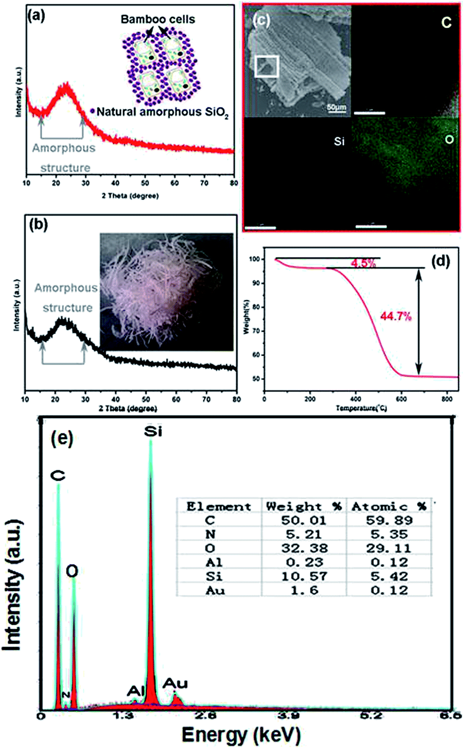 | ||
| Fig. 2 (a) XRD pattern of SiO2–C/NCs sample after heat treat at 700 °C in N2 atmosphere for 4 h. The inset schematic depicts the microstructure of BLs; (b) XRD pattern of BLs ash after heat treat at the oxygen atmosphere, showing its amorphous nature. The inset panel in (b) shows an optical image of the BLs ash; (c) SEM image and EDS elemental maps of SiO2–C/NCs sample, showing the distributions of C, Si and O in the micron range of white square area, which was scanned for C, Si and O elemental mapping, respectively; (d) TGA of SiO2–C/NCs sample at oxygen atmosphere; (e) EDX spectrum of SiO2–C/NCs sample. | ||
BLs can continuously absorb silica acid from soil and the silicic acid is stored in the form of amorphous silica, which mainly accumulates around bamboo cells and is responsible for mechanical strength in framework (the inset in Fig. 2a).34 The insert in Fig. 2b shows an optical image of the BLs ash, which demonstrates that the SiO2 still keeps the general shape of the BLs. The amorphous silica in BLs is a good insulator and has excellent heat resistant performance, corrosion resistance and no reaction with Li+. Thus, we speculated that the amorphous silica will enhance the structural stability of electrode during electrochemical process. After heat treatment at 700 °C in nitrogen atmosphere, the amorphous silica is retained and incorporated with carbon to form the 3D interconnected network structure of SiO2–C/NCs. To evaluate the SiO2 content in the SiO2–C/NCs sample, thermogravimetric analysis (TGA) was performed under oxygen atmosphere. As shown in Fig. 2d, a weight loss of 4.5% below 100 °C is attributed to the dissipation of physically absorbed water, while the mass loss of 44.7% above 600 °C is due to the burning of carbon. Therefore, the actual content of amorphous SiO2 and carbon in the SiO2–C/NCs sample is about 50.8 and 49.2%, respectively. The EDX spectrum of SiO2–C/NCs sample in Fig. 2e shows that the sample contains O, Si, N and C elements and other trace elements. These results further validates the results of XRD and TGA. There are also the doping elements of N, Al existing in the composite at the same time. The appearance of the element of Au is attributed to gold sputtering when the materials are examined.
SEM images of SiO2–C/NCs reveal that the natural hierarchical microstructure of the BLs have been retained after heat treatment (Fig. 3). Fig. 3a shows the tight arranged structure of BLs after thermal decomposition at 700 °C in N2 atmosphere. An enlarge SEM images of Fig. 3b reveals the 3D scalariform structure both on surfaces and fractured cross-sections. Fig. 3c shows the hierarchical porous structure with micron to nanometer holes from vessel and fiber bundles in bamboo cell, which are responsible for water transport and other nutrients. As is well known to all, BLs contain abundant organic matter. So we speculate that the formation of graphitizing C nanoparticle in SiO2–C/NCs sample may be attributable to the thermal decomposition of some biomacromolecules in bamboo cells. Fig. 3d shows the morphology features of SiO2–C/NCs anode after 190 cycles at the charging rate of 294.7 mA h g−1. It is seen that the SiO2–C/NCs particles is still intact although the surface become smooth because of growth of the passivated surface film layer on the particle surface and expansion pulverization loss of silica–carbon nanoparticles during cycling is smaller. The amorphous network structure of SiO2 can act as a buffer to accommodate the volume change of the graphitized carbon nanoparticles.
 | ||
| Fig. 3 SEM images of SiO2–C/NCs sample after heat treat at 700 °C in N2 atmosphere for 4 h, showing the 3D scalariform structure (a and b) and (c) hierarchical porous structure, which inherited the natural structure of xylem vessels in bamboo cell. (d) The morphology features of SiO2–C/NCs anode after 190 cycles at the charging rate of 294.7 mA h g−1. | ||
The unique nanostructure of SiO2–C/NCs was also characterized by HRTEM. Fig. 4a displays that the SiO2–C/NCs particle with 3D nanoporous structure is built with the SiO2–C nanosheets piece by piece. It is seen that the some C nanoparticle with a feature size of less than 10 nm were embedded (area containing the red ring) in the nanosheets. These C nanoparticles appear as oval particles and spread inside the SiO2–C nanosheets without aggregation. An enlarged image in Fig. 4b shows the nanostructure of a C nanoparticle with a spacing value of 0.209 nm, which belongs to the (100) diffraction facet of graphitic carbon.4 Ceder et al. demonstrated that most channels in small enough nanoparticles are unblocked, making all sites accessible by very rapid migration of Li+ through the channels.35 Thus, the Li ion in the SiO2–C/NCs can migrate more quickly along the C nanoparticles, resulting in effective kinetic energy transport. Furthermore, the C nanoparticles are evenly distributed in SiO2–C nanosheets, which further indicates the nanocomposite structure of SiO2–C/NCs sample. In other words, it can be clearly seen that the SiO2–C nanosheets have amorphous network structure, which is one reason why achieving high lithium-storage capacity (Fig. 4c).
 | ||
| Fig. 4 (a) A overview of the multi-level biosilicon–carbon nanosheets with QDs embedded; (b) an enlarged image of the red ring in (a), showing a carbon quantum dot; (c) the structure of SiO2 nanosheets; (d) pore size distribution for QDs SiO2–C nanosheets, and the inset is the corresponding nitrogen adsorption–desorption isotherm loop. | ||
The nano porous nature of SiO2–C/NCs is further validated by N2 physisorption measurements. The isotherm in Fig. 4e exhibit type IV N2-adsorption isotherms with H3-type hysteresis loops at 0.55–0.9, characteristic of mesoporous materials. The Brunauer–Emmett–Teller (BET) specific surface area of SiO2–C/NCs sample is about 49 m2 g−1. Fig. 4d depicts the pore size distribution for SiO2–C/NCs, which basically shows bimodal size distribution of mesoporous. Obviously, those pores are mainly caused by the natural hierarchical microstructure of the BLs. Such mesoporous structure is expected to facilitate Li+ diffusion at the electrolyte–electrode interface and provide freedom for volume change associated with Li+ intercalation. SiO2–C/NCs with good mechanical integrity can acts as “buffer area” to relax strain stress for volume expansion/contraction, leading to higher coulombic efficiency without fading during Li+ intercalation/deintercalation.36,37 Amorphous SiO2–C/NCs network with porous structure could provide large amount of active sites for Li storage and allow electrolytes to penetrate easily into SiO2–C/NCs during the electrochemical process.38
Cyclic voltammetry (Fig. 5a) was performed to characterize the electrochemical performance of SiO2–C/NCs electrode in the voltage range of 0–3 V vs. Li/Li+ at a scan rate of 0.1 mV s−1. All CV curves display the typical carbon anode behavior. The flat cathodic plate corresponds to Li ion intercalation into SiO2–C/NCs at 0–0.3 V and the formation of the solid electrolyte interphase (SEI) layer.31 The anodic peak at 0.2 V is attributed to the Li ion extraction. In the second cycle, the intensity of the CV cure is lower than 1st cycle and followed cycles. The difference is related to the irreversible growth of SEI resulting from kinetically activated electrolyte degradation and other side reaction.39 From 3rd cycle, the subsequent cycles of SiO2–C/NCs electrode are overlapped well, showing the good reversibility of the electrochemical reaction.
 | ||
| Fig. 5 (a) Cyclic voltammetry (CV) curves of SiO2–C/NCs for the first five cycles at 0.1 mV s−1; (b) charge–discharge curves of SiO2–C/NCs at 200 mA g−1; (c) cycle stability of SiO2–C/NCs and commercialized graphite anode at 200 mA g−1. | ||
The cycling performance of SiO2–C/NCs at 200 mA g−1 is shown in Fig. 5b and c, respectively. For comparing and highlighting the excellent energy density of SiO2–C/NCs at high density current, the charge–discharge capacity of the commercialized graphite anode at the same condition is also shown in Fig. 5c. The initial discharge capacity of SiO2–C/NCs anode is 586.2 mA h g−1, which exceeds the theoretical capacity of graphite (372 mA h g−1). From the 2nd cycle onward (Fig. 5b), SiO2–C/NCs electrode shows the satisfying cycling stability. As the cycle number increasing, the coulombic efficiency steadily reaches to nearly 100%, indicating good reversibility of the electrochemical reactions and high structure stability. At the end of the 180th cycle, a reversible capacity as high as 294.7 mA h g−1 can still be retained. In sharp contrast, the electrochemical performance of the commercialized graphite is far behind SiO2–C/NCs whether in initial discharge process (only 124.6 mA h g−1 in the first discharge process) or in followed cycles (49.6 mA h g−1 after 23 cycles). And most importantly, the impressive cycle life of SiO2–C/NCs is better than those previous reported nanostructured carbon materials, including nitrogen-containing carbon (251 mA h g−1 after 20 cycles at a current density of 100 mA g−1),40 carbon nanofibers (206 mA h g−1 after 50 cycles at 37.2 mA g−1)41 and CNTs (266 mA h g−1 at a current density of 100 mA g−1 after 100 cycles).42
Such an outstanding electrochemical performance in SiO2–C/NCs anode demonstrates the advantage of our work.
The rate capability and coulombic efficiency of SiO2–C/NCs are also explored, as shown in Fig. 6. From 200 mA g−1 to 1000 mA g−1, the charge–discharge capacity remains stable and decreases regularly with the increasing of current density. The corresponding coulombic efficiency still closes to 100% at different current density. It is striking that a high capacity of 106.7 mA h g−1 still be obtained (2 times than 49.6 mA h g−1 for commercialized graphite anode at 200 mA g−1) at a very high current density of 1000 mA g−1 after 20 cycles. With the current increased to 2000 mA g−1, the discharge capacities of the as-synthesized sample gradually decrease. The existence of nonconductive amorphous silica from BLs which can strengthen the structure stability as well as decrease the active sites for Li+ is suggested to be partly responsibly for this phenomenon. Another reason may be related to the irreversible decomposition of the binder and electrolyte, as well as the growth of the passivated SEI layer.43 However, when the current density reverses back to 200 mA g−1, a satisfactory reversible capacity of 117.4 mA h g−1 is recovered after another 20 cycles. Such remarkable rate capabilities demonstrate that our SiO2–C/NCs own superior structural stability and may bring significant impact on the development of new electrode materials.
 | ||
| Fig. 6 Charge–discharge capacities and coulombic efficiencies versus cycle number for SiO2–C/NCs. | ||
To exactly explain the kinetic differences among SiO2–C/NCs and graphite, the EIS were performed in Fig. 7. It is seen that SiO2–C/NCs demonstrates a lower charge transfer resistance (the diameter of the semicircle at the high frequency region, 137.5 Ω cm−2) than graphite (482 Ω cm−2). After 10 cycles at 200 mA g−1, the charge transfer resistance for SiO2–C/NCs is 111.5 Ω cm−2, which is smaller than that of before cycling. The reduction of resistance might be attributed to the activation of carbon structure.44 The results confirm that the SiO2–C/NCs posses high conductivity and enhanced conversion reaction kinetics. Through the above analysis, the excellent electrochemical performance of the SiO2–C/NCs could be attributed to its unique 3D interconnected network structure of nanocomposite and hierarchical porous structure. During the electrochemical process, on the one hand, the amorphous SiO2–carbon network induces more active sites, which provide additional Li storage and result in a high capacity; on the other hand, the C nanoparticles has unblocked transmission channels and elasticity, which not only could transport Li+ more quickly, but also could buffer the volume expansion/contraction. Additionally, the multi-level SiO2–C/NCs with high conductivity could greatly enhance electron transport to keep the charge balance during the Li+ rapid intercalation/deintercalation. Besides, incorporating the interconnected silica network with C nanoparticles ensures the structural stability of the electrode during the cycling process.
 | ||
| Fig. 7 Electrochemical impedance spectra (EIS) of the SiO2–C/NCs before and after 10 cycles at 200 mA g−1 and graphite. | ||
Compared with nanostructured carbon materials, amorphous SiO2–C composites with different structure and morphologies have many excellent electrochemical properties. Nguyen et al.44 prepared the carbon fiber-interwoven amorphous nano-SiOx/graphene electrode, which exhibits impressive cycling performance with capacity retention of 80% and coulombic efficiencies of 99% over 50 cycles. Lv et al.45 synthesized amorphous SiO2/C composite electrode, exhibiting high reversible capacity (∼600 mA h g−1), stable cycling performance and excellent rate-capability. Wang et al.46 designed and constructed a graphene-wrapped SiO2 nanotube network electrode, demonstrating an ultrahigh discharge capacity of 1145.3 mA h g−1 after 100 cycles (100 mA g−1). Although the reversible capacities of SiO2–C/NCs electrode are relatively lower compared with these reports, this simple, green and sustainable strategy will open a new avenue for large scale preparation and application of nanostructured electrode materials from biomass materials. The higher amorphous SiO2 content in the SiO2–C/NCs sample and its structural characteristics as possibly are its most main reasons of the lower reversible capacities.
The test results show that the SiO2–C/NCs inherited the natural hierarchical structure of the bamboo leaves. Compared with the commercialized graphite anode (Fig. 8a), the SiO2–C/NCs possess unique compositional and structural features that are beneficial to lithium storage. First, the 3D nanoporous interconnected network from natural biomaterials possesses the advantages of both nanosized building blocks and microsized assemblies, which are favorable for high reversible capacities.47,48 Second, the amorphous SiO2 network can improve the structural stability of SiO2–C/NCs and act as an efficient buffering for accommodating volume changes during lithium insertion/extraction (Fig. 8b).49 Finally, a large number of carbon nanoparticles with a feature size of less than 10 nm are embedded in the 3D interconnected network structure, which can improve the electrical conductivity of the SiO2–C hybrid network. Moreover, in situ nanocomposite of SiO2–C might activate and promote the lithium insertion/extraction. Therefore, the SiO2–C/NCs show remarkable lithium-storage performance, good capacity retention and rate capability, which make it an ideal anodic candidate for advanced LIBs.
 | ||
| Fig. 8 Schematic illustration of the volume change of the lithium insertion in graphite (a) and SiO2–C/NCs (b) before and after cycles. | ||
Conclusions
We are the earlier to prepare the amorphous SiO2–carbon nanocomposite (SiO2–C/NCs) with three-dimensional (3D) interconnected network and hierarchical porous structure and apply it to the anode material. SiO2–C/NCs derived from bamboo leaves was synthesized by a simple heat treatment at N2 atmosphere. A large number of carbon nanoparticles with a feature size of less than 10 nm are embedded in the 3D interconnected network structure of SiO2–C/NCs. Because of the good structural stability and the conductive pathway provided by the unique nanostructure, the as-formed SiO2–C/NCs exhibits more advantage than graphite and other artificial nanostructure carbon materials used as commercial Li-ion batteries. The excellent lithium storage performance of the SiO2–C/NCs can be ascribed to its fascinating characteristics, including (3D) interconnected network and hierarchical porous structure, dispersed carbon nanoparticles, and multi-level SiO2–C nanosheets. More importantly, this method is simple, low cost, and is easy to realize industrialization. The materials used are cheap renewable resources. We believe that this novel, green and sustainable strategy will open a new avenue for produce nanostructured electrode materials using biomass materials. This strategy can also be used for exploring other applications, such as catalysts, sensors and fluorescent.Acknowledgements
The authors thank Natural Science Foundation of China (Grant No. 51272144, 51472127 and 51172132) for the financial support. They also thank Shikun Liu for helpful experiments.Notes and references
- K. J. Stevenson, J. Solid State Electrochem., 2012, 16, 2017–2018 CrossRef CAS.
- T. F. Yi, C. Y. Li, Y. R. Zhu, J. Shu and R. S. Zhu, J. Solid State Electrochem., 2009, 13, 913–919 CrossRef CAS.
- M. Broussely, P. Biensan, F. Bonhomme, P. Blanchard, S. Herreyre, K. Nechev and R. J. Staniewicz, J. Power Sources, 2005, 146, 90–96 CrossRef CAS.
- Y. Xia, Z. Xiao, X. Dou, H. Huang, X. H. Lu, R. J. Yan, Y. P. Gan, W. H. Zhu, J. P. Tu and W. K. Zhang, ACS Nano, 2013, 7, 7083–7092 CrossRef CAS PubMed.
- P. Wu, N. Du, H. Zhang, J. Yu, Y. Qi and D. Yang, Nanoscale, 2011, 3, 746–750 RSC.
- M. Xia, Q. Liu, Z. Zhou, Y. Tao, M. F. Li, K. Liu, Z. Wu and D. Wang, J. Power Sources, 2014, 266, 29–35 CrossRef CAS.
- Y. Yang, M. T. McDowell, A. Jacksom, J. J. Cha, S. S. Hong and Y. Cui, Nano Lett., 2010, 10, 1486–1491 CrossRef CAS PubMed.
- Y. Xia, Z. Xiao, X. Dou, H. Huang, X. H. Lu, R. J. Yan, Y. P. Gan, W. H. Zhu, J. P. Tu and W. K. Zhang, ACS Nano, 2013, 7, 7083–7092 CrossRef CAS PubMed.
- Z. Li, Z. Xu, X. Tan, H. Wang, C. M. B. Holt, T. Stephenson, B. C. Olsen and D. Mitlin, Energy Environ. Sci., 2013, 6, 871–878 CAS.
- H. Xia, M. Lai and L. Lu, J. Mater. Chem., 2010, 20, 6896–6902 RSC.
- F. Han, W. C. Li, M. R. Li and A. H. Lu, J. Mater. Chem., 2012, 22, 9645–9651 RSC.
- A. V. Murugan, T. Muraliganth and A. Manthiram, J. Phys. Chem. C, 2008, 112, 14665–14671 CAS.
- L. W. Ji, Z. Lin, M. Alcoutlabi and X. W. Zhang, Energy Environ. Sci., 2011, 4, 2682–2699 CAS.
- B. Gao, C. Bower, J. D. Lorentzen, L. Fleming, A. Kleinhammes, X. P. Tang, L. E. McNeil, Y. Wu and O. Zhou, Chem. Phys. Lett., 2000, 327, 69–75 CrossRef CAS.
- X. X. Wang, J. N. Wang, H. Chang and Y. F. Zhang, Adv. Funct. Mater., 2007, 17, 3613–3618 CrossRef CAS.
- P. Guo, H. H. Song and X. H. Chen, Electrochem. Commun., 2009, 11, 1320–1324 CrossRef CAS.
- S. W. Woo, K. Dokko, H. Nakano and K. Kanamura, Electrochemistry, 2007, 75, 635–640 CrossRef CAS.
- Y. S. Hu, P. Adelhelm, B. M. Smarsly, S. Hore, M. Antonietti and J. Maier, Adv. Funct. Mater., 2007, 17, 1873–1878 CrossRef CAS.
- F. Cheng, Z. Tao, J. Liang and J. Chen, Chem. Mater., 2008, 20, 667–681 CrossRef CAS.
- L. Ji, Z. Lin, M. Alcoutlabi and X. Zhang, Energy Environ. Sci., 2011, 4, 2682–2699 CAS.
- S. Luo, K. Wang, J. P. Wang, K. L. Jiang, Q. Q. Li and S. S. Fan, Adv. Mater., 2012, 24, 2294–2298 CrossRef CAS PubMed.
- H. Ma, F. Cheng, J. Chen, J. Zhao, C. Li, Z. Tao and J. Liang, Adv. Mater., 2007, 22, 4067–4079 CrossRef.
- J. S. Jang, W. Li, S. H. Oh and J. S. Lee, Chem. Phys. Lett., 2006, 425, 278–282 CrossRef CAS.
- A. S. Aricò, P. Bruce, B. Scrosati, J. M. Tarascon and W. Schalkwijk, Nat. Mater., 2015, 4, 366–377 CrossRef PubMed.
- H. Yao, G. Zheng, W. Li, M. T. McDowell, Z. She, N. Liu, Z. Lu and Y. Cui, Nano Lett., 2013, 13, 3385–3390 CrossRef CAS PubMed.
- Z. Schnepp, W. Yang, M. Antonietii and C. Giordano, Angew. Chem., Int. Ed., 2010, 122, 6714–6716 CrossRef.
- Z. Chen, V. Augustyn, J. Wen, Y. W. Zhang, M. Q. Shen, B. Dunn and Y. F. Lu, Adv. Mater., 2011, 23, 791–795 CrossRef CAS PubMed.
- H. Yao, G. Zheng, W. Li, M. T. McDowell, Z. Seh, N. Liu, Z. Lu and Y. Cui, Nano Lett., 2013, 13, 3385–3390 CrossRef CAS PubMed.
- L. S. Chan, W. H. Cheung, S. J. Allen and G. McKay, Sep. Purif. Technol., 2009, 67, 166–172 CrossRef CAS.
- S. Jun, S. H. Joo, R. Ryoo, M. Kruk, M. Jaroniec, Z. Liu, T. Ohsuna and O. Terasaki, J. Am. Chem. Soc., 2000, 43, 10712–10713 CrossRef.
- F. Kleitz, S. H. Choi and R. Ryoo, Chem. Commun., 2003, 2136–2137 RSC.
- X. X. Xiang, Z. Z. Huang, E. H. Liu, H. J. Shen, Y. Y. Tian, H. Xie, Y. H. Wu and Z. L. Wu, Electrochim. Acta, 2011, 56, 9350–9356 CrossRef CAS.
- J. R. Dahn, W. Xing and Y. Gao, Carbon, 1997, 35, 825–830 CrossRef CAS.
- T. P. Ding, J. X. Zhou, D. F. Wan, Z. Y. Chen, C. Y. Wang and F. Zhang, Geochim. Cosmochim. Acta, 2008, 72, 1381–1395 CrossRef CAS.
- R. Malik, D. Burch, M. Bazant and G. Ceder, Nano Lett., 2012, 10, 4123–4127 CrossRef PubMed.
- Y. Ren, A. R. Armstrong, F. Jiao and P. G. Bruce, J. Am. Chem. Soc., 2010, 132, 996–1004 CrossRef CAS PubMed.
- Y. Wang, H. J. Zhang, L. Lu, L. P. Stubbs, C. C. Wong and J. Lin, ACS Nano, 2010, 24, 4753–4761 CrossRef PubMed.
- Y. L. Ding, C. Y. Wu, H. M. Yu, J. Xie, G. S. Cao, T. J. Zhu, X. B. Zhao and Y. W. Zeng, Electrochim. Acta, 2011, 56, 5844–5848 CrossRef CAS.
- Y. M. Liu, X. Y. Zhao, F. Li and D. G. Xia, Electrochim. Acta, 2011, 56, 6448–6452 CrossRef CAS.
- E. Liu, H. Shen, X. X. Xiang, Z. Z. Huang, Y. Y. Tian, Y. H. Wu, Z. L. Wu and H. Xie, Mater. Lett., 2012, 1, 390–393 CrossRef.
- A. Ramos, I. Cameán, N. Cuesta and A. B. García, Electrochim. Acta, 2014, 146, 769–775 CrossRef CAS.
- X. Li, J. Liu, Y. Zhang, Y. Li, H. Liu, X. Meng, J. Yang, D. Geng, D. Wang, R. Li and X. Sun, J. Power Sources, 2012, 197, 238–245 CrossRef CAS.
- X. Zhang, Z. Bi, W. He, G. Yang, H. Liu and Y. Yue, Energy Environ. Sci., 2014, 7, 2285–2294 CAS.
- D. T. Nguyen, C. C. Nguyen, J. S. Seon Kim, J. Y. Kim and S. W. Song, ACS Appl. Mater. Interfaces, 2013, 5, 11234–11239 CAS.
- P. Lv, H. Zhao, J. Wang, X. Liu, T. Zhang and Q. Xia, J. Power Sources, 2013, 237, 291–294 CrossRef CAS.
- H. Wang, P. Wu, M. Qu, L. Si, Y. Tang, Y. Zhou and T. Lu, ChemElectroChem, 2015, 2, 508–511 CrossRef CAS.
- Q. Zhu, P. Wu, J. Zhang, W. Zhang, Y. Zhou, Y. Tang and T. Lu, ChemSusChem, 2015, 8, 131–137 CrossRef CAS PubMed.
- J. Li, P. Wu, Y. Tang, X. Xu, Y. Zhou, Y. Chen and T. Lu, CrystEngComm, 2013, 15, 10340–10345 RSC.
- P. Wu, H. Wang, Y. Tang, Y. Zhou and T. Lu, ACS Appl. Mater. Interfaces, 2014, 6, 3546–3552 CAS.
| This journal is © The Royal Society of Chemistry 2016 |