A chitosan–Pt nanoparticles/carbon nanotubes-doped phosphomolybdate nanocomposite as a platform for the sensitive detection of nitrite in tap water

Zhenyuan Bai, Chunlei Zhou, Ning Gao, Haijun Pang and Huiyuan Ma*
Key Laboratory of Green Chemical Engineering and Technology of College of Heilongjiang Province, College of Chemical and Environmental Engineering, Harbin University of Science and Technology, Harbin 150040, China. E-mail: mahy017@163.com; Fax: +86-0451-86392716; Tel: +86-0451-86392716

Received 20th September 2015 , Accepted 28th November 2015

First published on 1st December 2015


Abstract

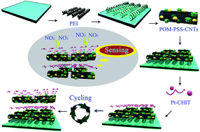
A composite multilayer film was fabricated based on H7P2Mo17V1O62 (P2Mo17V)–carbon nanotubes (CNTs) and Pt–chitosan (Pt–CHIT) nanoparticles using layer-by-layer self-assembly method. The incorporation of CNTs and Pt–CHIT nanoparticles into the P2Mo17V-based composite film endowed the modified electrode with a fast electron transfer rate and high electrocatalytic activity towards nitrite oxidation. The as-prepared sensor showed a fast response to nitrite with a good linear concentration range from 1.25 × 10⁻⁷ M to 4.167 × 10⁻³ M and a high sensitivity of 0.019 μA mM−1. Most noteworthy is its ultra-low detection limit of 3.8 × 10⁻⁹ M at the signal-to-noise ratio of 3 under optimal experimental conditions, which was below the detection limit of the vast majority of nitrite sensors reported before. The as-prepared sensor was able to distinguish the oxidation response of common interferences in solution mixtures under multiple potentials. It was successfully applied for determination of nitrite in tap water samples with satisfactory results.


1. Introduction

Nitrite is a highly toxic substance, which widely exists among environmental, food and physiological systems, such as in tap water, food additives and soil.1 As a hazardous substance, nitrite can react with dietary components to form toxic and carcinogenic nitrosamines in the stomach.2,3 Thus, there are specific requirements concerning nitrite levels; the maximum permissible amount of nitrite ions in drinking water is 50 mg L−1 according to the World Health Organization.2,4 Much concern has been shown about the level of nitrite in tap water, foods and beverages, and accurate determination of nitrite is essential to reduce the health risks. In recent years, many analytical techniques have been developed for the detection of nitrite, mainly involving spectrophotometry,5 fluorescence,6 flow injection analysis,7 chromatography,8 capillary electrophoresis chemiluminescence9 and so on. However, using these methods demands tedious and time consuming sample pretreatment and expensive reagents. Therefore, it is desirable to develop simple and low cost methods to determine nitrite. Electrochemical techniques, which are characterized by their low cost, easy operation and high sensitivity, have aroused widespread interest.10

New assembly techniques are required for creating advanced materials with enough structural flexibility to be tuned for specific applications, and to be practical, the techniques must be implemented at relatively low cost.11 Layer-by-layer (LBL) self-assembly is a ubiquitous phenomenon in nature at molecular and supramolecular levels, and it has been proven to be a promising and useful bottom-up approach to prepare functional materials. Compared with other available assembly methods, LBL is simplicity12,13 and the controllability of the thickness, composition, morphology, structure and physicochemical properties.14,15 To date, a variety of materials such as polyelectrolytes,16,17 nanoparticles,18–20 and inorganic materials21,22 have been employed for the construction of LBL films. This versatility has led to recent exceptional growth in the use of LBL-generated nanocomposites and is becoming a very attractive and powerful technology for the development of sensors because this technique is simple, universal, low cost and environmentally-friendly.

Polyoxometalates (POMs), as one of the most important inorganic building blocks, not only possess rich structural versatility such as Keggin, Wells–Dawson, Lindqvist types, etc., but also demonstrate superior redox activity.23,24 POMs can undergo reversible multielectron redox transformations without any significant structural alteration under rather mild conditions, which makes them play an important role as catalysts.25,26 In the recent study, polyoxotungstates and polyoxomolybdates have been extensively studied as electrocatalysts.27,28 However, the electrochemical and electrocatalytic properties of the vanadium-substituted polyoxometalates have been studied much less than those of the parent polyanions. The substitution of MoVI (or WVI) by VV allows the adjustment of the electrochemical properties of POMs since the oxidizing ability decreases in the following order: VV > MoVI > WVI. Moreover, substitution of vanadium into the POM framework can shift the stability of the species to higher pH values, which is a key factor in several (electro)catalytic processes.29–31 Meanwhile, the immobilization of POMs onto the variable electrode surface, while maintaining and enhancing their beneficial properties is important for their applications in areas of electrocatalyst and sensor.32

Multiwall carbon nanotubes (MWCNTs) with extraordinary mechanical strength, high surface to volume ratio, excellent electrical conductivity and high chemical stability33–35 have received considerable attention in recent years including the fabrication of electrochemical sensors and elevation the sensitivity and electrocatalytic activity of the corresponding sensor devices.36 Over the past few years, the investigation of POMs-modified CNTs has become a fascinating subject. Given the excellent structural and electronic properties of CNTs and POMs, the development of POMs-modified CNTs as electrode materials has attracted significant attention. Song et al. investigated the electrocatalytic properties of the composite film CNTs–SiW11.37 Chen et al. fabricated a composite film containing PMo12-modified CNTs and Pt or Pt–Ru, and then they studied its electrochemical properties towards methanol electro-oxidation.38 What's more, CNTs can not only serve as promising catalyst supports39 but also can integrate with metal nanoparticles to form a composite with remarkable activity for small molecules.

The uses of noble metal nanoparticles have gained considerable attention because of their size-dependent electrical, chemical and optical properties.40 We are interested in the catalytic and electrochemical properties of highly dispersed platinum nanoparticles. Polyelectrolyte chitosan (CHIT), as a typical biological macromolecule, possesses remarkable characteristics, such as excellent biodegradability, biocompatibility, and nontoxicity.41,42 Therefore, it can be used as cationic polyelectrolyte for the formation of LBL films when its functions are protonated in acid solutions. Many applications of chitosan depend on the interaction between amino groups and anionic surface-active substances, such as small molecule surfactants, phospholipids, or polyoxometalate.43 That is to say, the interaction between amino groups and anionic compounds can significantly enhance the stability of the immobilized system. So CHIT was chosen to simply produce stable Pt nanoparticles in this paper.

Based on the above description, this work focuses on fabrication of a composite film with high electrocatalytic activity for nitrite consisting of the Pt–CHIT NPs, P2Mo17V and CNTs using layer-by-layer self-assembly method. The procedure involved in the preparation of this composite film is very flexible and highly reproducible. The film growth was monitored by UV-vis spectroscopy, surface morphology and topography was examined by atomic force microscopy (AFM), and composition was assessed by X-ray photoelectron spectroscopy (XPS). The sensing performance of the composite film for the detection of nitrite at phosphate buffer solution (pH = 6.0) was investigated by cyclic voltammetry and amperometric response. This novel sensor offers a very good selectivity and ultra-low detection limit of nitrite. Furthermore, it was successfully used to determine nitrite in tap water.

2. Experimental

2.1. Materials and apparatus

H7P2Mo17V1O62 (P2Mo17V) was synthesized according to the literature method,44 and then identified by FT-IR and cyclic voltammetry analysis. Poly(ethylenimine) (PEI MW 750[thin space (1/6-em)]000) and poly(styrenesulfonate) (PSS MW 70[thin space (1/6-em)]000) were commercially purchased from Aldrich and used without further purification. H2PtCl6 (MW 232) and chitosan (CHIT) were purchased from Sigma-Aldrich China Co. Ltd. All other reagents were analytical grade and prepared with deionized water (18 MΩ cm). Multi-walled carbon nanotubes (MWCNTs, with >95% purity, diameter 15–20 nm) were obtained from College of Materials and Chemistry and Chemistry Engineering, Heilongjiang University (Harbin, China).

AFM images were obtained by Dimension TM3100 series of AFM produced from Digital Instruments (Santa Barbara, California, USA). TEM images were obtained on an H-7650 TEM. UV-vis absorption spectra were recorded on a quartz slide using a U-3900 UV-vis spectrophotometer from Hitachi (Japan). Cyclic voltammetry (CV) was conducted by a CHI760D Electrochemical Workstation (Shanghai Chenhua Instrument Corporation, China), with a conventional three-electrode system in which the modified ITO or bare ITO electrode was used as the working electrode, an Ag/AgCl as the reference electrode and a platinum wire as counter electrode.

2.2. Substrate preparation

ITO-coated glass was cleaned by immersing it in a series of ultrasonically agitated solvents (30% KOH alcohol solution, ethanol, H2O) at 40 °C for 20 min. A quartz or silicon substrate was immersed into fresh piranha solution (30% H2O2/98% H2SO4, v/v = 3[thin space (1/6-em)]:[thin space (1/6-em)]7; CAUTION: piranha solutions are very aggressive and corrosive; appropriate safety precautions should be utilized, including the use of acid-resistant gloves and adequate shielding.) and heated until no bubbles were released. The substrate was carefully rinsed with deionized water and dried with nitrogen. The substrates were then immersed in solution of PEI (0.1 mg mL−1) for one night to form the self-assembled monolayer terminated with –NH2 functional groups at the exposed surface. Subsequently, the quartz or silicon substrate was rinsed with deionized water and dried for further experiments. The amino units acted as the donor to construct the assembly by cooperating bonding.

2.3. Syntheses of the Pt–CHIT nanoparticles

According to the ref. 45, briefly, a 0.2 wt% CHIT solution was prepared by dissolving 40 mg of CHIT flakes into 10 mL of 1.0% acetic acid and stirred for 3 h at room temperature until complete dissolution. The pH of resulting solution was adjusted to 3.0–4.0 using a concentrated NaOH solution, followed by the addition of 2 mL of 0.01 M H2PtCl6, and the solution was sonicated for 15 min. Then the Pt(IV) was reduced by using 1 mL of freshly prepared 5% NaBH4. Reduction of PtCl6 by NaBH4 yields a clear yellow brown colored solution. The proposed Pt–CHIT nanoparticles were characterized by TEM (Fig. S1), which are highly monodispersed with an average diameter of ca. 6 nm. The prepared Pt–CHIT solution has very excellent stability, and no aggregation of nanoparticles was noticed after four months when the solution was stored in the dark at room temperature.

2.4. Fabrication of the composite films

The {PEI/[P2Mo17V-PSS-CNTs/Pt–CHIT]n/P2Mo17V-PSS-CNTs} multilayer films were assembled on different substrates due to their convenience for a particular analysis method: quartz slides for UV-vis spectroscopy, silicon wafers for AFM, and ITO-coated glass for XPS and cyclic voltammetry studies.

First, the PEI was deposited onto a cleaned substrate by immersing the substrate in PEI (10 mM) solutions for one night, in order to reduce possible interference from the substrate and to form a more uniformly charged surface,46–48 followed by rinsing with deionized water and drying under a nitrogen stream. And then, the pre-coated substrate was alternately immersed in the P2Mo17V-PSS-CNTs suspension (3.0 mg mL−1 of P2Mo17V mixed with 1.0 mg mL−1 PSS and 1.0 mg mL−1 of CNTs, w/w/w = 3[thin space (1/6-em)]:[thin space (1/6-em)]1[thin space (1/6-em)]:[thin space (1/6-em)]1) and Pt–CHIT aqueous solution for 30 min and rinsed with deionized water after each immersing. This procedure was repeated as many times as the number of bilayers needed, the composite film {PEI/[P2Mo17V-PSS-CNTs/Pt–CHIT]n/P2Mo17V-PSS-CNTs} was formed. The whole preparation process of the composite film is shown in Scheme 1. For comparison, the films {PEI/[P2Mo17V/Pt–CHIT]n/P2Mo17V}, {PEI/[PSS-CNTs/Pt–CHIT]n} and {PEI/[P2Mo17V-PSS-CNTs/CHIT]n/P2Mo17V-PSS-CNTs} were prepared according to the same procedure.


image file: c5ra19383d-s1.tif
Scheme 1 Schematic illustration of the as-prepared sensor.

3. Results and discussion

3.1. {P2Mo17V-PSS-CNTs/Pt–CHIT}n film growth

UV-vis spectroscopy was used to monitor the layer-by-layer self-assembling process of the composite film owing to its facility in evaluating the growth process of the multilayers film. Fig. 1 depicts the UV-vis spectra of {PEI/[P2Mo17V-PSS-CNTs/Pt–CHIT]n} films, with n = 1–10, in the range of 180–800 nm. The spectra showed three peaks at 194, 224, and 310 nm, respectively. As is reported, over our investigated wavelength range, Pt nanoparticles has one characteristic absorption at 195 nm,49 P2Mo17V has two characteristic absorptions in the polyoxometalate cage, Mo–O charge-transfer absorption at 226 nm and V–O charge-transfer absorption at 312 nm,50 PSS has two characteristic absorptions of pendent aromatic groups at 195 and 225 nm.51 So the absorption at 194 nm can be assigned to the overlap peak of Pt nanoparticles and PSS, the absorption at 224 nm can be assigned to the overlap peak of PSS and P2Mo17V, the absorption at 310 nm can be assigned to P2Mo17V. The absorbance in the wavelength range of 180–800 nm increases steadily with the bilayer number, which confirmed the irreversible adsorption of P2Mo17V-PSS-CNTs and Pt–CHIT on the composite film.
image file: c5ra19383d-f1.tif
Fig. 1 UV-vis absorption spectra of the composite film {PEI/[P2Mo17V-PSS-CNTs/Pt–CHIT]n/P2Mo17V-PSS-CNTs} for n = 1–10 adsorbed on a quartz slide. The inset shows the absorbance at λmax = 224 nm as a function of n.

As the numbers of bilayers increased there was an linear increment in the absorbance at λmax = 224 nm (the inset of Fig. 1), which this suggests that the amount of material deposited in each bilayer is constant and consistent with a stepwise growth process for this LBL hybrid film up to 10 layers. This is indicative of strong interaction between the charged components P2Mo17V-PSS-CNTs and Pt–CHIT, each deposition cycle is uniform and stable. From the UV-vis spectra, the surface coverage (Γ) of P2Mo17V can be calculated according to the following equation: Γ = (NAAλ)/2λ,52–54 where NA is Avogadro's constant, Aλ is the absorbance at a given wavelength λ (310 nm), ελ is the molar extinction coefficient (ε310 = 1.036 × 10⁶ M−1 cm−1) and m (10) is the number of layer. The average surface coverage calculated from UV-vis spectra is ca. 5.724 × 10¹⁵ P2Mo17V anions per cm−2 or 9.508 × 10⁻⁹ mol cm−2.

3.2. Surface morphology

Tapping mode AFM image of the {PEI/[P2Mo17V-PSS-CNTs/Pt–CHIT]10/P2Mo17V-PSS-CNTs} multilayer film was performed to find out its surface topography. Fig. 2 shows the AFM image of the composite film adsorbed on a silicon substrate.
image file: c5ra19383d-f2.tif
Fig. 2 Tapping mode 3D AFM image of {PEI/[P2Mo17V-PSS-CNTs/Pt–CHIT]10/P2Mo17V-PSS-CNTs} deposited on a silicon wafer.

From the typical three-dimensional AFM image of this composite film, it is clearly observed that the surface of the composite film was covered by a lot of protuberant peaks. The particle size and root-mean-square (RMS) roughness of the film is 85.9 and 6.34 nm calculated over an area of 10 × 10 μm2. It was reported that the low roughness and vertical grain structure with gap are particularly suitable for the fabrication of large-area and thin-film electronic devices.32

The presence of elemental composition in the multilayer film of {PEI/[P2Mo17V-PSS-CNTs/Pt–CHIT]10/P2Mo17V-PSS-CNTs} deposited on the silicon was verified by XPS, as indicated in Fig. S2. The spectrum of Pt reveals doublet peaks of Pt 4f7/2 and Pt 4f5/2 levels at 73.4 and 75.1 eV, respectively, which is identical to platinum in the zero-valent state in the film.55,56 From the Fig. S2(b)–(d), one peak of P 2p at 133.1 eV, two peaks of Mo 3d at 232.6 and 235.8 eV, and one V 2p peak at 533 eV were observed.28,57 Furthermore, the spectrum of C 1s (Fig. S2(e)) shows two peaks at approximately 284.5 and 286.1 eV. The peak at 284.5 eV is attributed to the aliphatic carbon (–C–C– or –C–H–), and that at 286.1 eV is attributed to carbon bonded to the amine groups (–C–NR2, where R is C or H) of the chitosan. The N 1s spectrum (Fig. S2(f)) displays double peaks at 399.3 and 401.5 eV, which are corresponding to amine and amide, respectively.58,59 These results substantiated that P2Mo17V, carbon nanotube and Pt–CHIT NPs were indeed incorporated into the composite film, which were consistent with UV spectra.

3.3. Electrochemical characterization of composite films

The CVs of the composite film {PEI/[P2Mo17V-PSS-CNTs/Pt–CHIT]10/P2Mo17V-PSS-CNTs} at different scan rates are shown in Fig. 3. It can be seen that when the scan rate increased from 100 to 800 mV s−1, the cathodic peak currents and the corresponding anodic peak currents increased simultaneously. The composite film {PEI/[P2Mo17V-PSS-CNTs/Pt–CHIT]10/P2Mo17V-PSS-CNTs} shows three pairs of strong redox peaks with mean peak potentials E1/2 of −0.579 V, −0.406 V and −0.249 V, and two pairs of very weak redox peaks with mean peak potentials E1/2 of −0.016 V, 0.558 V respectively vs. Ag/AgCl attributed to the immobilized P2Mo17V. In this context, the pair I is corresponding to one-electron of vanadium-center (VV → VIV), II and III correspond to two one-electron Mo-centered reduction processes (MoVI → MoV),31 while the other two peaks (IV and V) correspond to two two-electron Mo-centered (MoVI → MoV).60 From the inset of Fig. 3, we can see the dependence of the cathodic and anodic peak currents of the forth pair wave on scan rates, which is indicative of a surface confined electrochemical process.
image file: c5ra19383d-f3.tif
Fig. 3 Cyclic voltammograms of the composite film {PEI/[P2Mo17V-PSS-CNTs/Pt–CHIT]10 P2Mo17V-PSS-CNTs } in 0.2 M PBS (pH = 6.0) vs. Ag/AgCl at different scan rates (from inner to outer: 100, 200, 300, 400, 500, 600, 700 and 800 mV s−1). The inset: plots of the cathodic and anodic peak currents of the fifth couple waves against scan rate.

Electrochemical surface coverage (Γ) of electroactive species immobilized at electrode surfaces can be estimated from cyclic voltammetry according to the equation Γ = (4ipRT)/(n2F2νA), where ip is the peak current (amperes), n is the number of electrons transferred, ν is the scan rate (V s−1), A is the geometric area of the working electrode (square centimeters), R is the gas constant, T is the temperature (298 K), and F is Faraday's constant.61 The surface coverage of P2Mo17V can be directly calculated from the peak current ip because the polyelectrolyte matrix is not electroactive in the whole experiment. Taking the forth couple of redox peaks as example, the surface coverage of P2Mo17V per layer amounts to 1.66 × 10⁻⁸ mol cm−2 for the composite film {PEI/{P2Mo17V-PSS-CNTs/Pt–CHIT}10/P2Mo17V-PSS-CNTs} at scan rate of 0.1 V s−1. This result is in agreement with the surface coverage of 9.508 × 10⁻⁹ mol cm−2 determined by UV-vis spectra.

3.4. Electrochemical impedance spectra

The complex impedance can be presented as the sum of the real (Zre) and imaginary (Zim) components that originate mainly from the resistance and capacitance of the films. The semicircle diameter of EIS equals the electron transfer resistance (Ret), which controls the electron transfer kinetics of the redox probe at the electrode interface. In this work, Ret reflects the restricted diffusion of the redox probe through the multilayer system, which relates directly to the accessibility of the underlying electrode.62,63 The EIS figures of the {PEI/[P2Mo17V-PSS-CNTs/Pt–CHIT]10/P2Mo17V-PSS-CNTs}, {PEI/[CNTs-PSS/Pt–CHIT]10}, {PEI[P2Mo17V/Pt–CHIT]10/P2Mo17V} and {PEI/[P2Mo17V-PSS-CNTs/CHIT]10/P2Mo17V-PSS-CNTs} films are shown in Fig. 4. The frequency range was between 0.01 and 1[thin space (1/6-em)]000[thin space (1/6-em)]000 Hz with signal amplitude of 5 mV. The diameter of the semicircle increases in the order of the {PEI/[P2Mo17V-PSS-CNTs/Pt–CHIT]10/P2Mo17V-PSS-CNTs} < {PEI/[CNTs-PSS/Pt–CHIT]10} < {PEI[P2Mo17V/Pt–CHIT]10/P2Mo17V} < {PEI/[P2Mo17V-PSS-CNTs/CHIT]10/P2Mo17V-PSS-CNTs} < bare ITO. This result indicates a lower electron transfer resistance and larger electron transfer rate in the composite film {PEI/[P2Mo17V-PSS-CNTs/Pt–CHIT]10/P2Mo17V-PSS-CNTs}. That is to say, the synergy between P2Mo17V-PSS-CNTs and Pt–CHIT could accelerate the electron transfer in the composite film. The electrode system can be simplified by a equivalent circuit, and the inset of Fig. 4 shows a equivalent circuit obtained from the obtained impedance data of the composite film {PEI/[P2Mo17V-PSS-CNTs/Pt–CHIT]10/P2Mo17V-PSS-CNTs}. The equivalent circuit included the following parameters: Rs is the ohmic resistance of the electrolyte solution; Rp is the resistance to charge transfer. In this simple equivalent circuit, it was assumed that the resistance to charge transfer (Rp) was in parallel to the constant phase element (CPE), which was selected to replace the pure capacitor in the equivalent circuit by considering the surface roughness and inhomogeneity of the composite film. This parallel combination of Rp and CPE1 gave rise to a semicircle in the complex plane plot of Zim against Zre. According to the result of impedance spectrum, Rp was 1275 Ω and CPE was 8.0812 × 10⁻⁵ F. The electric charge transmission speed constant (k) can be calculated by the following formula:64,65 k = RT/RpCn2F2A, where Rp is the resistance to charge transfer, n is the number of electrons transferred per electroactive specie, C is the constant phase element (CPE), A is the geometric area of the electrode (square centimeters), and all other terms have their usual meaning. According to the formula, the electric charge transmission speed constant (k0) of the composite film {PEI/[P2Mo17V-PSS-CNTs/Pt–CHIT]10/P2Mo17V-PSS-CNTs} was estimated to be 1.72 × 10⁻⁶ cm s−1, while the electric charge transmission speed constant (k) of bare ITO, the {PEI/[P2Mo17V-PSS-CNTs/CHIT]10/P2Mo17V-PSS-CNTs}, the {PEI[P2Mo17V/Pt–CHIT]10/P2Mo17V} and {PEI/[CNTs-PSS/Pt–CHIT]10} films were estimated to be 9.81 × 10⁻¹¹ cm s−1, 6.64 × 10⁻⁹ cm s−1, 1.84 × 10⁻⁹ cm s−1 and 4.67 × 10⁻⁷ cm s−1, respectively. From the above results, it is clearly noted that the incorporation the CNTs, P2Mo17V and Pt–CHIT nanoparticle could improve the conductivity of the composite film.
image file: c5ra19383d-f4.tif
Fig. 4 The EIS figure of the composite film {PEI/[P2Mo17V-PSS-CNTs/Pt–CHIT]10/P2Mo17V-PSS-CNTs} (black dot), film {PEI/[CNTs-PSS/Pt–CHIT]10} (dark blue dot), {PEI/[P2Mo17V/Pt–CHIT]10/P2Mo17V} (green dot), {PEI/[P2Mo17V-PSS-CNTs/CHIT]10/P2Mo17V-PSS-CNTs} (light blue dot) and bare ITO (red dot) in 0.2 M PBS (pH = 6.0), the inset: a simple equivalent circuit about the composite film electrode.

3.5. Electrocatalytic studies

The electroactive LBL films usually can enhance the reduction/oxidation of many species by acting as mediators between the substrate and the bare electrode. In this work, the prepared {PEI/[P2Mo17V-PSS-CNTs/Pt–CHIT]n/P2Mo17V-PSS-CNTs} multilayer film was used as working electrode to improve the oxidation of nitrite to build a high sensitive sensor of nitrite.

A series of comparative experiments for the {PEI/[P2Mo17V-PSS-CNTs/Pt–CHIT]n/P2Mo17V-PSS-CNTs} film were performed to the oxidation of nitrite to seek the optimized bilayer number and pH. The parallel electrocatalytic experiments of the composite film {PEI/[P2Mo17V-PSS-CNTs/Pt–CHIT]n/P2Mo17V-PSS-CNTs} with different bilayer numbers (n = 2, 4, 6, 8, 10, 12 and 14) towards oxidation of nitrite were performed. The electrocatalytic activity of POM toward oxidation of nitrite can be evaluated by calculating the electrocatalytic efficiency (CAT) using the equation below:66

CAT = 100% × [ip(POMs, NO2) − ip(POMs)]/ip(POMs)
where ip(POMs) and ip(POMs, NO2) are the catalytic currents of the POMs in the absence and presence of NO2, respectively. When 0.3 mM nitrite was added, the electrocatalytic efficiencies of the composite film {PEI/[P2Mo17V-PSS-CNTs/Pt–CHIT]n/P2Mo17V-PSS-CNTs} with different bilayer numbers (n = 2, 4, 6, 8, 10, 12 and 14) are 210%, 478%, 372%, 286%, 961%, 156% and 169%, respectively (see Fig. S3(a)). It can be seen that 10-bilayer composite film has the highest catalytic efficiency, the reason of the 12 and 14 bilayer composite films having lower catalytic efficiency probably is that electron transport and reactant mass transport become limiting in thicker films.67 Meanwhile, the oxidation of nitrite by the {PEI/[P2Mo17V-PSS-CNTs/Pt–CHIT]10/P2Mo17V-PSS-CNTs} composite film in different pH solutions ranging from 5.0 to 7.0 was carried out. As shown in Fig. S3(b), the pH has a large effect on the electrocatalytic efficiencies, and the electrocatalytic efficiencies of the {PEI/[P2Mo17V-PSS-CNTs/Pt–CHIT]10/P2Mo17V-PSS-CNTs} composite film toward oxidation of nitrite is the largest (961%) in pH = 6.0 phosphate buffer solution. So, to give the as-prepared sensor with higher catalytic efficiency, 10-bilayers composite film was chosen as the working electrode and pH = 6.0 phosphate buffer solution as the buffer solution in follow experiments.

Fig. 5 shows the CVs obtained from the {PEI/[P2Mo17V-PSS-CNTs/Pt–CHIT]10/P2Mo17V-PSS-CNTs} composite film in the absence and in the presence of increasing concentrations of nitrite in the interval from 0.1 to 0.5 mM, which recorded in the potential range of 0.3 V to 1.0 V at scan rate 50 mV s−1. It can be seen that the anodic peak current at about +0.9 V of the composite film gradually increased with the addition of NO2, which indicates an electrocatalytic effect in the electrooxidation of NO2. The inset of Fig. 5 shows the relationship between the concentrations of NO2 and catalytic currents. From the inset, it can be seen that with the increase of the concentrations of NO2, the corresponding catalytic currents of the anodic peak enhanced linearly, which indicates different concentrations of NO2 can be quantified. The analysis of the corresponding current intensities leads to CAT = 961% for the electrooxidation of nitrite (3 × 10⁻⁴ mol dm−3). In comparison, oxidation of NO2 by three different composite films {PEI/[CNTs-PSS/Pt–CHIT]10}, {PEI/[P2Mo17V/Pt–CHIT]10/P2Mo17V} and {PEI/[P2Mo17V-PSS-CNTs/CHIT]10/P2Mo17V-PSS-CNTs} were investigated respectively. When 0.3 mM NO2 is catalyzed, the catalytic efficiency of the film {PEI/[CNTs-PSS/Pt–CHIT]10} is 510%, the film {PEI/[P2Mo17V/Pt–CHIT]10/P2Mo17V} is 110%, and that of the film {PEI/[P2Mo17V-PSS-CNTs/CHIT]10/P2Mo17V-PSS-CNTs} is 154% (calculated from Fig. S4(a)–(c), respectively), which are significantly lower than that of the composite film {PEI/[P2Mo17V-PSS-CNTs/Pt–CHIT]10/P2Mo17V-PSS-CNTs} (961%). The high CAT indicated that this composite film offers a significant electrocatalysis of nitrite.


image file: c5ra19383d-f5.tif
Fig. 5 Cyclic voltammograms of the composite film {PEI/[P2Mo17V-PSS-CNTs/Pt–CHIT]10/P2Mo17V-PSS-CNTs} in 0.2 M PBS (pH = 6.0) obtained with different concentrations of NO2: (0, 0.1, 0.2, 0.3, 0.4 and 0.5 mM); scan rate 50 mV s−1. The inset shows the catalytic peak current vs. analyte concentration at 0.9 V.

The significant enhancement in the catalytic efficiency of the composite film {PEI/[P2Mo17V-PSS-CNTs/Pt–CHIT]10/P2Mo17V-PSS-CNTs} may be arising from the change of the electron transport pathway in the composite film ascribed to the combination of the electroactive CNTs, P2Mo17V and Pt–CHIT in the composite film. Polyoxometalate acts as a proton and electron reservoir in the electrocatalytic process, and the good electrical conductivity and large surface area of the CNTs and Pt–CHIT nanoparticles facilitate the electron transfer and provide active sites. And the coexistence of CNTs, Pt–CHIT and P2Mo17VVO627− improve significantly the kinetics process of nitrite oxidation. Chitosan group nanoparticles have more advantages than normal nanoparticles, since they are allowed to fabricate more permeable films. As a result, remarkable catalytic efficiency enhancement of the composite film electrode was observed. That is to say, the incorporation of CNTs into the V-substituted POM and introduction the Pt nanoparticles into CHIT solution could constitute a useful strategy for the preparation of electrochemical sensors with improved sensing performance.

In the neutral conditions (PBS, pH = 6.0), the nitrite is oxidized to nitrate according to the following reaction:68

NO2 + H2O → NO3 + 2H+ + 2e

The vanadium is the reactive site for this catalytic reaction.10 The NO2 is oxidized by P2Mo17VVO627− to yield NO3 and the one-electron reduced species P2Mo17VIVO627−.

3.6. Analytical evaluation of the nitrite sensor

3.6.1. Amperometric detection of the NO2. Under optimal conditions, amperometric responses upon successive addition of NO2 were detected on {PEI/[P2Mo17V-PSS-CNTs/Pt–CHIT]10/P2Mo17V-PSS-CNTs} in 0.2 M PBS (pH = 6.0) at +0.9 V through 1200 s at 50 s interval. The typical amperometric response curves of different concentrations of NO2 are shown in Fig. 6(a). A well-defined, stable and fast amperometric response increasing step-wise with the level of NO2 can be observed, demonstrating highly efficient catalytic ability of composite film for NO2 electrooxidation. The response time (defined as the time to achieve 95% steady-state current) of this as-prepared sensor was estimated to be 2 s from the inset of Fig. 6(a), which is far less than what has been achieved by Malika Ammam (<6 s),69 Malcolm R. Smyth (5 s)70 and Pei-Hui Yang (5 s),71 and is some less than that achieved by Shan-Shan Li (3 s).2 As a potential candidate for routine nitrite analysis in tap water, improving the response time is very necessary. The current responses are linear with NO2 concentrations from 1.25 × 10⁻⁷ to 4.167 × 10⁻³ M, with the regression equation as: INO2 (μA) = 0.435 + 0.019CNO2 (mM), R2 = 0.99809 (see Fig. 6(b)). The limit of detection assessed as 3 × S/D is estimated to be 3.8 × 10⁻⁹ M (S/N = 3) and the sensitivity is 0.019 μA mM−1. As we know, many NO2 sensors have been developed by using POMs or other materials. A comparison of the performance of our newly as-prepared sensor with those already reported in literatures regarding the performance of the NO2 assay was listed in Table 1. It can be seen from Table 1 that this sensor has competitive performance among these sensors. Specially, linear response range and detection limit of this as-prepared sensor are superior to those sensors reported before. These results indicated that this as-prepared nitrite sensor could be an excellent platform for the detection of nitrite.
image file: c5ra19383d-f6.tif
Fig. 6 (a) Typical amperometric responses curve of the composite film {PEI/[P2Mo17V-PSS-CNTs/Pt–CHIT]10/P2Mo17V-PSS-CNTs} to successive additions of NO2 at applied potential of 0.9 V vs. Ag/AgCl in 0.2 M PBS (pH = 6.0). The insets show the enlargement of parts of curve. (b) Calibration plot of steady-state currents obtained at the composite film {PEI/[P2Mo17V-PSS-CNTs/Pt–CHIT]10/P2Mo17V-PSS-CNTs} against concentrations of NO2.
Table 1 Comparative characteristics of this as-prepared sensor and some other sensors for the determination of nitrite
Electrode Method Working potential (V) Linear range (mol L−1) LOD (μM) Ref.
K6[P2W18O62]/ITO CV −0.22 1.0 × 10 −4 to 1.5 × 10⁻³ 0.96 72
POMs(PFS-POMs)4/cysteamine/Au CV −0.07 2.0 × 10 −5 to 2.0 × 10 −2 1.56 73
PDDA/P2W17V–CNTs/ITO CV 0.9 0.05 × 10 −6 to 2.13 × 10 −3 0.0367 10
CDP–GS–MWCNTs CV 0.7 5 × 10 −6 to 6.75 × 10 −3 1.65 74
Au–RGO/PDDA DPV 0.89 0.05 × 10 −6 to 8.5 × 10 −3 0.04 75
La–MWCNTs/GCE CA 0.75 0.4 × 10 −6 to 0.71 × 10 −3 0.13 76
P2Mo17V-PSS-CNTs/Pt–CHIT/ITO CV 0.9 1.25 × 10⁻⁷ to 4.167 × 10⁻³ 0.0038 This work


3.6.2. Sensitivity and selectivity. It is well-known that the sensitivity and selectivity of the electrochemical sensors are markedly dependent on the applied potential. In order to determine whether this sensor is affected by the presence of common interferent in food system, the possible interference for the detection of NO2 at the as-prepared electrode was investigated by adding various foreign chemical species into the solution, including C2H5OH, acetic acid, phosphoric acid, glucose, Na2SO4, KBrO3, citric acid, Na2CO3, KCl and KNO3. From Fig. 7, it can be obtained that catalytic current of NO2 oxidation by the composite film {PEI/[P2Mo17V-PSS-CNTs/Pt–CHIT]10/P2Mo17V-PSS-CNTs} increased with the positive shift of the applied potential, showing the sensitivity of this sensor is dependent on the applied potential, and the more positive the applied potential is the higher sensitivity of this sensor shows. On the other hand, it can be concluded that at all applied potentials the interferences of these ten species toward this electrochemical sensor is nearly negligible, demonstrating that this as-prepared sensor has very superior selectivity. It was also noted that at the applied potentials of +0.9 and +1.0 V, the sensitivity of the sensor is superior and suitable, but in order to give this as-prepared sensor lower energy consumption, +0.9 V (vs. Ag/AgCl) is chosen as the optimized applied potential for determination of NO2.
image file: c5ra19383d-f7.tif
Fig. 7 The selectivity profile of the composite film obtained with 0.5 mM NO2, C2H5OH, acetic acid, phosphoric acid, glucose, Na2SO4, KBrO3, citric acid, Na2CO3, KCl and KNO3 at different applied potentials 0.6, 0.7, 0.8, 0.9 and 1.0 V in 0.2 M PBS (pH = 6.0).
3.6.3. Reproducibility and stability. The reproducibility and stability of the proposed nitrite sensor were further evaluated. As shown in Fig. 8, the stability of the sensor was checked by cyclic voltammetry scanning for 100 cycles from −0.6 V to 1.2 V at 50 mV s−1 in 0.2 M PBS (pH = 6.0), which testifies the good stability of the as-prepared sensor. In order to study the reproducibility of the sensor, the catalytic efficiency of ten identically modified electrodes in 0.2 M PBS which contained 0.3 mM NO2 were investigated under the same experimental conditions (see Table S1 and Fig. S5). According to the Table S1 and Fig. S5, the relative standard deviation (RSD) can be calculated to be only 3.56% by the following formula: RSD = SD/[x with combining macron], which demonstrated excellent reproducibility. Good reproducibility and high stability of the as-prepared sensor make it possible for durable and reliable sensing nitrite in practical application.
image file: c5ra19383d-f8.tif
Fig. 8 Cyclic voltammograms of the composite film {PEI/[P2Mo17V-PSS-CNTs/Pt–CHIT]10/P2Mo17V-PSS-CNTs} curves for 100 cycles in 0.2 M PBS (pH = 6.0). Scan rate: 50 mV s−1.

3.7. Nitrite sample analysis

In order to verify the practicality of the proposed sensor, it was applied to detect nitrite in real sample (tap water). Typically, added 4 mL real sample into the stirring solution containing 40 mL, 0.2 M PBS (pH = 6.0). Then the standard nitrite (0.01 mol L−1) solution was added into above mentioned solution six times to accomplish the detection. By standard addition method (see Fig. S6), a linear relation was obtained for the catalytic current I (μA) versus NO2 concentration C (mM) (I = −3.212 + 226.278 × C, R2 = 0.997). The concentration of NO2 can be obtained from the above-mentioned linear regression equation and then the recovery was calculated (see Table 2), all these results indicated that the proposed sensor could be used for the direct detection of nitrite in real sample with a good average recovery of 100.83%.
Table 2 Results of the determination of nitrite in different samplesa
Sample NO2 added (mM) NO2 found (mM) Recovery (%)
a The average recovery (%) was 100.83%.
1 0.02273 0.0305 134.18
2 0.06818 0.07356 107.89
3 0.1136 0.11662 102.66
4 0.1591 0.15968 100.36
5 0.2273 0.22428 98.67
6 0.2727 0.26734 98.03
7 0.3409 0.33193 97.37


4. Conclusions

In summary, a composite film for sensing nitrite was fabricated by incorporating Dawson-type polyoxometalate P2Mo17V, CNTs and Pt–CHIT NPs using LBL self-assembly method. By integrating P2Mo17V, CNTs and Pt–CHIT NPs into the composite film, the synergistic effect among all components was realized and maximized, leading to superior performance in electrocatalysis compared to any component alone. The significant advantages of this novel composite film include: (1) unique 3-D structure of CNTs facilitated high loading of active P2Mo17V and Pt–CHIT NPs on to the electrode surface and enhanced the electrocatalysis efficiency for nitrite. (2) CNTs maximized the dispersion of Pt–CHIT and P2Mo17V, and increased robustness of the whole composite film. (3) High electrical conductivity and active surface area of CNTs and Pt–CHIT NPs relieved the surface fouling effect, and enhanced the electron transfer rate between the molecules and electrode. Compared to other pre-reported nitrite sensors, this sensor possesses wide liner range, ultra-low detection limit, excellent selectivity, and highly reproducible as well as easy fabrication. It could be used practically in tap water. This investigation provided a methodology to promote the development of highly sensitive, selective, and durable and reproducible composite electrodes to be used in electrochemical sensor devices.

Acknowledgements

This work was financially supported by the NSF of China (21371041, 51572063), the science and technology innovation foundation of Harbin (2014RFXXJ076).

References

  1. X. Li, C. T. Wei, Y. H. Song, H. Z. Zhu, S. H. Chen, P. Li, L. L. Sun and L. Wang, Environ. Eng. Sci., 2015, 32, 185 CrossRef CAS.
  2. S. S. Li, Y. Y. Hu, A. J. Wang, X. X. Weng, J. R. Chen and J. J. Feng, Sens. Actuators, B, 2015, 208, 468 CrossRef CAS.
  3. Y. L. Zhao, D. A. Zhao and D. L. Li, Int. J. Electrochem. Sci., 2015, 10, 1144 Search PubMed.
  4. J. Y. Qu, Y. Dong, Y. Wang and H. H. Xing, Sens. Biosensing Res., 2015, 3, 74 CrossRef.
  5. V. Vishnuvardhan, R. Kala and T. P. Rao, Anal. Chim. Acta, 2008, 623, 53 CrossRef CAS PubMed.
  6. C. C. Ju, H. J. Yin, C. L. Yuan and K. Z. Wang, Spectrochim. Acta, Part A, 2011, 79, 1876 CrossRef CAS PubMed.
  7. M. Yaqoob, B. F. Biot, A. Nabi and P. J. Worsfold, Luminescence, 2012, 27, 419 CrossRef CAS PubMed.
  8. I. M. P. L. V. O. Ferreira and S. Silva, Talanta, 2008, 74, 1598 CrossRef CAS PubMed.
  9. H. X. Zhang, L. J. Zhang, C. Lu, L. X. Zhao and Z. X. Zheng, Spectrochim. Acta, Part A, 2012, 85, 217 CrossRef CAS PubMed.
  10. D. Zhang, H. Y. Ma, Y. Y. Chen, H. J. Pang and Y. Yu, Anal. Chim. Acta, 2013, 792, 35 CrossRef CAS PubMed.
  11. S. Srivastava and N. A. Kotov, Acc. Chem. Res., 2008, 41, 1831 CrossRef CAS PubMed.
  12. G. Decher, M. Eckle, J. Schmitt and B. Struth, Curr. Opin. Colloid Interface Sci., 1998, 3, 32 CrossRef CAS.
  13. S. T. Dubas and J. B. Schlenoff, Macromolecules, 1999, 32, 8153 CrossRef CAS.
  14. N. Ferreyra, L. C. Guérente, J. Fatisson, M. L. Teijelo and P. Labbé, Chem. Commun., 2003, 2056 RSC.
  15. F. X. Zhang and M. P. Srinivasan, Langmuir, 2007, 23, 10102 CrossRef CAS PubMed.
  16. N. Anwar, M. Vagin, R. Naseer, S. Imar, M. Ibrahim, S. S. Mal, U. Kortz, F. Laffir and T. McCormac, Langmuir, 2012, 28, 5480 CrossRef CAS PubMed.
  17. H. Paloniemi, M. Lukkarinen, T. Ääritalo, S. Areva, J. Leiro, M. Heinonen, K. Haapakka and J. Lukkari, Langmuir, 2006, 22, 74 CrossRef CAS PubMed.
  18. A. Devadoss, J. W. Lee, C. Terashima, A. Fujishima, Y. P. Kim, J. K. Kang and U. Paik, Sens. Actuators, B, 2015, 208, 204 CrossRef CAS.
  19. J. P. Wang, H. Gao, F. L. Sun and C. X. Xu, Sens. Actuators, B, 2014, 191, 612 CrossRef CAS.
  20. M. Li, X. J. Bo, Z. C. Mu, Y. F. Zhang and L. P. Guo, Sens. Actuators, B, 2014, 192, 261 CrossRef CAS.
  21. D. M. Fernandes, C. M. A. Brett and A. M. V. Cavaleiro, J. Solid State Electrochem., 2011, 15, 811 CrossRef CAS.
  22. S. Q. Liu and Z. Y. Tang, Nano Today, 2010, 5, 267 CrossRef CAS.
  23. S. Khadempir, A. Ahmadpour, M. T. H. Mosavian, N. Ashraf, F. F. Bamoharram, S. G. Mitchell and J. M. de la Fuente, RSC Adv., 2015, 5, 24319 RSC.
  24. M. Vasilopoulou, A. M. Douvas, L. C. Palilis, S. Kennou and P. Argitis, J. Am. Chem. Soc., 2015, 137, 6844 CrossRef CAS PubMed.
  25. D. Y. Du, J. S. Qin, S. L. Li, Z. M. Su and Y. Q. Lan, Chem. Soc. Rev., 2014, 43, 4615 RSC.
  26. S. Herrmann, C. Ritchie and C. Streb, Dalton Trans., 2015, 44, 7092 RSC.
  27. D. Zhang, Y. Y. Chen, H. J. Pang, Y. Yu and H. Y. Ma, Electrochim. Acta, 2013, 105, 560 CrossRef CAS.
  28. D. M. Fernandes, A. Teixeira and C. Freire, Langmuir, 2015, 31, 1855 CrossRef CAS PubMed.
  29. M. Sadakane and E. Steckhan, Chem. Rev., 1998, 98, 219 CrossRef CAS PubMed.
  30. P. Mialane, J. Marrot, E. Rivière, J. Nebout and G. Hervé, Inorg. Chem., 2001, 40, 44 CrossRef CAS PubMed.
  31. D. M. Fernandes, A. D. S. Barbosa, J. Pires, S. S. Balula, L. C. Silva and C. Freire, ACS Appl. Mater. Interfaces, 2013, 5, 13382 CAS.
  32. C. L. Zhou, S. Li, W. Zhu, H. J. Pang and H. Y. Ma, Electrochim. Acta, 2013, 113, 454 CrossRef CAS.
  33. X. L. Xia, D. W. Fan, B. H. An, Y. Y. Cai and Q. Wei, J. Mol. Liq., 2015, 206, 335 CrossRef CAS.
  34. E. T. Thostenson, Z. F. Ren and T. W. Chou, Compos. Sci. Technol., 2001, 61, 1899 CrossRef CAS.
  35. N. Butwong, L. Zhou, W. Ng-eontae, R. Burakham, E. Moore, S. Srijaranai, J. H. T. Luong and J. D. Glennon, J. Electroanal. Chem., 2014, 717–718, 41 CrossRef CAS.
  36. M. M. Barsan, M. E. Ghica and C. M. A. Brett, Anal. Chim. Acta, 2015, 881, 1 CrossRef CAS PubMed.
  37. W. Chen, L. J. Huang, J. Hu, T. F. Li, F. F. Jia and Y. F. Song, Phys. Chem. Chem. Phys., 2014, 16, 19668 RSC.
  38. D. W. Pan, J. H. Chen, W. Y. Tao, L. H. Nie and S. Z. Yao, Langmuir, 2006, 22, 5872 CrossRef CAS PubMed.
  39. K. Mukhopadhyay, S. Phadtare, V. P. Vinod, A. Kumar, M. Rao, R. V. Chaudhari and M. Sastry, Langmuir, 2003, 19, 3858 CrossRef CAS.
  40. C. L. Tan, X. Huang and H. Zhang, Mater. Today, 2013, 16, 29 CrossRef CAS.
  41. P. Sorlier, A. Denuzière, C. Viton and A. Domard, Biomacromolecules, 2001, 2, 765 CrossRef CAS PubMed.
  42. M. Rinaudo, Prog. Polym. Sci., 2006, 31, 603 CrossRef CAS.
  43. B. Krajewska, Enzyme Microb. Technol., 2004, 35, 126 CrossRef CAS.
  44. D. R. Park, H. Kim, J. C. Jung, S. H. Lee and I. K. Song, Catal. Commun., 2008, 9, 293 CrossRef CAS.
  45. M. H. Yang, Y. Yang, H. F. Yang, G. L. Shen and R. Q. Yu, Biomaterials, 2006, 27, 246 CrossRef CAS PubMed.
  46. C. Q. Peng, Y. S. Thio and R. A. Gerhardt, Langmuir, 2012, 28, 84 CrossRef CAS PubMed.
  47. D. Lee, Z. Gemici, M. F. Rubner and R. E. Cohen, Langmuir, 2007, 23, 8833 CrossRef CAS PubMed.
  48. J. Bravo, L. Zhai, Z. Z. Wu, R. E. Cohen and M. F. Rubner, Langmuir, 2007, 23, 7293 CrossRef CAS PubMed.
  49. S. Li, H. Y. Ma, K. P. O'Halloran, H. J. Pang, H. R. Ji and C. L. Zhou, Electrochim. Acta, 2013, 108, 717 CrossRef CAS.
  50. H. Y. Ma, Y. Gu, Z. J. Zhang, H. J. Pang, S. Li and L. Kang, Electrochim. Acta, 2011, 56, 7428 CrossRef CAS.
  51. H. Y. Ma, J. Peng, Y. H. Chen, Y. H. Feng and E. B. Wang, J. Solid State Chem., 2004, 177, 3333 CrossRef CAS.
  52. C. X. Li, K. P. O'Halloran, H. Y. Ma and S. L. Shi, J. Phys. Chem. B, 2009, 113, 8043 CrossRef CAS PubMed.
  53. I. Creaser, M. C. Heckel, R. J. Neitz and M. T. Pope, Inorg. Chem., 1993, 32, 1573 CrossRef CAS.
  54. F. Caruso, D. G. Kurth, D. Volkmer, M. J. Koop and A. Müller, Langmuir, 1998, 14, 3462 CrossRef CAS.
  55. Z. J. Mei, Y. Li, M. H. Fan, L. Zhao and J. Zhao, Chem. Eng. J., 2015, 259, 293 CrossRef CAS.
  56. X. Zhang, X. J. Zan and Z. H. Su, J. Mater. Chem., 2011, 21, 17783 RSC.
  57. Y. Y. Bao, L. H. Bin and L. X. Wu, J. Solid State Chem., 2011, 184, 546 CrossRef CAS.
  58. G. Lawrie, I. Keen, B. Drew, A. C. Temple, L. Rintoul, P. Fredericks and L. Grøndahl, Biomacromolecules, 2007, 8, 2533 CrossRef CAS PubMed.
  59. L. Ning, W. J. Zhang, H. Yan, H. J. Pang, H. Y. Ma and Y. Yu, J. Colloid Interface Sci., 2013, 403, 91 CrossRef CAS PubMed.
  60. H. M. Ji, L. D. Zhu, D. D. Liang, Y. Liu, L. L. Cai, S. W. Zhang and S. X. Liu, Electrochim. Acta, 2009, 54, 7429 CrossRef CAS.
  61. D. M. Fernandes and C. Freire, J. Appl. Electrochem., 2014, 44, 655 CrossRef CAS.
  62. S. Y. Gao, Z. X. Wu, D. M. Pan, Z. Lin and R. Cao, Thin Solid Films, 2011, 519, 2317 CrossRef CAS.
  63. M. F. Shao, X. Y. Xu, J. B. Han, J. W. Zhao, W. Y. Shi, X. G. Kong, M. Wei, D. G. Evans and X. Duan, Langmuir, 2011, 27, 8233 CrossRef CAS PubMed.
  64. E. Sabatani and I. Rubinstein, J. Phys. Chem., 1987, 91, 6663 CrossRef CAS.
  65. L. Chen, L. Tian, L. Liu, X. F. Tian, W. B. Song, H. D. Xu and X. H. Wang, Sens. Actuators, B, 2005, 110, 271 CrossRef CAS.
  66. D. M. Fernandes, J. G. Vos and C. Freire, J. Colloid Interface Sci., 2014, 420, 127 CrossRef CAS PubMed.
  67. C. J. Campbell, C. K. Njue, B. Nuthakki and J. F. Rusling, Langmuir, 2001, 17, 3447 CrossRef CAS.
  68. W. L. Sun, F. Yang, H. Z. Liu, J. L. Kong, S. L. Jin, G. Y. Xie and J. Q. Deng, J. Electroanal. Chem., 1998, 451, 49 CrossRef CAS.
  69. M. Ammam, B. Keita, L. Nadjo and J. Fransaer, Talanta, 2010, 80, 2140 CrossRef PubMed.
  70. X. L. Luo, A. J. Killard and M. R. Smyth, Chem.–Eur. J., 2007, 13, 2143 Search PubMed.
  71. H. Wang, P. H. Yang, H. H. Cai and J. Y. Cai, Synth. Met., 2012, 162, 331 Search PubMed.
  72. F. Cao, S. Guo, H. Ma and J. Gong, Electroanalysis, 2012, 24, 418 CrossRef CAS.
  73. C. Chen, Y. Song and L. Wang, Electroanalysis, 2008, 20, 2543 CrossRef CAS.
  74. Y. Zhang, R. Yuan, Y. Q. Chai, W. J. Li, X. Zhong and H. A. Zhong, Biosens. Bioelectron., 2011, 26, 3977 CrossRef CAS PubMed.
  75. S. F. Jiao, J. Jin and L. Wang, Sens. Actuators, B, 2015, 208, 36 CrossRef CAS.
  76. W. Zhang, R. Yuan, Y. Q. Chai, Y. Zhang and S. H. Chen, Sens. Actuators, B, 2012, 166–167, 601 CrossRef CAS.

Footnote

Electronic supplementary information (ESI) available: The characterization of Pt–CHIT nanoparticle and XPS spectra of the composite film, additional electrochemical investigations. See DOI: 10.1039/c5ra19383d

This journal is © The Royal Society of Chemistry 2016
Click here to see how this site uses Cookies. View our privacy policy here.