
 Open Access Article
 This Open Access Article is licensed under a
 Creative Commons Attribution 3.0 Unported Licence
Correction: Supramolecularly engineered phospholipids constructed by nucleobase molecular recognition: upgraded generation of phospholipids for drug delivery
Dali
Wang
a,
Chunlai
Tu
a,
Yue
Su
a,
Chuan
Zhang
a,
Udo
Greiser
b,
Xinyuan
Zhu
*a,
Deyue
Yan
a and
Wenxin
Wang
*b
aSchool of Chemistry and Chemical Engineering, State Key Laboratory of Metal Matrix Composites, Shanghai Jiao Tong University, 800 Dongchuan Road, Shanghai 200240, People's Republic of China. E-mail: xyzhu@sjtu.edu.cn; Fax: +86-21-54741297; Tel: +86-21-34203400
bCharles Institute of Dermatology, School of Medicine and Medical Science, University College Dublin, Belfield, Dublin 4, Ireland. E-mail: wenxin.wang@ucd.ie
First published on 1st July 2015
Abstract
Correction for ‘Supramolecularly engineered phospholipids constructed by nucleobase molecular recognition: upgraded generation of phospholipids for drug delivery’ by Dali Wang et al., Chem. Sci., 2015, 6, 3775–3787.
DMA and DOA were displayed incorrectly in the graphical abstract and Fig. 1 and 3. The corrected figures are shown below.
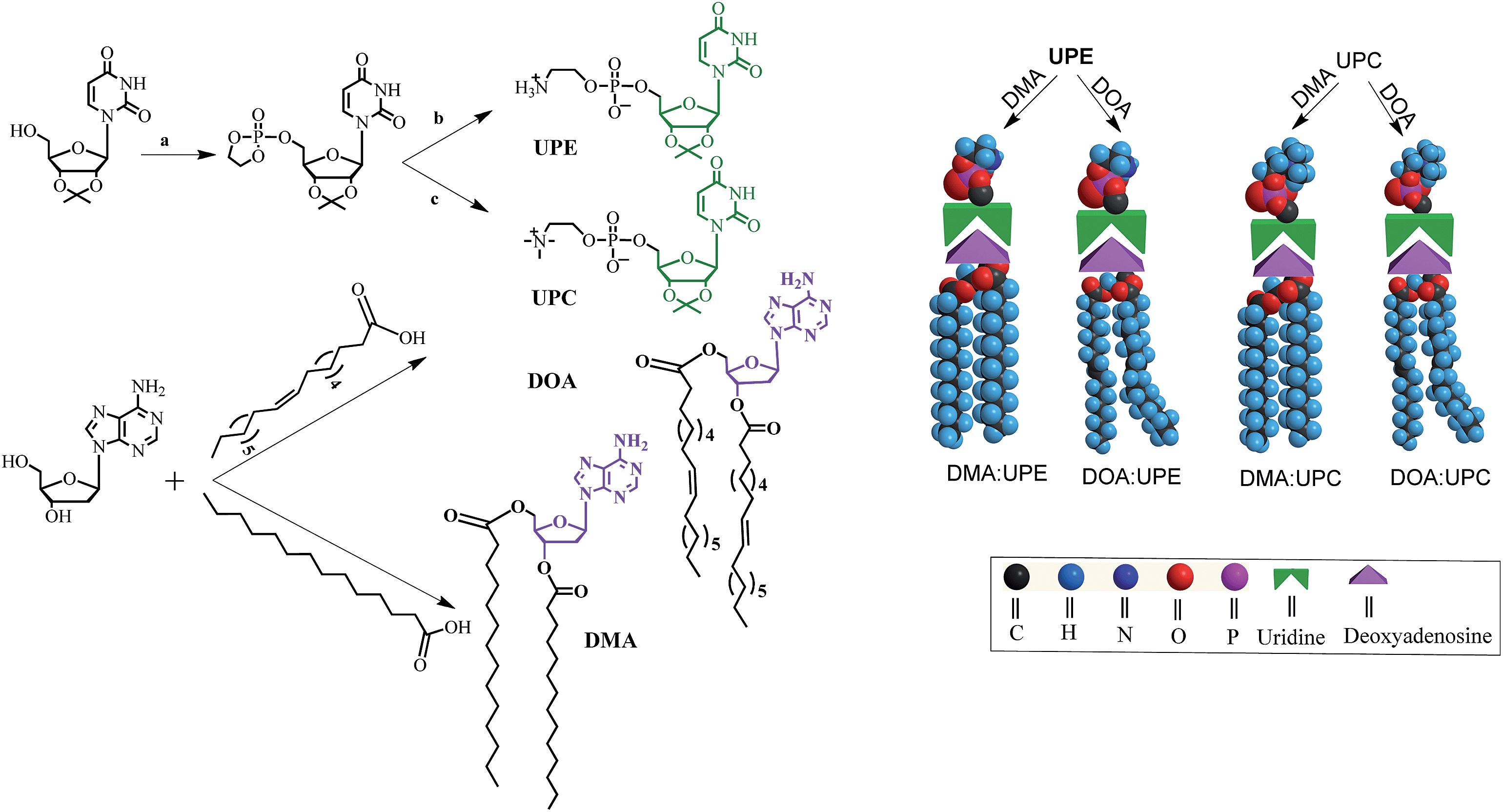 | ||
Fig. 1 Synthetic route, chemical structures of nucleoside phospholipids and schematic representation for the formation of supramolecular phospholipids. Reagents and conditions: (a) chlorooxodioxaphospholane, TEA, THF, 0 °C, 15 h; (b) trimethylamine, acetonitrile, THF, 60 °C, 24 h. (c) Ammonia, acetonitrile, THF, 65 °C, 48 h. UPE and UPC are uridine-functionalized PE and PC as hydrophilic phospholipid head, respectively. DMA and DOA are adenosine-functionalized myristic acid and oleic acid as hydrophobic tails, respectively. Through the molecular recognition between adenosine and uridine, these two components form four different types of supramolecular nucleoside phospholipids (DMA![[thin space (1/6-em)]](https://www.rsc.org/images/entities/char_2009.gif) :UPE, DOA:UPE, DMA:UPC and DOA:UPC) by mixing a uridine-terminated head and an adenosine-terminated tail. :UPE, DOA:UPE, DMA:UPC and DOA:UPC) by mixing a uridine-terminated head and an adenosine-terminated tail. | ||
 | ||
| Fig. 3 Characterization of molecular self-assembly of supramolecular nucleoside phospholipids DOA:UPC. (a) Schematic representation of a supramolecular liposome self-assembled from the DOA:UPC nucleoside phospholipids. Supramolecular nucleoside phospholipids self-assemble into liposome-like bilayer structures in aqueous solution. (b) Representative TEM images of negatively stained supramolecular DOA:UPC liposomes. The liposome wall thickness is about 6.5 nm. (c) Representative SEM image of supramolecular DOA:UPC liposomes (scale bars are 500 nm). (d) DLS profile for the supramolecular liposomes. (e) Relationship of the absorbance and the concentration of DOA:UPC in aqueous solutions (λ = 313 nm, 25 °C). (f) Estimation of the length of an extended DOA:UPC molecule according to the Chem3D results. | ||
Graphical abstract:

The Royal Society of Chemistry apologises for these errors and any consequent inconvenience to authors and readers.
| This journal is © The Royal Society of Chemistry 2015 |