Sensitive and selective detection of mercury ions based on papain and 2,6-pyridinedicarboxylic acid functionalized gold nanoparticles

Cui Lai*ab, Lei Qinab, Guangming Zeng*ab, Yunguo Liuab, Danlian Huangab, Chen Zhangab, Piao Xuab, Min Chengab, Xiangbin Qinc and Manman Wangab
aCollege of Environmental Science and Engineering, Hunan University, Changsha 410082, Hunan, PR China. E-mail: laicui@hnu.edu.cn; zgming@hnu.edu.cn; Fax: +86-731-88823701; Tel: +86-731-88822754
bKey Laboratory of Environmental Biology and Pollution Control (Hunan University), Ministry of Education, Changsha 410082, Hunan, PR China
cSchool of Mathematics and Statistics, Central South University, Changsha 410075, Hunan, PR China

Received 4th November 2015 , Accepted 17th December 2015

First published on 22nd December 2015


Abstract

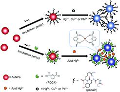
Here we demonstrate the rational design of a sensitive and selective colorimetric method for mercury ion (Hg2+) detection by using papain and 2,6-pyridinedicarboxylic acid (PDCA) functionalized gold nanoparticles (AuNPs). Papain is a protein with seven cysteine residues and 212 amino acid residues, which can combine with Hg2+, and PDCA is a chelating ligand, which has a strong affinity for Hg2+. Selectivity measurements reveal that the sensor is specific for Hg2+ even with interference by high concentrations of other metal ions. This sensor was also used to detect Hg2+ ions from real samples of tap water, river water, and pond water spiked with Hg2+ ions, and the results showed good agreement with the found values determined by atomic fluorescence spectrometry. The absorbance ratio (A650/A520) was linear with the Hg2+ concentration in the range of 0.01 μM to 14 μM. Under the optimum conditions (1.0 × 10⁻⁷ M papain, 5.0 × 10⁻⁴ M PDCA, pH 6.0 at 30 °C), the detection limit of Hg2+ was as low as 9 nM, which met the maximum allowable standard of the Environmental Protection Agency (EPA) set in drinking water. In conclusion, the P-PDCA-AuNPs sensor can be used to detect the concentration of Hg2+ with high sensitivity and good selectivity, and it can lead to dramatically improved colorimetric sensors.


1. Introduction

Nowadays, the development of industry is increasing rapidly. The pollution of toxic heavy metal ions in the environment is an important issue, especially in aquatic ecosystems.1–3 As a very important natural element, mercury is widely used in industry, agriculture and manufacture. Due to its strong affinity with fatty tissue in animals, mercury ions tend to biomagnify and bioaccumulate more easily than other metal ions.4–6 Especially, methyl mercury is extensively present in fish species, and most of its occurrence in human originates from food through the food chain.7,8 Mercury ions, as an extremely stable form of inorganic mercury, cause the damage of kidneys, brain, and the nervous and endocrine systems.9,10 Therefore, the detection of mercury levels in water samples is obviously significant.

To date, a series of analytical methods have been developed to detect mercury ions. These analytical methods include flame atomic absorption spectrometry (FAAS),11 atomic fluorescence spectrometry (AFS),12 inductively coupled plasma mass spectrometry (ICP-MS),13 inductively coupled plasma emission spectroscopy,14 electrochemical methods15 and fluorescent methods,16,17 and so on. Although these ways can detect mercury accurately and sensitively, these methods are partly expensive, time-consuming, rely on bulky instrumentation, ungreen, and, in some cases, are intolerant to interferences. To extend the application field of detection, varieties of colorimetric methods based on functionalized gold nanoparticles (AuNPs) have been developed for the simple, rapid and on-line determination of Hg2+.18–23 In recent years, rapid colorimetric sensing methods have attracted more and more interests because of their simple and inexpensive construction, rapid sensing capabilities, and high sensitivity. On the other hand, the aggregation of AuNPs can cause a range of rapid color change from ruby red to deep purple, even blue due to the coupling of interparticle Surface Plasmon Resonance (SPR), with very high extinction coefficients (108–1010 M−1 cm−1).24–26 Consequently, several groups had developed colorimetric sensor for the identification of mercury ions.

Colorimetric sensors based on AuNPs have been extensively used as the detection of small concentrations of heavy metal ions such as cadmium, lead, arsenic and mercury.27–33 In recently, the formation of thymine–Hg2+–thymine (T–Hg2+–T) was revealed very stably, Lee et al.18 reported method for colorimetric detection of Hg2+ based on DNA-functionalized AuNPs. This method was highly selectivity and sensitivity, but the requirement of electronic heating and read-out unit for detection at elevated temperatures made this sensor costly and impractical for rapid on-site sample analysis. To overcome these challenges, Xue et al.34 developed a colorimetric sensor system for detection of Hg2+ using DNA/nanoparticle conjugates. This sensor could operate within an adjustable operating temperature range, but the detection of real samples was not given. Most of aforementioned methods determining mercury used DNA functionalized AuNPs, but few of them used enzymes. Guo et al.35 synthesized P-AuNPs through the Au–S between papain and AuNPs to detect Hg2+, the interferences of Pb2+ and Cu2+ were still inevitably. Thus, a highly sensitivity and selectivity method to detect Hg2+ with optics is still needed.

Darbha et al.36 developed the method for detection of Hg2+ using functionalized AuNPs based on the strong affinity between 2,6-pyridinedicarboxylic acid (PDCA) and Hg2+. Yu et al.37 used PDCA to improve the selectivity toward Hg2+. Inspired by these studies, in this paper, we developed a rapid colorimetric sensor to detect Hg2+ using papain and 2,6-pyridinedicarboxylic acid (PDCA) functionalized gold nanoparticles (P-PDCA-AuNPs). In the presence of Hg2+, the functionalized AuNPs aggregated through metal–ligand interactions. The PDCA ligands bound to the AuNPs species via Au–N bounds, which improved the selectivity towards Hg2+ through a cooperative effect.36 Hg2+ could be detected by the colorimetric response of AuNPs and UV-Vis spectrophotometer, even by the naked eyes. In the real samples analysis, the results were still satisfactory. The approach allows for sensitive and selective detection of Hg2+, which has potential application in real time detection.

2. Materials and methods

2.1. Materials

Chloroauric acid hydrated (HAuCl4·4H2O) and sodium citrate were purchased from Sinopharm Chemical Reagent Co., Ltd (Beijing, China). Papain, extracting from the Caria papaya, was purchased from Sigma-Aldrich (USA). 2,6-Pyridinedicarboxylic acid (PDCA) was obtained from J&K Scientific Ltd (Beijing, China). Mercuric nitrate was purchased from Xiya Chemical Industry Co., Ltd (Shandong, China). All the reagents were used without further purification. Other chemicals involving metal ions salts were all of analytical grade and were obtained from Damao Reagent of Tianjin (Tianjin, China). Ultrapure water (18.2 Ω, Milli-Q Millipore) was used in each experimental process. All of the solutions were prepares in Milli-Q water, and Milli-Q water was also used for further dilution.

2.2. Instruments

Colorimetric measurement was recorded by a UV-2007 spectrometer (Shimadzu corporation, Kyoto, Japan) to obtain the absorption spectra of reaction solution using a quartz cuvette (1 cm path length), and the transmission electron microscopy (TEM) images of AuNPs were performed on a TECNAI T20 G2 electron microscope instrument operated at an accelerating voltage of 200 kV (FEI, Netherland). The magnification of TEM is 71[thin space (1/6-em)]000×. The contents of Hg2+ in the real samples were measured using the Atomic Fluorescence Spectrometry (AFS).

2.3. Preparation of AuNPs

The approximately 13 nm diameter AuNPs were prepared according to reported method with a little alteration.19,38 All glassware was soaked with freshly prepared 3[thin space (1/6-em)]:[thin space (1/6-em)]1 (v/v) HNO3–HCl (aqua regia) for 30 min, rinsed with ultrapure water, and dried for several hours before use. An aqueous solution (100 mL) containing 1 mL HAuCl4 (0.1%) was brought to boil in a conical flask (250 mL) with rapid stirring. Upon boiling, a 38.8 mM freshly prepared sodium citrate solution (10 mL) was added quickly and left to continue boiling under stirring, within this time, the color of the solution changed from light yellow, blue, purple, finally to ruby red. When the solution turned ruby red from light yellow, the resulting solution was stirred for another 15 min at room temperature. Then, stirring was stopped, and the solution was left allowed to cool to room temperature, filtered through a 0.22 μm syringe filter. The resulting spherical AuNPs were stored at 4 °C. The concentration of the prepared AuNPs was about 8.48 nM according to Beer–Lambert's low with an extinction coefficient of 2.78 × 10⁸ M−1 cm−1 at 520 nm for the 13 nm AuNPs.37,39

2.4. Preparation of P-PDCA-AuNPs

To detect Hg2+ selectively, the surface of the AuNPs was functionalized with papain and 2,6-pyridinedicarboxylic acid (PDCA). For the preparation of P-PDCA-AuNPs, solid papain was dissolved in ultrapure water to 1.0 × 10⁻⁷ M and stored in the dark at −20 °C firstly. Then, 2,6-pyridinedicarboxylic acid (PDCA) was dissolved in ultrapure water to 5.0 × 10⁻⁴ M. The pH of AuNPs solutions was adjusted to 6.0 by NaOH or HCl solutions. An excess amount of papain and PDCA was added to ensure the functionalizing completely. The papain solution (0.2 mL, 1.0 × 10⁻⁷ M) was added to AuNPs solutions (2 mL, 2.12 nM). The mixture was shaken for 30 min and incubated for 12 hours in the dark. The P-AuNPs were centrifuged at 12[thin space (1/6-em)]000 rpm for 20 min. After removing the supernatants, the sediment was resuspended in ultrapure water. PDCA solution (0.2 mL, 5 × 10⁻⁴ M) was added to the prepared P-AuNPs solutions. Finally, the mixture was shaken for another 30 min and was incubated for 6 hours in the dark. The final concentration of P-PDCA-AuNPs solutions was about 2.23 nM.

2.5. Colorimetric detection of Hg2+

Colorimetric detection of Hg2+ was used to evaluate the range of determination concentration of Hg2+ with the P-PDCA-AuNPs system. The pH value was adjusted to 6.0, and then a series of Hg2+ solutions (0.2 mL) with different concentrations (0, 0.01, 0.1, 2, 4, 6, 8, 14, 20, 50, 100, 500, 1000, 2000 μM) was added into the P-PDCA-AuNPs solution in a 5 mL sample bottle. After thorough shaking, the solutions were incubated at 30 °C for 15 min. The color change of the mixture was observed and recorded. Then the resulting solution was measured by the UV-Vis absorption spectra over the wavelength range from 200 nm to 800 nm and the absorbance values at 520 nm (A520) and 650 nm (A650) were also measured. The ratio of A650/A520 was calculated to indicate the aggregation degree of AuNPs. During the detection process, the final concentration of P-PDCA-AuNPs was 2.23 nM. To explore the selectivity, several commonly ions were tested using the sensing systems including Al3+, Ca2+, Cd2+, Cu2+, Fe2+, Fe3+, K+, Mg2+, Mn2+, NH4+, Pb2+, Zn2+ and Na+ ions. The concentration of these ions in the control experiments was 20 mM and carried out for 15 min at 30 °C. The concentration of Hg2+ was 1 mM. UV-Vis spectra of P-PDCA-AuNPs solutions were recorded 15 min after the addition of different kinds of competing ions.

2.6. Analysis of real samples

Analysis of real samples was used to test the applicability of P-PDCA-AuNPs in detection of Hg2+. Samples of river water, tap water and pond water were collected from the Hsiang River (Changsha, China), our laboratory and Lake of Peach, respectively. Firstly, the water samples were filtered through a 0.22 μm syringe filter. And then, the pH of the water was adjusted to 6.0. Finally, the samples were prepared by spiking with the standard solutions of Hg2+. The final concentration was 5 and 10 μM, respectively. For further verifying the application of this system, 0.01 μM of Hg2+ were also added to the real samples.

3. Results and discussion

3.1. Characterization of functionalized AuNPs

Papain, getting from the Caria papaya, is a plant endoprotease belonging to the cysteine endopeptidase family, which has a single polypeptide chain including 212 amino acid residues, including seven cysteine residues, with a molecular weight of 23[thin space (1/6-em)]400.40,41 The advantages of wonderful pH span, highly sensitivity, temperature stability, low price and short response time make papain can be used for detection of Hg2+.42,43 Structurally, papain can coat with AuNPs on the surfaces through sulfhydryl group, and it may interact with Hg2+ through amino group.41 Herein, papain can be used to detect Hg2+. Guo et al.35 used papain to determine Hg2+, but the sensitivity and selectivity was still need to improve. Papain has seven cysteine residues, except for Hg2+, Pb2+ and Cu2+ can bind with cysteine residues. Moreover, as a protein, papain has many functional groups like other proteins, such as amino, carboxyl, hydroxyl and so on. Pb2+ and Cu2+ can interact with these groups inducing the aggregation of AuNPs. On the other hand, PDCA, as a chelating ligand, has strong affinity with Hg2+.37 The stability constants of heavy-metal ions with PDCA are log[thin space (1/6-em)]K = 10.0 (Cd), 8.2 (Pb), 20.2 (Hg), and 8.5 (Mn).16,44 PDCA can be used as a chelating agent to achieve good selectivity because it can form much more stable complex with Hg2+ ions than other metal ions. Moreover, the carboxylic group of PDCA can combine with AuNPs on the surfaces. It could be used to improve the selectivity of Hg2+ detection (Scheme 1). As previous studies reported,37 the use of PDCA as co-additive in a sensor system can improve the selectivity and sensitivity for Hg2+. Thus, we suppose that if functionalized AuNPs with PDCA and papain, the level of Hg2+ could be determined by naked eyes and the sensitivity and selectivity also could be improved.
image file: c5ra23157d-s1.tif
Scheme 1 Schematic of aggregating process of P-PDCA-AuNPs induced by Hg2+.

To demonstrate the feasibility of this assumption, the design was applied to detect Hg2+ using papain and PDCA functionalized AuNPs. After centrifuging, a little difference about the color of AuNPs was observed. The UV-Vis absorption spectra of P-PDCA-AuNPs revealed broadening and slight red shift (Fig. 1). P-PDCA-AuNPs had slight polydisperse when comparing with unmodified AuNPs. It is possibly that AuNPs grew in the process of preparation. It has reported that in the citrate protected AuNPs solutions, there may exists Au3+ and some amino acids of papain, such as tyrosine, would reduce Au3+ to Au (0) in the alkaline aqueous solution. Furthermore, some reported literatures revealed that protein can reduce Au3+ into Au (0).45,46 Therefore, the color change indicated that papain and PDCA were modified on the surface of AuNPs. Moreover, control experiments using the papain were carried out, and 1 mM Hg2+ was added (Fig. 1). Upon addition of 1 mM Hg2+ to the mixture solution, its color rapidly changed from red to blue (existing in insert of the Fig. 1). Hg2+ was combined with papain and PDCA, decreased the distance of AuNPs, which result a shift of the surface plasma resonance peak and red-to-blue color changes. The UV-Vis absorption spectra of this sensor system became broader and shifted to much longer wavelength (650 nm). Transmission electron microscope (TEM) images of functionalized AuNPs (Fig. 2) also proved the aggregation of P-PDCA-AuNPs. The results proved that the papain and PDCA were sensitively responded to Hg2+.


image file: c5ra23157d-f1.tif
Fig. 1 UV-Vis absorption spectra of the AuNPs (13 nm), P-PDCA-AuNPs (13 nm), and P-PDCA-AuNPs (13 nm) in the presence of 1 mM of Hg2+. Inset shows the color change versus the (1) AuNPs (13 nm), (2) P-PDCA-AuNPs (13 nm), and (3) P-PDCA-AuNPs (13 nm) in the presence of 1 mM of Hg2+.

image file: c5ra23157d-f2.tif
Fig. 2 TEM images of the (A) AuNPs (13 nm), (B) P-PDCA-AuNPs (13 nm) and (C) P-PDCA-AuNPs (13 nm) in the presence of 1 mM of Hg2+.

3.2. Optimal of papain concentration

To obtain the optimum concentration of the sensor system, we studied the effect of papain concentration on the stability of this system. To get excellent sensitivity, we obtained the AuNPs solutions at pH 6.0 and 30 °C. Concentrations of 1.0 × 10⁻⁵, 1.0 × 10⁻⁶, 1.0 × 10⁻⁷, 1.0 × 10⁻⁸ M papain were added to 2.12 nM AuNPs solutions. In order to make the PDCA combine with AuNPs closely, excessive amount of PDCA (5.0 × 10⁻⁴ M) was added to the solutions. When PDCA and papain were added to the solution, the color of AuNPs had a little change. After adding 1 mM Hg2+, the color became purple gradually along with the increase of concentration in 15 min (Fig. S-3 and insert of Fig. S-1). With the decrease of the concentration, the absorbance of P-PDCA-AuNPs increases gradually at 520 nm and correspondingly decreases at 650 nm. But it is opposite to the concentration of 1.0 × 10⁻⁸ M. We use the ratio of the absorbance of P-PDCA-AuNPs at 650 and 520 nm (A650/A520) to prove the degree of AuNPs aggregation (Fig. S-2). A higher A650/A520 value is bound up with the degree of AuNPs aggregation, and a lower A650/A520 indicates that the AuNPs disperse well in the solution. When the concentration of papain is 1.0 × 10⁻⁷ M, the value of A650/A520 comes to the highest, which indicates that the aggregation of P-PDCA-AuNPs is completely. Taking into account the response sensitivity and background signal, 1.0 × 10⁻⁷ M papain was selected as the optimized concentration.

3.3. Temperature and pH effects to functionalized AuNPs

Temperature and pH are two crucial factors of the sensitivity in this research. In order to investigate the pH effect of colorimetric responses, different pH (6, 7, 8 and 9) of functionalized AuNPs was obtained. Firstly, the pH of AuNPs solution (0.2 mL) was adjusted to 6, 7, 8 and 9, respectively, then 0.2 mL papain and PDCA were added. The P-PDCA-AuNPs was easily aggregated at acid aqueous solutions (pH ≤ 6.0). When the pH was high (≥6.0), the P-PDCA-AuNPs were stable.35,40 With the increase of pH values, the P-PDCA-AuNPs were more stable, which was possibly attributed that the electrostatic repulsion between the detection systems increased with the pH value.35 Thus, in the presence of Hg2+, the detection solutions exhibited obvious color change and the P-PDCA-AuNPs were easily induced aggregation at pH 6. The ratio value of A620/A520 at pH 6 was also much greater than that at pH 7, 8, and 9 (Fig. S-4), which demonstrated the pH of 6 was the optimal pH for this sensor system. Hence, we chose the pH 6 as the optimal condition in this work. In order to demonstrate the temperature effect of functionalized AuNPs on the detection of Hg2+, nine different temperatures (10, 20, 30, 40, 50, 60, 70, 80, 90 °C) of P-PDCA-AuNPs were obtained through setting the AuNPs solutions to the refrigerator and water bath, respectively. Then 0.2 mL Hg2+ (1 mM) were added. After incubation 15 min, the absorbance at 650 nm of the solutions was measured on a UV-2700 (Shimadzu) spectrophotometer. The ratio of absorbance of the UV-Vis absorption spectra at 650 and 520 nm (A650/A520) was used to demonstrate the amount of functionalized AuNPs aggregation (Fig. S-5). In the addition of Hg2+ (1 mM), with the increase of temperature to 30 °C, the ratio of A650/A520 increased too, then it declined very slowly. When it reached 90 °C, the ratio of A650/A520 decreased sharply. That is because papain has highly sensitivity temperature stability and starts losing its activation at 90 °C.41,42 Results showed that the best temperature was range from 30 °C to 60 °C. Therefore, 30 °C was selected as the optimal temperature in this work. As a result, the P-PDCA-AuNPs sensor could be used as a colorimetric sensor for Hg2+ in a broad temperature range, which is convenient for a practical application.

3.4. Colorimetric detection of Hg2+

It is well known that Hg2+ tends to interact with carboxylic group and amino such as PDCA19,47,48 and cysteine.49 Thus, a rational strategy to determine Hg2+ is to combine P-PDCA-AuNPs and Hg2+. Inspired by this theory and under the best optimized detection condition (pH 6.0 for 15 min at 30 °C), the different concentrations of Hg2+ were added to P-PDCA-AuNPs to investigate the sensitivity of this sensor. As shown in Fig. 3A, with the increase of Hg2+ concentration, a decrease in the intensity of the absorbance at 520 nm and an increase at 650 nm can be observed. It revealed that the P-PDCA-AuNPs aggregation induced deposition with the rising of Hg2+ concentration. As envisioned, when different amounts of Hg2+ were added to an aqueous solution of P-PDCA-AuNPs, the color change of the solution (from red to blue) was observed by naked eye inspection (Fig. 3B). The lowest detection concentration with the unaided eye is 2 μM. The ratio of A650/A520 as a function of the concentration of Hg2+ is shown in Fig. 4, which increases gradually with the rise of the concentration of Hg2+. A linear correlation exists in insert of the Fig. 4. Under the optimum conditions, the detection of Hg2+ was linear with A650/A520 values in the range of 0.01 μM to 14 μM. The calibration equation was obtained by fitting the experimental data obtained:
y = (0.0252 ± 0.0002)x + (0.1139 ± 0.0005)
where y was the ratio of spectral absorbance A650/A520, and x was the concentration of Hg2+ (μM). Additionally, the correlation coefficient of this equation was 0.9959. The sensor exhibited a detection limit of 9 nM (1.8 ppb), which met the MAL (maximum allowable level) of ∼2 ppb in drinking water set by the Environmental Protection Agency (EPA). In the presence of high concentration of Hg2+, the UV-Vis absorption spectra became broader and showed red shift and the A650/A520 value was not in a linear range. Compared with other sensors (Table 1), the sensitivity of this method was also impressive.

image file: c5ra23157d-f3.tif
Fig. 3 (A) UV-Vis absorption spectra of P-PDCA-AuNPs were in the presence of different concentrations of Hg2+ (A to N express 2000, 1000, 500, 100, 50, 20, 14, 8, 6, 4, 2, 0.1, 0.01, 0 μM, respectively). (B) Images of P-PDCA-AuNPs containing different concentrations of Hg2+ (0, 0.01, 0.1, 2, 4, 6, 8, 14, 20, 50, 100, 500, 1000, 2000 μM).

image file: c5ra23157d-f4.tif
Fig. 4 Ratio values of A650/A520 for P-PDCA-AuNPs versus the concentrations of Hg2+ (0, 0.01, 0.1, 2, 4, 6, 8, 14, 20, 50, 100, 500, 1000, 2000 μM). Image of insert is the linear correlation. Error bars were obtained from three experiments.
Table 1 Functionalized AuNPs probes for detection of Hg2+
Technique Capping agent LOD (nM) Linear range (μM) Reference
a BSA, bovine serum albumin. R6G, rhodamine 6G. MPA, 3-mercaptopropionate acid.b RB, rhodamine B.c NAC-QDs, N-acetyl-L-cysteine functionalized quantum dots.d AMP, adenosine monophosphate.e MSD, 5′-SH(CH2)6A10TTCTTTCTTCCCCTTGTTTGTT-FAM-3′.f CEQC, carboxylethyl quaternized cellulose.g FRET, fluorescence resonance energy transfer.
Fluorescence aBSA, R6G and MPA 10 Not given Chang et al.50
bRB and PDCA 10 0.015–0.25 Huang and Chang47
gFRET cNAC-QDs and R6G 249 24.9–124.6 Hu et al.51
RB6, MPA and dAMP 50 0.05–1.0 Yu and Tseng52
Colorimetry eDNA (MSD) 30 0.02–1.0 Tan et al.53
fCEQC 40 3.0–24 You et al.49
Papain 4000 Not given Guo et al.35
DNA 1000 Not given Xue et al.23
DNA oligonucleotides 500 0–5 Xu et al.54
MPA and AMP 500 0.5–3.5 Yu and Tseng52
Thrombin-binding aptamer, PDCA 200 0.39–8.89 Wang et al.55
Papain and PDCA 9 0.01–14 This work


3.5. Selectivity and interference study

The results above indicate that the P-PDCA-AuNPs sensor is sensitive to the determination of Hg2+. As a functional sensor system, there must be high selectivity for Hg2+ over other ions. A variety of competitive ions, including Al3+, Ca2+, Cd2+, Cu2+, Fe2+, Fe3+, K+, Mg2+, Mn2+, NH4+, Pb2+, Zn2+ and Na+ ions, were added to the sensing system, the ratios of A650/A520 were calculated. When only papain was functionalized on the surface of AuNPs, several commonly ions were tested using the papain-AuNPs (P-AuNPs) system. The assay showed negligible responses toward Al3+, Ca2+, Cd2+, Fe2+, Fe3+, K+, Mg2+, Mn2+, NH4+, Zn2+ and Na+, but a red-to-blue color change and red shift in the plasmon band energy to longer wavelengths were noted in the presence of Hg2+, Cu2+ as well as Pb2+. It agreed with the findings of Guo et al.35 Further higher selectivity of our sensor toward Hg2+ was achieved by adding PDCA. As shown in Fig. 5, except for Hg2+, none of these competitive ions can induce the apparent aggregation of P-PDCA-AuNPs. Even though the concentrations of these ions were 20 times higher than that of Hg2+, these ions induced negligible changes in comparison with Hg2+. When all the other competitive ions were present in a water sample solely, the P-PDCA-AuNPs solutions showed no obvious response. In contrast, when Hg2+ existed in the sample, the P-PDCA-AuNPs solutions aggregated and exhibited apparent color change simultaneously. Aggregation in the presence of Hg2+ is because of binding with chelating ligands, inducing both shift in the plasmon band energy to longer wavelength and a red-to-blue color change (Fig. S-6B). In particular, PDCA ligands bound to the functionalized AuNPs system through Au–N bonds can improve the selectivity toward Hg2+ via a cooperative effect.19,36 Hg2+ can cause the aggregation of P-PDCA-AuNPs with the obvious increase of A650/A520 value. The plasma absorbance peak of Hg2+ has apparent red shift (Fig. S-6A). Furtherly, Hg2+ and other ions were mixed to form a mixture solution as a sample for the interference study of the sensor. The A650/A520 value was obviously higher than other samples without Hg2+. The results indicated that this sensor system could be utilized to detect Hg2+ with high selectivity. The selectivity in the presence of only PDCA was also developed, the results exhibited the selectivity was still not satisfactory. From all the results, it is concluded that functionalized of AuNPs surface with papain and PDCA is very important to increase the selectivity of Hg2+. The PDCA ligands bound to the AuNPs species improved selectivity toward Hg2+ through the cooperative effect, while it formed complexes with other ions in the bulk solutions, suppressing their interference with the sensors. Moreover, in Fig. S-6B, a purple-to-red color change can be observed in the presence of Fe2+. That is because the PDCA ligands in the solution form complexes with Fe2+ and induce the shift in plasmon band energy to shorter wavelength.36
image file: c5ra23157d-f5.tif
Fig. 5 Ratio values of A650/A520 for P-PDCA-AuNPs versus the different kinds of ions. Concentration of Hg2+ was 1 mM, while others were 20 mM. Error bars were obtained from three experiments.

3.6. Analytical applications

To evaluate our sensor for the detection of Hg2+ in real samples, tap water samples from our laboratory, pond water from the Lake of Peach (Changsha, China) and river water samples from Hsiang River (Changsha, China) were used for this sensor. Firstly, we filtered the water samples through 0.22 μm membrane. Then, river water, tap water and pond water were spiked with standard Hg2+ solutions (0.01 μM, 5 μM and 10 μM, respectively). The results were summarized in Table 2. As can be observed in Table 2, the recoveries of standard addition were between 0.01 μM and 10 μM. The data were then compared by the AFS measurement. The concentrations of Hg2+ in the spiked water samples detected by the P-PDCA-AuNPs sensor system with an external calibration revealed good agreement with those, which was determined with a standard addition calibration. It confirmed that there was no interference encountered in the analysis of these water samples.
Table 2 Levels of Hg2+ in real samples were measured using our method and AFS measurement with the range of relative standard deviation from 0.10% to 2.52%
Samples Added (μM) Found (μM) Recovery (%) RSDa (%) AFSb
a RSD, relative standard deviation.b AFS, atomic fluorescence spectrometry.
Tap water 0.01 0.012 112.00 0.17 0.014
5 5.03 100.60 1.00 5.13
10 10.07 100.70 1.53 10.21
River water 0.01 0.013 113.00 0.15 0.016
5 5.14 102.80 2.08 5.20
10 10.27 102.70 2.00 10.40
Pond water 0.01 0.011 110.00 0.10 0.013
5 5.10 102.00 1.75 5.15
10 10.16 101.60 2.06 10.23


4. Conclusions

In conclusion, we describe here a fast, sensitive and selective colorimetric detection of Hg2+ based on the principle that Hg2+ can induce the aggregation of the P-PDCA-AuNPs. The color of the P-PDCA-AuNPs solutions change from red to blue when different concentrations of Hg2+ ions were added, which can be monitored by UV-Vis spectra for the detection of Hg2+ in river water, tap water and pond water samples. Different kinds of ions make this detection system show good selectivity to Hg2+. Under the optimum conditions (2.23 nM P-PDCA-AuNPs, 15 min, 30 °C to pH 6.0), the detection limit of this system was 9 nM (1.8 ppb), which is lower than the maximum allowable level of ∼10 nM (2 ppb) in drinking water in the USA. The linear range of this sensor was 0.01 μM to 14 μM with a correlation coefficient of 0.9959. The sensor system herein presented has demonstrated to be sensitive and selective for the detection of Hg2+. For its sensitivity and selectivity, we believe the detection system will have potential application in the development for monitoring Hg2+ in environmental samples. The results demonstrate fundamental understanding of the enzyme biochemistry, and it can lead to dramatically improved colorimetric sensors.

Acknowledgements

We would like to thank the Program for the National Natural Science Foundation of China (51378190, 51278176, 51408206, 51579098, 51521006), the Fundamental Research Funds for the Central Universities, Hunan University Fund for Multidisciplinary Developing (531107040762), the Program for New Century Excellent Talents in University (NCET-13-0186), the Program for Changjiang Scholars and Innovative Research Team in University (IRT-13R17), Scientific Research Fund of Hunan Provincial Education Department (No. 521293050) support of this study.

References

  1. W.-W. Tang, G.-M. Zeng, J.-L. Gong, J. Liang, P. Xu, C. Zhang and B.-B. Huang, Sci. Total Environ., 2014, 468, 1014–1027 CrossRef PubMed.
  2. P. Xu, G. M. Zeng, D. L. Huang, C. L. Feng, S. Hu, M. H. Zhao, C. Lai, Z. Wei, C. Huang and G. X. Xie, Sci. Total Environ., 2012, 424, 1–10 CrossRef CAS PubMed.
  3. C. Zhang, G. Zeng, D. Huang, C. Lai, C. Huang, N. Li, P. Xu, M. Cheng, Y. Zhou and W. Tang, RSC Adv., 2014, 4, 55511–55518 RSC.
  4. L. Campbell, D. Dixon and R. Hecky, J. Toxicol. Environ. Health, Part B, 2003, 6, 325–356 CAS.
  5. T. J. Dickerson, N. N. Reed, J. J. LaClair and K. D. Janda, J. Am. Chem. Soc., 2004, 126, 16582–16586 CrossRef CAS PubMed.
  6. G. Zeng, M. Chen and Z. Zeng, Science, 2013, 340, 1403 CrossRef CAS PubMed.
  7. C. J. Watras and J. W. Huckabee, Mercury pollution integration and synthesis, CRC Press, 1994 Search PubMed.
  8. G. Zeng, M. Chen and Z. Zeng, Nature, 2013, 499, 154 CrossRef CAS PubMed.
  9. R. K. Zalups, Pharmacol. Rev., 2000, 52, 113–144 CAS.
  10. T. W. Clarkson, L. Magos and G. J. Myers, N. Engl. J. Med., 2003, 349, 1731–1737 CrossRef CAS PubMed.
  11. V. N. Mehta and S. K. Kailasa, RSC Adv., 2015, 5, 4245–4255 RSC.
  12. Y. Xiang, A. Tong and Y. Lu, J. Am. Chem. Soc., 2009, 131, 15352–15357 CrossRef CAS PubMed.
  13. M. Ravichandran, Chemosphere, 2004, 55, 319–331 CrossRef CAS PubMed.
  14. R. Suddendorf, J. Watts and K. Boyer, J. - Assoc. Off. Anal. Chem., 1981, 64, 1105–1110 CAS.
  15. C. M. Welch and R. G. Compton, Anal. Bioanal. Chem., 2006, 384, 601–619 CrossRef CAS PubMed.
  16. C.-C. Huang, Z. Yang, K.-H. Lee and H.-T. Chang, Angew. Chem., 2007, 119, 6948–6952 CrossRef.
  17. A. Chatterjee, M. Santra, N. Won, S. Kim, J. K. Kim, S. B. Kim and K. H. Ahn, J. Am. Chem. Soc., 2009, 131, 2040–2041 CrossRef CAS PubMed.
  18. J. S. Lee, M. S. Han and C. A. Mirkin, Angew. Chem., Int. Ed. Engl., 2007, 46, 4093–4096 CrossRef CAS PubMed.
  19. D. Li, A. Wieckowska and I. Willner, Angew. Chem., Int. Ed. Engl., 2008, 47, 3927–3931 CrossRef CAS PubMed.
  20. Z. Chen, C. Zhang, H. Ma, T. Zhou, B. Jiang, M. Chen and X. Chen, Talanta, 2015, 134, 603–606 CrossRef CAS.
  21. L. Cui, Z. Guang-Ming, D.-L. Huang, F. Chong-Ling, H. Shuang, S. Feng-Feng, Z. Mei-Hua, C. Huang and W. Zhen, Chin. J. Anal. Chem., 2010, 38, 909–914 Search PubMed.
  22. J.-L. Gong, B. Wang, G.-M. Zeng, C.-P. Yang, C.-G. Niu, Q.-Y. Niu, W.-J. Zhou and Y. Liang, J. Hazard. Mater., 2009, 164, 1517–1522 CrossRef CAS PubMed.
  23. X. Xue, F. Wang and X. Liu, J. Am. Chem. Soc., 2008, 130, 3244–3245 CrossRef CAS PubMed.
  24. Z. Wang and L. Ma, Coord. Chem. Rev., 2009, 253, 1607–1618 CrossRef CAS.
  25. S. S. Agasti, S. Rana, M. H. Park, C. K. Kim, C. C. You and V. M. Rotello, Adv. Drug Delivery Rev., 2010, 62, 316–328 CrossRef CAS PubMed.
  26. C. Zhang, L. Liu, G.-M. Zeng, D.-L. Huang, C. Lai, C. Huang, Z. Wei, N.-J. Li, P. Xu and M. Cheng, Biochem. Eng. J., 2014, 91, 149–156 CrossRef CAS.
  27. A. Baghel, B. Singh, P. Pandey and K. Sekhar, Anal. Sci., 2007, 23, 135–137 CrossRef PubMed.
  28. Y. M. Guo, Y. Zhang, H. W. Shao, Z. Wang, X. F. Wang and X. Y. Jiang, Anal. Chem., 2014, 86, 8530–8534 CrossRef CAS PubMed.
  29. H. N. Kim, W. X. Ren, J. S. Kim and J. Yoon, Chem. Soc. Rev., 2012, 41, 3210–3244 RSC.
  30. Y. W. Lin, C. C. Huang and H. T. Chang, Analyst, 2011, 136, 863–871 RSC.
  31. J. Du, S. Yin, L. Jiang, B. Ma and X. Chen, Chem. Commun., 2013, 49, 4196–4198 RSC.
  32. Y. G. Wu, L. Liu, S. S. Zhan, F. Z. Wang and P. Zhou, Analyst, 2012, 137, 4171–4178 RSC.
  33. Y. G. Wu, F. Z. Wang, S. S. Zhan, L. Liu, Y. F. Luo and P. Zhou, RSC Adv., 2013, 3, 25614–25619 RSC.
  34. X. J. Xue, F. Wang and X. G. Liu, J. Am. Chem. Soc., 2008, 130, 3244–3245 CrossRef CAS PubMed.
  35. Y. Guo, Z. Wang, W. Qu, H. Shao and X. Jiang, Biosens. Bioelectron., 2011, 26, 4064–4069 CrossRef CAS PubMed.
  36. G. K. Darbha, A. K. Singh, U. S. Rai, E. Yu, H. Yu and P. Chandra Ray, J. Am. Chem. Soc., 2008, 130, 8038–8043 CrossRef CAS PubMed.
  37. C. J. Yu, T. L. Cheng and W. L. Tseng, Biosens. Bioelectron., 2009, 25, 204–210 CrossRef CAS PubMed.
  38. C. Lai, G.-M. Zeng, D.-L. Huang, M.-H. Zhao, Z. Wei, C. Huang, P. Xu, N.-J. Li, C. Zhang and M. Chen, Spectrochim. Acta, Part A, 2014, 132, 369–374 CrossRef CAS PubMed.
  39. M. Kalita, S. Balivada, V. P. Swarup, C. Mencio, K. Raman, U. R. Desai, D. Troyer and B. Kuberan, J. Am. Chem. Soc., 2014, 136, 554–557 CrossRef CAS PubMed.
  40. I. B. Kim and U. H. F. Bunz, J. Am. Chem. Soc., 2006, 128, 2818–2819 CrossRef CAS PubMed.
  41. P. Kaul, H. Sathish and V. Prakash, Food Nahrung, 2002, 46, 2–6 CrossRef CAS PubMed.
  42. X. Y. Liu, H. Y. Zeng, M. C. Liao, B. Feng and B. F. C. A. Gohi, Biochem. Eng. J., 2015, 97, 125–131 CrossRef CAS.
  43. C. Lai, G.-M. Zeng, D.-L. Huang, M.-H. Zhao, H.-L. Huang, C. Huang, Z. Wei, N.-J. Li, P. Xu and C. Zhang, Int. Biodeterior. Biodegrad., 2013, 82, 180–186 CrossRef CAS.
  44. J. R. Kalluri, T. Arbneshi, S. A. Khan, A. Neely, P. Candice, B. Varisli, M. Washington, S. McAfee, B. Robinson, S. Banerjee, A. K. Singh, D. Senapati and P. C. Ray, Angew. Chem., Int. Ed., 2009, 48, 9668–9671 CrossRef CAS PubMed.
  45. J. M. Slocik, M. O. Stone and R. R. Naik, Small, 2005, 1, 1048–1052 CrossRef CAS PubMed.
  46. J. Xie, Y. Zheng and J. Y. Ying, J. Am. Chem. Soc., 2009, 131, 888–889 CrossRef CAS PubMed.
  47. C. C. Huang and H. T. Chang, Anal. Chem., 2006, 78, 8332–8338 CrossRef CAS PubMed.
  48. R. Dominguez-Gonzalez, L. G. Varela and P. Bermejo-Barrera, Talanta, 2014, 118, 262–269 CrossRef CAS PubMed.
  49. J. You, H. Hu, J. Zhou, L. Zhang, Y. Zhang and T. Kondo, Langmuir, 2013, 29, 5085–5092 CrossRef CAS PubMed.
  50. H. Y. Chang, T. M. Hsiung, Y. F. Huang and C. C. Huang, Environ. Sci. Technol., 2011, 45, 1534–1539 CrossRef CAS PubMed.
  51. B. Hu, L.-L. Hu, M.-L. Chen and J.-H. Wang, Biosens. Bioelectron., 2013, 49, 499–505 CrossRef CAS PubMed.
  52. C. J. Yu and W. L. Tseng, Langmuir, 2008, 24, 12717–12722 CrossRef CAS PubMed.
  53. D. Tan, Y. He, X. Xing, Y. Zhao, H. Tang and D. Pang, Talanta, 2013, 113, 26–30 CrossRef CAS PubMed.
  54. X. Xu, J. Wang, K. Jiao and X. Yang, Biosens. Bioelectron., 2009, 24, 3153–3158 CrossRef CAS PubMed.
  55. Y. Wang, F. Yang and X. Yang, Biosens. Bioelectron., 2010, 25, 1994–1998 CrossRef CAS PubMed.

Footnote

Electronic supplementary information (ESI) available. See DOI: 10.1039/c5ra23157d

This journal is © The Royal Society of Chemistry 2016
Click here to see how this site uses Cookies. View our privacy policy here.