DOI:
10.1039/C5RA22976F
(Paper)
RSC Adv., 2015,
5, 107839-107846
Stability and controlled antibiotic release from thin films embedded with antibiotic loaded mesoporous silica nanoparticles
Received
2nd November 2015
, Accepted 28th November 2015
First published on 2nd December 2015
Abstract
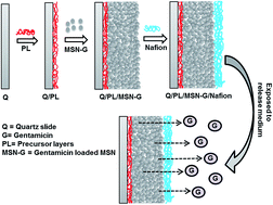
Herein we report the preparation of antibiotic thin film coatings with excellent stability and well-regulated drug release profile. These films were prepared by pre-loading of the antibiotic gentamicin into mesoporous silica nanoparticles (MSN–G) to form drug nanoreservoirs, which were then coated onto glass substrates followed by adding a polymer (Nafion) protecting layer. Fourier transform infrared (FTIR) spectroscopy and scanning electronic microscopy (SEM) confirmed the successful coating of MSN–G particles on slides. Factors such as the thickness and number of Nafion layers, the thickness of the MSN–G layer, as well as the pH of the surrounding medium were found to affect the stability of the films in solution. A typical film with 7.5 mg MSN–G particles and a thin layer of Nafion coating remained intact more than 80 days in a pH 7.4 simulated body fluid at 37 °C, which is highly promising for further biomedical application. The loaded gentamicin was found to be slowly released from thin films and the release profile could be tuned by varying the pH of the release media. For example, a steady and sustained release of gentamicin (total 95% of the loading amount) was achieved over up to 38 days in a mildly acidic solution (pH 5.5) or 56 days at the physiologic condition (pH 7.4). These films with outstanding stability and controlled release profile are expected to find promising applications in many fields, such as antibiotic coatings for biomedical devices.
1. Introduction
Implanted or indwelling medical devices represent important and revolutionary achievements of modern medical science as they can save patients' lives or substantially improve quality of life. To achieve this, a variety of medical devices, such as cardiac pacemakers, prosthetic joints, heart valves, intravascular catheters and cerebrospinal fluid shunts, are inserted surgically into the human body for managing different health crises.1,2 Unfortunately, infections associated with these life-saving indwelling devices may lead to hospitalization, prolonged courses of antibiotics, further surgical intervention, and even lethal consequences.3,4 In the United States, approximately one million cases of nosocomial infections occur annually that are related with indwelling devices.5 Bacterial colonization of (or adherence to) the surface of implanted devices causes infections as well as failure of the device to function appropriately.6 Therefore, such medical device associated infections have become a great concern to patients' wellbeing and health outcomes.
Scientists around the world have already attempted to overcome this life-threatening issue by altering the surface topography, dimensions, and material composition of indwelling devices.7–11 Recently, researchers are utilizing nanoscience in order to tackle implant-associated infections. In 2015, Taresco et al. synthesized core/shell magnetic nanoparticles consisting of a manganese iron oxide core and a polymeric shell (either polyacrylamide or polycaprolactone) to combat biofilm growth on medical device surfaces. A naturally occurring antimicrobial agent named usnic acid was used as model drug.12 Similarly, magnetite-based core–shell nanoparticles loaded with cephalosporin were developed by Grumezescu et al. These nano-reservoirs retained anti-biofilm activity and are therefore expected to protect the implanted device by preventing bacterial colonization.13 In recent times, metal ion-assisted self-assembly of nano-complexes was studied for local delivery of minocycline hydrochloride (MH) with possible future implementation on implanted devices.14 All these examples illustrate that the indwelling device-related infections have become an alarming problem that urgently needs to be addressed. Therefore, our present study has been designed to address this critically important health issue.
Mesoporous silica nanoparticles (MSNs) are widely used in nanomedicine. Different types of MSNs were applied for immediate, sustained, controlled and targeted drug delivery systems.15 Scientist explored MSNs as a promising nano vehicle for delivering various drugs i.e. antibiotic, anti-hypertensive, analgesic, anti-cancer, anti-hypercholesterolemia drugs and so on.16–20 However, drug delivery from MSNs particles are being studied for long. But, comparatively less study has been carried out to involve thin film embedded with MSNs as drug delivery system. In 2014, Zhao and co-workers introduced ibuprofen loaded MSNs (IB–MSN) in chitosan hydrogel and developed IB–MSNs/chitosan hydrogel film of 2 mm thickness. They developed a pH and electro-responsive film for tunable drug release from the titanium implants.21 However, antibiotic loaded MSNs was used as filler in poly(methyl methacrylate) (PMMA) based bone cements for obtaining a sustained release of antibiotic in order to reduce the risk of post-operative joint infection.22 Developing thin film with drug loaded nanoparticles is associated with some challenges mainly stability of film, inhibition of early burst release and controlled drug release for long period. Our present study has been designed to establish thin film incorporated with gentamicin loaded MSNs (MSN–G) by addressing the above mentioned challenges.
In this study, we aimed to prepare an antibiotic thin film coating with high drug loading and the ability to slowly release the antibiotic over time in a controlled manner. An important application of such a thin film could be the protection of implanted medical devices from bacterial contamination, as discussed above. Our strategy was to pre-load antibiotic (gentamicin, G) into mesoporous silica nanoparticles (MSN) to form drug nanoreservoirs (MSN–G), which are then coated onto glass substrates. MSNs were chosen due to their valuable intrinsic properties such as high surface area, chemical stability, and biocompatibility.23 In addition, the porous nature (∼2.8 nm) of the MSNs used in this study has favoured an optimal loading as well as holding of entrapped antibiotic for a longer time period. In order to improve the stability of the particles on the substrate (quartz or glass slides), the substrate surface is initially coated with precursor multilayers of polyelectrolytes. And a protecting layer of Nafion is also applied outside the MSN–G layer to ensure good retention of the entire film. Factors that possibly affect the stability of the film are then studied in detail. In addition, controlled gentamicin release from the thin film is also investigated by varying the Nafion layer and the pH of the release media.
2. Experimental
2.1. Chemicals and materials
Tetraethylorthosilicate (TEOS), cetyltrimethyl-ammonium bromide (CTAB), triblock copolymer F127, ammonium hydroxide (28% NH3 basis), polystyrene sulfonate (PSS, average Mw ∼ 70
000), poly(allylamine hydrochloride) (PAH, average Mw ∼ 58
000), polyethylenimine (PEI, branched, ∼25
000), Nafion perfluorinated resin solution (5 wt% in lower aliphatic alcohols and water, contains 15–20% water), and H2O2 (30%, w/w) were purchased from Sigma-Aldrich. Gentamicin sulphate (active fraction: 590 μg gentamicin per mg) was purchased from AK Scientific Inc. Phosphate buffer solutions (PBS) of various pH were prepared by mixing 0.1 M potassium dihydrogen phosphate and 0.1 M dipotassium hydrogen phosphate, and the final pH was adjusted by using 1 M hydrochloric acid (HCl) or 1 M sodium hydroxide (NaOH). Simulated body fluid (SBF) was prepared as reported previously.24 Ultrapure water was obtained from a Millipore water purification system (Milli-Q) and used throughout all experiments.
2.2. Synthesis of MSNs and antibiotic loading
Synthesis of MSNs and gentamicin loading were accomplished by following our previous work.25 MSNs were synthesized based on the modified Stöber method.26 Briefly, 0.5 g cetyltrimethyl-ammonium bromide (CTAB), 2.05 g triblock copolymer F127, 43.1 mL ethanol, 11.2 mL concentrated NH4OH solution (28 wt%) and 96 mL Milli Q water were mixed by vigorous stirring at room temperature (RT). Then, 1.93 mL tetraethyl orthosilicate (TEOS) was added quickly into the homogenous mixture under vigorous stirring followed by further stirring for 1.5 min. The mixture was kept static for next 24 hours at RT. Then, the precipitate was collected by centrifugation and three times washing. The final product (MSNs) was collected via calcination at 550° for 6 hours. Subsequently, MSNs were loaded with gentamicin by dispersing MSNs (60 mg) in 1 mL of gentamicin solution (20 mg mL−1 gentamicin in PBS at pH 9.2). Drug loading was quantified via UV-Vis spectrophotometric assay at 247 nm and loading efficiency was calculated as mass (μg) of drug loaded per mg of MSNs.
2.3. Preparation of thin films containing MSN–G
Prior to film preparation, quartz slides were cleaned via immersion into ‘piranha solution’ (3
:
1 solution of concentrated sulphuric acid and hydrogen peroxide) for 2 min followed by thorough washing with water.
Precursor layers were immobilized on the substrate surface via the layer-by-layer (LbL) self-assembly technique. Slides were first immersed into a solution of branched PEI (1 mg mL−1 in 0.5 M NaCl) for 15 min followed by rinsing with three sequential dips into Milli Q water (1 min duration each time). Subsequently, precursor multilayers were developed by sequential alternating immersions into solutions of PSS (1 mg mL−1 in 0.5 M NaCl) and then PAH (1 mg mL−1 in 0.5 M NaCl) following the same immersion duration and rinsing protocol as for PEI. These sequential adsorption processes resulted in six layers of polyelectrolytes i.e. PEI/(PSS/PAH)2/PSS coated slides. Then, gentamicin loaded MSNs (MSN–G) were deposited as a layer by adding the aqueous suspension of MSN–G drop by drop onto pre-treated slides (surface area of 1.5 × 2 cm). After drying, one or multiple layers of Nafion (varying in concentration) were applied around the MSN–G layer and dried thoroughly. The schematic diagram in Fig. 1 illustrates the process of film preparation.
 |
| Fig. 1 Schematic diagram of thin film preparation. | |
2.4. Stability testing of thin films
The stability of thin films was studied by immersing the coated slides into 2 mL of SBF at pH 7.4. For the pH stability test, buffer solutions of pH 1.4, 5.5, 7.4, and 9.5 were used. The temperature was maintained at 37 °C throughout the stability testing.
2.5. Drug release from MSN–G thin film
Slides with loaded MSN–G were immersed into 2 mL of buffer solution at various pH (pH 5.5, 7.4 and 9.5) and the release profile was monitored. Gentamicin released from thin films was quantified via UV-Vis spectroscopy. For each release study, 0.5 mL of release medium was sampled at multiple time points to measure the gentamicin concentration via UV-Vis assay at 247 nm. Following the analysis, the sampling aliquot was returned to the release medium to maintain a constant volume.
2.6. Film characterization
Scanning electronic microscope (SEM) images were obtained on a field emission scanning electron microscope (FeSEM, ZEISS SUPRA 40VP, Germany) at an acceleration voltage of 3 kV. UV-Vis spectroscopy for the analysis of gentamicin loading and release was carried out via a Halo RB-10 UV-Vis spectrophotometer (Australia). The successful deposition of MSN–G particles and Nafion layer was confirmed through Fourier transform infrared (FTIR) spectroscopy. The FTIR spectra were analysed utilising a Thermo Scientific Nicolet iD5 spectrometer (USA).
3. Result and discussion
3.1. Preparation and characterization of MSN–G films
Spherical MSNs with an average pore diameter of ∼2.8 nm and particle size of 192 ± 7 nm were synthesised via a sol–gel process using a binary surfactant system.25 These MSNs were loaded with gentamicin at pH 9.2 through electrostatic interaction between the anionic MSNs and cationic drug molecules.25 The loading amount was calculated to be 219 μg gentamicin per mg MSNs. Gentamicin encapsulation efficiency was calculated to be 65.3 ± 0.8% according to the following equation:
Thin films with embedded gentamicin loaded MSNs (MSN–G) were then prepared on quartz slides (Q) via the LbL self-assembly technique, as illustrated in Fig. 1. SEM images in Fig. 2 show the surface morphology and the thickness of the film using 7.5 mg MSN–G. A few cracks in the film were observed, as shown in Fig. 2a, which resulted from drying during the SEM sample preparation. Spherical nanoparticles of ∼200 nm in diameter were noticed (Fig. 2b) while focussed closely on the apparent crack in Fig. 2a. Nonetheless, the existence of such spherical nanoparticles confirmed the successful incorporation of MSN–G particles in the film. The thickness of the film was measured to be approximately 4.4 μm from the cross sectional view of the film (Fig. 2c).
 |
| Fig. 2 SEM images of (a) the surface of the thin film (b) incorporated MSN–G particles in the film and (c) cross-sectional view of the film. | |
FTIR analysis (Fig. 3) confirmed the successful development of the thin film. The FTIR spectrum of a bare quartz slide (Q) and MSNs are shown in Fig. 3a and b respectively. It is noted that the typical Si–O–Si bond shows an infrared band in the 1130–1000 cm−1 region.27 The FTIR spectrum of MSNs provided the characteristic peak of siloxane bond at 1055 cm−1 (Fig. 3b). A film of MSN–G without Nafion coating, i.e. Q/PL/MSN–G, was analysed via FTIR to ensure the deposition of MSN–G particles over the precursor layers (PL). Gentamicin contains five amino groups and three hydroxyl groups in its chemical structure. The FTIR spectrum of Q/PL/MSN–G (Fig. 3c) showed a strong peak at 1055 cm−1 and a medium peak at 1628 cm−1 due to the C–N stretching and N–H bending vibration of the amino groups present in the loaded gentamicin. However, a broad band at 3550–3200 cm−1 range was noticed in Fig. 3c due to the stretching vibration of O–H from gentamicin too. The characteristic band of Si–O–Si from MSNs was overlapped with the C–N stretching vibration of gentamicin in Fig. 3c. In addition, no noticeable peaks were observed from the underlying precursor layers as those layers of PEI, PSS and PAH were very thin to account. Fig. 3d depicts the FTIR spectrum of a completed thin film i.e. MSN–G (Q/PL/MSN–G/N). The bands at 1203 and 1153 cm−1 were observed because of the asymmetric and symmetric stretching vibration of F–C–F in the CF2 group.28 Furthermore, the symmetric stretching of the O–S bond from the –SO3− group provided a band at 1056 cm−1.29 All these peaks were characteristic to Nafion. All these results indicated that the Nafion coated MSN–G thin film was prepared successfully.
 |
| Fig. 3 FTIR spectra of (a) untreated quartz slide, (b) MSNs, (c) Q/PL/MSN–G, and (d) Q/PL/MSN–G/N. | |
3.2. Stability of the MSN–G contained thin films
The development of films that encompass drug loaded nanoparticles and at the same time provide a sustainable controlled release profile can be considered as a highly promising effort. An important requirement for potential future use in medical applications is that the film is stable over a relevant period of time. In this regard, the surface of the substrate was treated with precursor multilayers of polyelectrolytes [PEI/(PSS/PAH)2/PSS] to facilitate a firm fixation of the MSN–G film. In addition, an outer Nafion layer was applied to provide a further improvement in the retention. Parameters that might affect the stability and/or the firmness of films include the thickness and/or number of protective Nafion layer(s), the thickness of the MSN–G layer, as well as the pH of the medium. These parameters were investigated and the results are presented below.
3.2.1. Effect of the Nafion layer (s) on the stability of thin films. The MSN–G particles as deposited on the precursor layers were eventually coated by an additional, outer, and protective layer of Nafion. Although Nafion was applied as an outer coating, it is believed that it also diffused through the void spaces among MSN–G particles. Thereby, Nafion provides crosslinking of the individual particles to each other along with the outermost protective shielding. This extra outermost protective layer was found to be essential to prevent the leakage of particles and ensure the firmness of the entire film for extended periods of time.We first investigated the effect of the thickness of the Nafion layer on the stability profile. In this study, three films loaded with 15 mg MSN–G were covered with 100 μL Nafion of three different concentrations (1, 3, and 5%). The film with the thickest Nafion layer (5%) was peeled off just after immersing into SBF at pH 7.4. Conversely, MSN–G films wrapped up by 1 or 3% Nafion layers provided enhanced stability compared to the former one. Moreover, the film with the thinnest Nafion layer was kept compact for up to 12 days whereas the film with a moderately thick Nafion layer was intact for 23 days. These results illustrated that neither very thin nor very thick coating of Nafion was advantageous compared with a moderately thick layer of Nafion.
It was thought that multi-layer Nafion protection would further increase the stability of the film; bilayers and trilayers of Nafion (1%) were thus applied as the outermost shields on 15 mg MSN–G layer and the stability was monitored as described above. However, the results were not as expected. MSN–G films with bilayers and trilayers of Nafion started peeling off very quickly (by one day) and were destroyed at the eighth day. In contrast, MSN–G film covered by a single layer of 1% Nafion was stable for 12 days. The amount of desorbed drug detected in solution also supported that the film with a monolayer of Nafion had better stability than films with bilayers and trilayers. As seen in Fig. 4, at the eighth day of immersion, approximately 38% (Fig. 4b) and 51% (Fig. 4c) of the entrapped drug was desorbed through the bilayers and trilayers of Nafion respectively, whereas only approximately 20% of the drug was allowed to discharge through the monolayer (Fig. 4a). Generally, the release of drug would be expected to be faster from a monolayer compared to bilayers and trilayers. However, the monolayer provided slow and sustained release because of upholding an intact film. In contrary, bilayers and trilayers of Nafion provoked a more rapid escape of drug through the damaged film.
 |
| Fig. 4 Leakage of loaded gentamicin through a (a) monolayer, (b) bilayers and (c) trilayers of Nafion. | |
3.2.2. Effect of the thickness of MSN–G layer on the stability of thin films. Not only the thickness of the Nafion layer but also the thickness of the MSN–G layer imparted a vital role in the stability profile of the thin film. A number of thin films were prepared by varying the amount of incorporated MSN–G and the thickness of the Nafion layer. Amounts of 15 or 7.5 mg MSN–G were immobilized onto the precursor layers and finally wrapped up via Nafion coating (1 or 3% Nafion). Fig. 5 shows the stability profiles of thin film based on the thickness of the MSN–G layer. Fig. 5a demonstrates that films incorporated with 15 mg MSN–G were stable for 12 and 23 days when covered by 1 and 3% of Nafion layer respectively. In contrary, comparably thin layers of MSN–G consisting of 7.5 mg MSN–G particles were unbroken for 10 days (1% Nafion) and even for 80 days while wrapped up via 3% Nafion (Fig. 5b). By using 3% Nafion, the stability increased more than two fold when reducing the thickness of MSN–G layer to 7.5 mg MSN–G.
 |
| Fig. 5 Effect of the thickness of the MSN–G layer encompassing (a) 15 mg and (b) 7.5 mg MSN–G particles on the stability of thin film. | |
3.2.3. Effect of pH on the stability of thin films. In addition to the characteristics of thin films, the utilised release medium can strongly influence the stability profile. For this study, films embedded with 7.5 mg MSN–G and covered by 3% Nafion layers were prepared and exposed to a wide pH range of pH 1.4, 5.5, 7.4, and 9.5. Films were found to be very stable in slightly acidic pH 5.5 and physiologic pH 7.4. The firmness of the entire film was maintained for approximately 50 days at pH 5.5 and 80 days in physiologic pH 7.4. In contrast, neither strong acidic (pH 1.4) nor alkaline (pH 9.5) conditions could support an intact film for a long period of time. Thin films were completely damaged by the 4th day of immersion at pH 1.4. This instability was likely related to the properties of the MSN. The isoelectronic point of silica is ∼2.30,31 Thereby, silica turns to neutral (no charge) when the pH is below 2. This phenomenon caused a weakening of the interaction between the loaded gentamicin and the MSNs. Eventually, an abrupt and quick outflow of entrapped gentamicin occurred which led to the damage of the entire film at pH 1.4. However, no visible crack was noticed but the release medium became blurred when exposed to the alkaline pH 9.5. A noticeable amount of incorporated MSN–G particles was dislodged from the films into the release medium and led to the blurry appearance. The observed instability at alkaline pH was mainly due to the rupture of the Nafion layer at basic pH. Generally, Nafion requires an acidic medium to maintain its stability and becomes ruptured in the alkaline condition. Specifically, Nafion undergoes swelling when the covalent bond [OCF2CFOCF2CF2] –SO3H− (i.e. the bond between carbon and sulphur atoms) is cleaved.32
3.3. Gentamicin release profile from MSN–G thin films
Both the properties of the thin film and the pH of the release medium were considered as important factors during the analysis of the release pattern of gentamicin. The pH of the release medium was kept constant (i.e. pH 7.4) while studying the features of thin films as governing factors of drug release. The characteristics of both the MSN–G layer and the Nafion layer were found to considerably impact on the release pattern.
In contrast, the properties of the thin film were kept unchanged while evaluating the effect of the pH of the release medium on the release profile. Herein, films with 7.5 mg MSN–G which were covered with a moderately thick Nafion layer were used for release studies at different pH of 5.5, 7.4 and 9.5.
3.3.1. Controlling drug release via Nafion coating. An effective drug delivery system should comply with a controlled, slow and steady outflow of drug molecules over a prolonged period of time. Initial burst release is the common and crucial problem in developing such drug delivery systems. A quick and sharp release profile was noticed from MSN–G particles in Fig. 6A (a). The initial burst release of loaded gentamicin from MSN–G particles was measured as 98% by 3 days. This initial intense outflow of drug was overcome via embedding the MSN–G particles in a film and producing an additional polymeric Nafion layer over the MSN–G thin film. This outermost protective layer shielded the MSN–G from the direct expose to the release medium, thus resulting in complete inhibition of the initial burst release. Moreover, a slow and continuous release over several weeks was obtained via varying the thickness of the Nafion layer (i.e. by applying a 1 or 3% Nafion solution). Films with a thin Nafion monolayer demonstrated approximately 25% drug release after 15 days as shown in Fig. 6A (b). Whereas, moderately thick layers of Nafion (3%) provided an even more controlled and slower release of gentamicin. Fig. 6A (c) depicts that thicker Nafion layers allowed for approximately 15% of drug release by 15 days. As Nafion layer holds negative charge, thereby, release of gentamicin was hindered by the charge density of Nafion. Generally, an increased concentration of Nafion in the protective layer would be expected to provide a high charge density. Thereby, such a high negative charge density eventually slowed down the release of positively charged gentamicin through the protective Nafion layer. Thus, a thin layer of Nafion provided slow release while a thick layer of Nafion provided even less and slower gentamicin release.
 |
| Fig. 6 (A) Gentamicin release profiles from (a) MSN–G particles, thin films embedded with 15 mg MSN–G layer and covered via (b) 1% and (c) 3% Nafion coating. (B) Gentamicin release profiles from thin films incorporated with 7.5 mg MSN–G layer covered via (a) 1% and (b) 3% Nafion coating. | |
Nonetheless, a limitation was witnessed from this release study (Fig. 6A). A bulk of the loaded gentamicin was trapped inside the films. The maximum release was observed to be approximately 25% and 15% via 1 and 3% Nafion coating, respectively. Those films were incorporated with 15 mg of MSN–G. The thickness of the MSN–G layer was suspected as the underlying reason. MSN–G particles deposited at the bottom could not release the gentamicin because of other MSN–G particles stacked on top of them. Alternatively, the release medium could not penetrate deeply inside the layer and thus the immobilized gentamicin was restrained.
Therefore, the thickness of the MSN–G layer was reduced to ensure the maximum release of loaded drug. Fig. 6B demonstrates that ∼95% release could be achieved from thin films embedded with 7.5 mg MSN–G. Again, the thickness of the Nafion layer had shown a great impact on the release behaviour. The 1% Nafion layer could not support a stable film, therefore intensive release (66%) was observed by 9 days of immersion as shown in Fig. 6B (a). However, film embedded with 7.5 mg MSN–G and 3% Nafion layer provided a remarkably slow release over 60 days as seen in Fig. 6B (b). Conclusively, this film prevented an initial burst release, ensured maximum ∼95% release of the incorporated antibiotic and maintained a slow release profile over several months. Such long term stability and controlled release profile of thin film are clinically beneficial. Usually, late infection of the implanted medical devices can occur even days/months after implantation.33,34 Thereby, prolonged stability and slow release of antibiotic is required clinically for biomedical application. Antibiotic coverage during the first few days after implantation can be provided via intravenous antibiotic therapy.
3.3.2. Controlling the release profile via pH change. A pH controlled release profile was established since pH plays a significant role in tuning the film property. At alkaline pH 9.5, the film was unsuccessful to maintain a slow and orderly release due to the rupture of the Nafion layer. Such a damaged Nafion layer facilitated the escape of entrapped gentamicin. Therefore, an outburst (96%) of loaded gentamicin was noticed by only 5 days of the release study (Fig. 7).
 |
| Fig. 7 Gentamicin release profile from thin films in various pH. | |
On the other hand, a sustained release of gentamicin was observed at both pH 5.5 and 7.4. Slightly acidic pH provided a slower and steadier release when compared to the alkaline pH 9.5. A maximum of 95% of the entrapped gentamicin was released by day 38 and thereafter the release profile reached the plateau phase. Compared to pH 5.5, the physiologic pH 7.4 slowed down the initial release and controlled the entire release profile for 56 days with maximum 93% release of loaded drug. The reason that gentamicin released at pH 5.5 is faster than at pH 7.4 is mainly due to the gentamicin loading condition which was at pH 9.2. The larger pH difference between the loading and release condition, the faster the drug is released.25 Therefore, the release of embedded drug from the thin film at pH 7.4 was observed to be the slowest when compared to other pHs.
4. Conclusions
In summary, we have successfully developed an antibiotic coating with high-capacity drug loading and outstanding stability. Notably, this thin film provides a control over the release profile of the entrapped antibiotic. The stability of the film was optimized efficiently by altering the physical properties of the unit layer(s), i.e. the thickness of the MSN–G layer and the Nafion layer. Outstandingly, the best retention (over approximately 80 days) of film was achieved at the physiologic condition (i.e. SBF of pH 7.4 at 37 °C). A pH controlled drug release was achieved by implementing the unique properties of Nafion polymer. The favourable retention of the film as well as the complete control of antibiotic release are promising results that may support its future implementation for biomedical applications. Prolonged stability (nearly 3 months) is one of the remarkable achievements which may assist such a film to protect implanted medical devices from bacterial infections. Moreover, the synthesis of this film is well reproducible, less complex and economical which further benefits the potential application in clinical practice. To conclude with, an efficient and stable drug loaded thin film was demonstrated to achieve a steady, sustained and slow release of antibiotic by implementing the advantages of nanoscience.
Acknowledgements
T. Tamanna acknowledges the Swinburne University Postgraduate Research Award (SUPRA) for supporting this work. CB Landersdorfer is an Australian National Health and Medical Research Council (NHMRC) Career Development fellow (APP1062509).
References
- C. Von Eiff, B. Jansen, W. Kohnen and K. Becker, Drugs, 2005, 65, 179–214 CrossRef CAS PubMed.
- S. Hogan, N. Stevens, H. Humphreys, J. O'Gara and E. O'Neill, Curr. Pharm. Des., 2015, 21, 100–113 CrossRef CAS PubMed.
- J. An, A. Sun, Y. Qiao, P. Zhang and M. Su, J. Mater. Sci.: Mater. Med., 2015, 26, 1–6 CrossRef CAS PubMed.
- B. H. Trachtenberg, A. M. Cordero-Reyes, M. Aldeiri, P. Alvarez, A. Bhimaraj, G. Ashrith, B. Elias, E. E. Suarez, B. Bruckner and M. Loebe, J. Card. Failure, 2015, 21, 119–125 CrossRef PubMed.
- R. O. Darouiche, N. Engl. J. Med., 2004, 350, 1422–1429 CrossRef CAS PubMed.
- R. A. Weinstein and R. O. Darouiche, Clin. Infect. Dis., 2001, 33, 1567–1572 CrossRef PubMed.
- S. R. Shah, A. M. Tatara, R. N. D'Souza, A. G. Mikos and F. K. Kasper, Mater. Today, 2013, 16, 177–182 CrossRef CAS.
- D. S. Trentin, D. B. Silva, A. P. Frasson, O. Rzhepishevska, M. V. da Silva, E. L. Pulcini, G. James, G. V. Soares, T. Tasca, M. Ramstedt, R. B. Giordani, N. P. Lopes and A. J. Macedo, Sci. Rep., 2015, 5, 8287 CrossRef CAS PubMed.
- M. L. W. Knetsch and L. H. Koole, Polymers, 2011, 3, 340–366 CrossRef CAS.
- S. B. Goodman, Z. Yao, M. Keeney and F. Yang, Biomaterials, 2013, 34, 3174–3183 CrossRef CAS PubMed.
- J. Valle, S. Burgui, D. Langheinrich, C. Gil, C. Solano, A. Toledo-Arana, R. Helbig, A. Lasagni and I. Lasa, Macromol. Biosci., 2015, 15, 1060–1069 CrossRef CAS PubMed.
- V. Taresco, I. Francolini, F. Padella, M. Bellusci, A. Boni, C. Innocenti, A. Martinelli, L. D'Ilario and A. Piozzi, Mater. Sci. Eng., C, 2015, 52, 72–81 CrossRef CAS.
- A. Grumezescu, R. Cristescu, M. Chifiriuc, G. Dorcioman, G. Socol, I. Mihailescu, D. Mihaiescu, A. Ficai, O. Vasile and M. Enculescu, Biofabrication, 2015, 7, 015014 CrossRef CAS PubMed.
- Z. Zhang, Z. Wang, J. Nong, C. A. Nix, H.-F. Ji and Y. Zhong, Biofabrication, 2015, 7, 015006 CrossRef PubMed.
- Y. Wang, Q. Zhao, N. Han, L. Bai, J. Li, J. Liu, E. Che, L. Hu, Q. Zhang, T. Jiang and S. Wang, Nanomedicine, 2015, 11, 313–327 CrossRef CAS PubMed.
- D. L. Clemens, B.-Y. Lee, M. Xue, C. R. Thomas, H. Meng, D. Ferris, A. E. Nel, J. I. Zink and M. A. Horwitz, Antimicrob. Agents Chemother., 2012, 56, 2535–2545 CrossRef CAS PubMed.
- Y. Zhang, J. Wang, X. Bai, T. Jiang, Q. Zhang and S. Wang, Mol. Pharm., 2012, 9, 505–513 CrossRef CAS PubMed.
- N. Lai, C. Lin, P. Ku, L. Chang, K. Liao, W. Lin and C. Yang, Nano Res., 2014, 7, 1439–1448 CrossRef CAS.
- Q. Zhang, X. Wang, P. Z. Li, K. T. Nguyen, X. J. Wang, Z. Luo, H. Zhang, N. S. Tan and Y. Zhao, Adv. Funct. Mater., 2014, 24, 2450–2461 CrossRef CAS.
- Z. Zhao, C. Wu, Y. Zhao, Y. Hao, Y. Liu and W. Zhao, Int. J. Nanomed., 2015, 10, 1691–1701 CAS.
- P. Zhao, H. Liu, H. Deng, L. Xiao, C. Qin, Y. Du and X. Shi, Colloids Surf., B, 2014, 123, 657–663 CrossRef CAS PubMed.
- S.-C. Shen, W. K. Ng, Z. Shi, L. Chia, K. G. Neoh and R. B. H. Tan, J. Mater. Sci.: Mater. Med., 2011, 22, 2283–2292 CrossRef CAS PubMed.
- S. Kwon, R. K. Singh, R. A. Perez, E. A. A. Neel, H.-W. Kim and W. Chrzanowski, J. Tissue Eng., 2013, 4, 2041731413503357 Search PubMed.
- M. R. Marques, R. Loebenberg and M. Almukainzi, Dissolution Technol., 2011, 18, 15–28 CrossRef CAS.
- T. Tamanna, J. Bulitta and A. Yu, J. Mater. Sci.: Mater. Med., 2015, 26, 1–7 CrossRef CAS PubMed.
- T. W. Kim, P. W. Chung and V. S. Y. Lin, Chem. Mater., 2010, 22, 5093–5104 CrossRef CAS.
- P. J. Launer, in Silicone Compounds Register and Review, ed. B. Arkles, et al., 1987, pp. 100–103 Search PubMed.
- K. Kunimatsu, T. Yoda, D. Tryk, H. Uchida and M. Watanabe, Phys. Chem. Chem. Phys., 2010, 12, 621–629 RSC.
- N. P. Cele, S. S. Ray and L. Sikhwivhilu, J. Nanomater., 2012, 1–7, DOI:10.1155/2012/717913.
- I. Larson, C. J. Drummond, D. Y. C. Chan and F. Grieser, Langmuir, 1997, 13, 2109–2112 CrossRef CAS.
- X. Huang, Z. Tao, J. C. Praskavich, A. Goswami, J. F. Al-Sharab, T. Minko, V. Polshettiwar and T. Asefa, Langmuir, 2014, 30, 10886–10898 CrossRef CAS PubMed.
- N. Bunkin, P. Ignatiev, V. Kozlov, A. Shkirin, S. Zakharov and A. Zinchenko, Water, 2013, 4, 129–154 Search PubMed.
- L. G. Rubin and B. Papsin, Pediatrics, 2010, 126, 381–391 CrossRef PubMed.
- H.-J. Trappe, P. Pfitzner, H. Klein and P. Wenzlaff, Br. Heart J., 1995, 73, 20–24 CrossRef CAS.
|
This journal is © The Royal Society of Chemistry 2015 |
Click here to see how this site uses Cookies. View our privacy policy here.