Free radical nano scavenger based on amphiphilic novolacs

Yue Wanga, Qian Zhang*a, Chao Xieb, Xin Zhaoc, Jinfen Niua, Jie Zhaoa, Jung Kwon Ohd, Peng Lic and Pengfei Li*c
aDepartment of Applied Chemistry, Xi'an University of Technology, No. 5 South Jinhua Road, Xi'an, Shaanxi, 710048 P. R. China. E-mail: qzh@xaut.edu.cn
bDepartment of Oral Implantology, State Key Laboratory of Military Stomatology, School of Stomatology, the Fourth Military Medical University, No. 169 West Changle Road, Xi'an, Shaanxi, 710032 P. R. China
cFrontier Institute of Science and Technology (FIST), Xi'an Jiaotong University, No. 99 Yanxiang Road, Xi'an, Shaanxi, 710054 P. R. China. E-mail: lipengfei@mail.xjtu.edu.cn
dDepartment of Chemistry and Biochemistry and Center for Nanoscience Research (CENR), Concordia University, 7141 Sherbrooke St. West, Montreal, Quebec, H4B 1R6 Canada

Received 2nd October 2015 , Accepted 27th October 2015

First published on 28th October 2015


Abstract

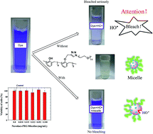
As the first synthetic plastic, bakelite, which was developed 100 years ago, is still being used nowadays. Beyond its conventional applications as engineering plastics till today, novolacs, the soluble form of bakelite, are modified by partly PEGylation via “Click” chemistry to construct a novel amphiphilic polymer in this research. The resulting novolacs-PEG is capable of forming micellar nano-aggregates in aqueous solution at concentrations above the critical micellar concentration = 10.5 μg mL−1. The remaining phenolic moities on the backbone of novolacs provide a unique chance to scavenge hydroxyl radicals, impersonating natural poly-phenolic antioxidants from food extract. As a result, the novolacs-PEG proves highly effective to protect crystal blue from being bleached by hydroxyl radicals generated by Fenton reagents. In addition, the novel micelles are nontoxic to cells in in vitro experiment, and so are very promising in anti-ROS (reactive oxygen species) applications. This new application of novolacs reveals a promising nano-platform of synthetic research to act against ROSs which can highly damage human health.


1. Introduction

Molecular oxygen is a critical component for living systems to generate energy; however, it can be detrimental as another form of reactive oxygen species (ROSs).1 When the balance between ROS production and antioxidant defense is disturbed, undesired oxidative stress (OS) is caused in the body.2 This undesired OS is known to result in oxidative DNA mutation3 and cell damage,4,5 which can be linked to cancers,6,7 ageing,8,9 ischemia injury,10 trauma and infection,11,12 brain13 and cardiovascular disease14 and so on. To suppress these medical challenges, the use of antioxidant defenders (or radical scavengers) is critical since they can scavenge the excess of ROS.

Nature is abundant with antioxidants. A number of natural antioxidants are available; examples include vitamin E from food15,16 as well as β-caronoids,17,18 lycopene,19 capsaicin,20 resveratrol,21 curcumin,22 piceatannol,23 catechin,24 rutin,25 myricetin,26 puerarin,27 artemisinin,28 gallic acid,29 ferulic acid,30 quercetin,31 apigenin,32 chrysin32 from plant extracts. Interestingly, most of these natural molecules are composed of multiple phenol moieties. In fact, synthetic phenol has been used as an effective radical inhibitor in free radical polymerizations.33–35

Bakelite is a common phenolic resin that has been used as important industrial plastics over the last 100 years since being developed.36 We have aimed to use such an old well known polymer to scavenge ROS, shedding light on the possible biomedical applications. However, one critical challenge of bakelite is its poor solubility in aqueous solution. We have hypothesized that a possible solution to circumvent the challenge can be constructing amphiphilic macromolecules which can self-assemble into nano-aggregate,37,38 so-called radical nano-scavenger (RNS). Most approaches to develop RNS involve the encapsulation of small molecules of antioxidants in nanoparticles such as liposome,39–41 solid lipid,42–44 micelle45–47 or crosslinked nanogel.48 A more promising approach is the synthesis of “intrinsically pure RNS” that can scavenge the generated radicals by materials themselves with no need of the encapsulation of small antioxidants. However, most accessible nanoparticles except ceria49 and fullerene50–52 (though some contradictory results still exist53) are lack of antioxidant properties and generate OS, thus being toxic to cells.54 Recently, few reports described the synthesis of block copolymers covalently attached with radical-scavenging groups as effective RNSs. A nitroxide-labeled amphiphilic block copolymer was synthesized and self-assembled to form micelle-like RNSs.55–58 The copolymer was responsive to pH56 or temperature55 to suppress inflammation. And recently, indomethacin was encapsulated in the micelles to enhance anti-inflammatory effect.58 Other efforts to make “intrinsically pure RNS” include modifying the commercially available antioxidant to make them surface-active. For example, alkyl chain was attached to ascorbic acid to make a new surfactant which can form antioxidant micelles,59 similar example can be found for p-hydroxyphenylacetic acid.60

Herein we report the synthesis and aqueous self-assembly of a novel bakelite-based amphiphilic block copolymer containing hydrophilic PEG blocks and to study their ability to scavenge ROS as an effective RNS. As illustrated in Scheme 1, novolacs, the soluble form of bakelite, were synthesized by condensation polymerization of phenol and formaldehyde, catalyzed by oxalic acid. Then, phenolic hydroxyl groups reacted with propargyl bromide to afford to the formation of alkyne-labeled novolacs (ALN). Separately, PEG monomethyl ether was converted to the corresponding azido-PEG (PEG-N3). The resulting PEG-N3 reacted with ALN through a click-type alkyne–azide reaction to form amphiphilic novolacs-PEG block copolymer. Our design possesses a number of advantages: (1) the chemistry is simple and straightforward. Synthesis of novolacs has been well established in bakelite industry. PEGylation through “Click” chemistry has also been well studied in the recent decade, and PEGylating reagents have been commercialized and easily accessed; (2) the content of Ar-OH which is critical in radical scavenging can be conveniently regulated mainly by partly substitution of the propargyl moities; (3) most importantly, amphiphilicity of the polymer leading to self-assembly into nano-aggregate allows the polymer a new research platform against ROS.


image file: c5ra20440b-s1.tif
Scheme 1 Illustration of the synthetic route of amphiphilic novolacs-PEG by “click” reaction between azide and alkyne moities.

2. Experimental section

2.1. Materials

Phenol (99.0%), formaldehyde (37 wt% in H2O), oxalic acid (99.5%), potassium carbonate (99.0%, anhydrous), p-toluenesulfonyl chloride (98.5%), hydrogen peroxide (30 wt% in water) and triethylamine (99.0%) were purchased from Sinopharm Chem. Ltd. Propargyl bromide (98%, stabilized by propylene oxide), benzyltriethylammonium chloride (TEBAC, 98%), methoxypolyethylene glycol (mPEG500, Mn = 500) and cuprous bromide (99.0%) were from Aladdin Industrial Corp. Potassium iodide (reagent grade), sodium azide (99.0%), acetone (HPLC grade), tetrahydrofuran (HPLC grade), N,N-dimethylformamide (HPLC grade) were from Sigma Aldrich. Column chromatography was performed with silica gel (SANPONT, 120 A, 400 mesh). Crystal blue (99.0%), H2O2 (30% aqueous solution) and FeSO4·7H2O (99%) and sodium azide (99%) were from Sinopharm Chem. Ltd. Dialysis membrane was from Spectra/por, MWCO = 1000.

2.2. Instrumentations

1H NMR (400 MHz) spectra were obtained using a Bruker Avance spectrometer. Molecular weight (MW) and molecular weight distribution were determined by gel permeation chromatography (GPC) using Malvern Viscotek 270MAX equipped with a refractive index (RI) detector. Two Viscotek columns (LT3000L and LT6000L, designed to determine MW up to 2[thin space (1/6-em)]500[thin space (1/6-em)]000 g mol−1) were used with THF as eluent at 30 °C at a flow rate of 1 mL min−1. Shodex polystyrene (PS) standards with MW 1.3 K to 2500 K were used for calibration. Aliquots of polymer samples were dissolved in THF and the resulting clear solutions were filtered using a 0.22 μm PTFE filter to remove any insoluble prior to injection. A drop of anisole was added as a flow rate marker. Attenuated Total Reflectance Fourier Transform Spectroscopy (ATR-FTIR) was collected on a Shimadzu Prestige-21 spectrophotometer. Spectra were recorded between 4000 and 650 cm−1 for 16 scans at 4 cm−1 resolution. Particle size was measured by dynamic light scattering (DLS) on DelsaNano C from Beckman Coulter. UV/vis spectrograph was recorded on MAPADA UV-3200 spectrophotometer using 1 cm quartz cuvette. Transmission Electron Microscopy (TEM) images were taken using a JEOL JEM 2100 HR-TEM, operated at 200 kV. TEM samples were prepared by dropping diluted micelle solution onto 230 mesh copper grid, and drying in air prior to observation.

2.3. Synthesis

Synthesis of novolacs. Novolac phenolic polymer was prepared in a traditional way. To a 250 mL three-necked flask were added phenol (14.12 g, 135 mmol, 1.0 eq.), formaldehyde (10.96 g aq. solution, 150 mmol, 0.9 eq.) and oxalic acid (1.097 g, 8.7 mmol, 0.06 eq.), stirred mechanically, bubbled with argon for 20 min, and heated to 85 °C, and kept for a given time in argon atmosphere. The resulting viscous precipitant at the bottom was collected, quickly dried in reduced pressure and precipitated twice in 150 mL of petroleum ether/ethanol = 8[thin space (1/6-em)]:[thin space (1/6-em)]2 v/v, then dried in vacuum at 70 °C for 18 h to give a colorless solid. 1H NMR (DMSO-d6, ppm): 9.08 (1H, Ar-OH), 7.15–6.77 (3H, Ar-H), 3.69 (2H, Ar-CH2–).
Synthesis of alkyne-labeled novolacs (ALN). Novolac (0.5 g, 4.72 mmol Ar-OH) was dissolved in 3 mL acetone in Schlenk flask. Then anhydrous K2CO3 (0.3252 g, 2.35 mmol) was ground, and added with TEBAC (5.37 mg, 0.02 mmol), KI (15.60 mg, 0.09 mmol) and propargyl bromide (262.7 μL, 2.36 mmol) to the novolac solution. The resulting mixture was deoxygenated by three freeze–pump–thaw cycles and the flask was backfilled with N2. Then the mixture was thawed, and the flask was immersed in an oil bath preheated to 60 °C for 20 h. The reaction was stopped by cooling the flask in ice water. Solvent was removed by evaporation in reduced pressure. The polymer was isolated by precipitation from 20 mL mixed solvent (petroleum ether/ethanol = 9[thin space (1/6-em)]:[thin space (1/6-em)]1 v/v) and 20 mL × 2 petroleum ether, then dried in vacuum for 15 h to give a (0.74 g) light yellow solid. 1H NMR (DMSO-d6, ppm): 9.08 (1H, Ar-OH), 7.19–6.97 (3H, Ar-H), 4.71–4.48 (2H, –O–CH2–), 3.69 (2H, –CH2–), 3.61 (1H, –C[triple bond, length as m-dash]H).
Synthesis of tosylated PEG (PEG-Ts). Methoxypolyethylene glycol (mPEG500, 8 g, 0.016 mol –OH) was dissolved in 28 mL THF, and added TEA (6.7 mL, 0.048 mol) in magnetic stirring. Separately, p-toluenesulfonyl chloride (9.2 g, 0.048 mol) was dissolved in 28 mL THF, then added dropwise to the flask with mPEG500. The resulting mixture was stirred for 45 h at room temperature, filtered to remove insolubles, and evaporated to get the crude product. It was further purified by column chromatography with ethyl acetate eluent. The final product was a colorless oil (yield 6.8 g). 1H NMR (CDCl3, ppm): 7.80 (2H, Ar-H), 7.35 (2H, Ar-H), 3.60 (2H, –(O–CH2–CH2) chain), 3.39 (3H, –O–CH3), 3.39 (3H, Ar-CH3).
Synthesis of azide-terminated PEG (PEG-N3). PEG-Ts (0.501 g, 0.764 mmol) was dissolved in 2 mL dry DMF with magnetic stirring, and added NaN3 (0.149 g, 2.293 mmol) under the protection of argon. The resulting mixture was heated to 110 °C and kept for 9 h. The reaction was stopped by cooling to room temperature. The insolubles were filtered off. The product was isolated by precipitation in 20 mL × 2 petroleum ether as a colorless oil (yield 0.36 g). 1H NMR (CDCl3, ppm): 3.66 (–(O–CH2–CH2)– chain), 3.39 (3H, –O–CH3).
Synthesis of PEG-grafted novolacs (novolacs-PEG). A typical procedure of grafting PEG-N3 onto the alkyne functionalized novolac backbone is as follows. To a Schlenk flask were added PEG-N3 (79.04 mg, 0.146 mmol of –N3), ALN (50.30 mg, 0.191 mmol of alkyne) and copper(II) bromide (6.2 mg, 0.0278 mmol) and 2 mL DMF were added to a Schlenk flask. The resulting mixture was again deoxygenated by two freeze–pump–thaw cycles and the flask was backfilled with N2. Separately, ascorbic acid (48.92 mg, 0.278 mmol) was dissolved in 0.2 mL deoxygenated DMF, and added to the previous mixture, followed by another two freeze–pump–thaw cycles and sealed in N2. The reaction was carried out in an oil bath preheated to 50 °C for 7.5 h. The resulting mixture was dialyzed in 2 L deoxygenated deionized water for 5 days to completely eliminate PEG-N3 of relatively low molecular weight (dialysis membrane from Spectra/por, MWCO = 1000). The product was isolated and dried in vacuum for 48 h. 1H NMR (DMSO-d6, ppm): 9.10 (1H, Ar-OH), 8.13 (1H, triazole H), 7.25–6.65 (3H, Ar-H), 5.07 (2H, –N–CH2–CH2–O–), 4.71 (2H, –O–CH2–C–), 4.51 (2H, –O–CH2–C–), 3.75 (2H, Ar-CH2–), 3.49 (–(O–CH2–CH2)– chain), 3.23 (3H, –O–CH3).

2.4. Micellar dispersion and characterization

Preparation of micellar dispersion. The micellization was the same as reported. Novolacs-PEG (4.00 mg) was dissolved in THF (0.5 mL), added dropwise to deionized water (10 mL). The resulting dispersion was kept under stirring for 24 h to remove THF, allowing for a stable micellar dispersion. The dispersion was much diluted for DLS and TEM characterization.
Determination of critical micellar concentration (CMC) using a Nile Red (NR) probe. A stock solution of Nile Red (NR) in THF at 1 mg mL−1 and a stock solution of novolacs-PEG in THF at 1 mg mL−1 were prepared. Water (10 mL) was added dropwise into a series of mixtures consisting of the same amount of the stock solution of NR (0.5 mL, 0.5 mg NR) and various amounts of the stock solution of novolacs-PEG in 20 mL vials. The resulting dispersions were stirred for 24 h to evaporate THF. The dispersions were then filtered using 0.45 μm PES filters to remove excess NR. A series of NR-loaded micelles at various concentrations of novolacs-PEG ranging from 2.0 × 10⁻⁴ to 0.2 mg mL−1 were formed. Their fluorescence spectra were recorded at λmax = 653 nm.

2.5. Study the property of radical scavenging

The efficacy of novolacs-PEG dispersion on radical scavenging was evaluated by its inhibition of the oxidation of hydroxyl radical generated from Fenton reagent. Crystal violet was chosen as the indicator. Quickly oxidation by hydroxyl radical can eliminate the color from crystal violet, so addition of effective hydroxyl radical scavenger will result in a retention of blue color. Experimentally, crystal violet (1.0 × 10⁻³ mol L−1), FeSO4·7H2O (1.43 × 10⁻² mol L−1), H2O2 (4.32 × 10⁻³ mol L−1) aqueous solution and novolacs-PEG micellar dispersion (0.4 mg mL−1) were prepared in advance as stock solutions. Then, crystal violet (0.2 mL), FeSO4·7H2O (0.4 mL), H2O2 (0.4 mL except in control) and novolacs-PEG micelle (0 mL in bleaching test, 0.6, 1.2 and 1.8 mL) were combined quickly in volumetric flask with addition of deionized water to form a dispersion of constant volume (25 mL) and mixed vigorously on vortex. The absorbance at 590 nm corresponding to the peak of crystal violet was tracked by UV-vis spectrometer at time intervals.

2.6. Cytotoxicity assay

The Dulbecco's modified Eagle medium (DMEM) (Gibco) supplemented with 10% fetal bovine serum (Gibco), 1.0 × 10⁵ U L−1 penicillin (Hyclone) and 100 mg L−1 streptomycin (Hyclone) was used as the complete growth medium. Mouse osteoblastic cell line (MC3T3-E1) were seeded in 96-well plate (Costar) at a density of 104 cells per well. After culturing for 24 h in a humidified incubator containing 5% CO2 at 37 °C, the culture medium was removed and then 100 μL of complete growth medium containing the micelles at different doses (0, 10, 15, 23, 52, 100 μg mL−1) was added into the plates. When the plate was incubated for another 24 h, the medium was removed and 10 μL of alamar Blue® reagent in 100 μL complete growth medium was then added into each well. The plate was incubated for 4 hours. After that, 100 μL of the medium in each well was transferred into a 96-well black plate (Costar). Fluorescence was read using 530 nm as the excitation wavelength and 600 nm as the emission wavelength using a microplate reader (Molecular Devices) according to the manufacturer's instructions. Tests were repeated four times for each group. The cell viability was estimated according to the following equation: cell viability was calculated as the percent ratio of fluorescent intensity of mixtures with micelles to control.

3. Results and discussion

Novolacs were synthesized by a traditional method with the mole ratio of formaldehyde/phenoly < 1/1 in the presence of oxalic acid as a catalyst.61 They were then purified by precipitation from a mixture of ether/ethanol = 8/2 (v/v) to remove low MW species and unreacted phenols. Then, the resulting novolacs reacted with propargyl bromide to yield ALN. The successful incorporation of alkyne groups was confirmed by the appearance of a new peak at 2119 cm−1 on FTIR (Fig. 1A) and 4.7 ppm from methylene protons adjacent to the alkyne group on 1H-NMR (Fig. 1C). In another set, PEG monomethyl ether with MW = 500 g mol−1 was tosylated, and then replaced by azido group. The molecular structure was confirmed by the intensive azido vibration appeared at 2102 cm−1 on FTIR and related peaks on 1H-NMR (Fig. 1C). The resulting alkyne-labeled novolacs and PEG-N3 reacted through click-type alkyne–azido reaction in the presence of Cu(I) to synthesize novolacs-PEG amphiphilic block copolymer. The reported procedure62 was slightly modified as ascorbic acid was used to reduce Cu(II) to Cu(I). The successful “clicking” was firstly confirmed by FTIR with the evidence of disappearance of azido peak (2102 cm−1) and decrease in alkyne peak (2119 cm−1, not magnified to enhance demonstration on Fig. 1A). In addition, molecular weight as the number average MW (Mn) was 609 g mol−1 for PEG-N3, and 1032 g mol−1 for alkyne-labeled novolacs. After “clicking”, the Mn increased to 1519 g mol−1 with an evidently shift of GPC curve to higher molecular weight on Fig. 1B. Such increase suggests the successful addition of roughly one PEG-N3 chain onto one novolacs backbone. Further, the molecular structure of novolacs-PEG was confirmed by 1H-NMR, as shown in Fig. 1C. The degree of alkyne substitution was 47% calculated by the proton at 4.71 ppm before “clicking” reaction. After “clicking”, it shifted to 5.07 ppm. At the same time, the methylene proton adjacent to the formed triazole moiety appeared at 4.51 ppm. The results indicated the successful “clicking” reaction between alkyne and azide which accounted to a conversion of about 35% on the reaction as designed. With the normalized integration of protons at 6.5–7.5 ppm, the grafting ratio of PEG was found to be 13% on each backbone polymer chain averagely, well corresponding to the results from GPC measurement by approximation. It is worth noting that the substitution degree of alkyne can be conveniently tuned by varying the feed ratio of alkyne/phenol, for example, from 20% to 100% successfully in follow-up experiments. And as a result, the remaining phenol groups which scavenge free radicals and the hydrophilic PEG side chains which govern the dispersibility in aqueous solutions can be balanced in composition in the design of the amphiphilic novolacs-PEG.
image file: c5ra20440b-f1.tif
Fig. 1 The comparison of FTIR spectra (A) and GPC chromatograms (B) before and after “clicking” reaction and NMR spectra in DMSO-d6 or CDCl3.

The combination of hydrophobic novolacs and hydrophilic PEG allows for the amphiphilicity of novolacs-PEG, and as a result, the ability to form micelle in aqueous solution via evaporation method.63 Using NR as the probe,64 the fluorescence intensity tended to deflect from the horizontal baseline at 10.0 μg mL−1 with the increase of concentration, and the CMC was determined to be 10.5 μg mL−1 by linear extrapolation (Fig. 2A). At concentrations above CMC, novolacs-PEG self-assembled into micellar nano-aggregates consisting of hydrophobic novolac cores surrounded with hydrophilic PEG coronas. DLS results (Fig. 2B) indicated a hydrodynamic diameter of ∼110 nm with a monomodal size distribution, which corresponded well to an lower value of average diameter of 98.5 ± 10.3 nm by TEM due to dehydration.64,65


image file: c5ra20440b-f2.tif
Fig. 2 Evolution of fluorescence intensity of NR (A) at 653 nm over concentration of novolacs-PEG to determine CMC; DLS diagram (B) and TEM images (B inset) of self-assembled micellar aggregates.

To study the property of novolacs-PEG to scavenge radicals, Fenton reagents66 (hydrogen peroxide and ferrous sulfate) were used to generate hydroxyl radical, a typical ROS in the body.67,68 Due to the fact that hydroxyl radical is so bleaching to be used in treating fabrics,69 removing dyes70 and pollutants,71 it would be expected to see a quickly fading out of color of crystal violet aqueous solution as designed in this experiment when Fenton reagents are added. By scavenging the culprit hydroxyl radicals, the color would be retained, the higher concentration of scavengers, the deeper the color remained. This is the fundamental of the evaluation essay in this experiment. Five parallel experiments were conducted, including the test of control (no addition of H2O2), bleaching (no addition of novolacs-PEG), and tests of no. 1 to 3 differing in the concentration of novolacs-PEG. Shown in Fig. 3, when there was no novolacs-PEG added in bleaching experiment, a very quick elimination of blue color was observed with absorbance deceasing from 0.83 to 0.22 within 5 min. When 0.6 mL novolacs-PEG were added (equivalent to 9.6 μg mL−1 slightly lower than CMC), a big increase of absorbance to 0.6 was found, indicating a considerable effect of successful scavenging of hydroxyl radicals, and so saving much of the blue color. When more novolacs-PEGs were added in separate tests, 1.2 mL (19.2 μg mL−1, no. 2 expt.) and 1.8 mL (27.6 μg mL−1, no. 3 expt.) both higher than CMC, the remaining absorbance increased further to 0.65 and 0.70 which had little difference from the control experiment, indicating almost complete elimination of hydroxyl radical, so protection of crystal blue from being bleached. It may be noticed a slowly color fading in control sample with time, it was basically caused by photo-bleaching in daytime. It's no doubt that poly-phenolics played a critical role in removing hydroxyl radicals from this research. The contribution may lie in two aspects, one from the free novolacs-PEG molecules especially when the concentration was lower than CMC, and the other from micellar nano-aggregates when the concentration was higher than CMC. In both cases, hydrophilic PEG moieties were important to increase the solubility of novolacs in aqueous solution. The formation of micelles above CMC can enhance the ability of radical scavenging, leaving the color almost unchanged compared to the control experiment.


image file: c5ra20440b-f3.tif
Fig. 3 Evolution of absorbance at 590 nm tracked by UV-vis spectrometer to show the ability to scavenge hydroxyl radical by novolacs-PEG. The inset table indicates the amount of H2O2 and novolacs-PEG used. The color change of solution in cuvettes is shown as images on the right.

To preliminarily assess the novolacs-PEG micelles toward biomedical applications, in vitro cytotoxicity with mouse osteoblastic cells was examined using alamarBlue assay (a fluorescent method to measure cell toxicity). Novolacs-PEG of different concentrations were cultured with cells. Cells without polymers were also tested simultaneously as controls. After 24 h incubation, cell viability was calculated based on the ratio of fluorescent intensity with micelles to control. Fig. 4 suggested >94% viability of cells in the presence of micelles up to 0.10 mg mL−1, ten times higher than CMC. The results suggests that novolacs-PEG is nontoxic to cells.


image file: c5ra20440b-f4.tif
Fig. 4 Viability of mouse osteoblastic cells cultured with various amounts of novolacs-PEG micelles for 24 h.

4. Conclusions

Amphiphilic novolacs synthesized by partly PEGylation via “click” chemistry provided a chance to scavenge ROS in this contribution. The successful “clicking” reaction was confirmed by FTIR, GPC and NMR. The overall PEGylation ratio was found to be 13%, suggesting one PEG chain on each novolacs backbone by approximation. The resulting novolacs-PEG formed micelle in aqueous solution by evaporation process. CMC was determined to be 10.5 μg mL−1 based on the fluorescence of NR via encapsulation method. The micellar particle size was found to be 110.1 nm by DLS and 98.5 nm by TEM in diameter. The effectiveness of scavenging hydroxyl radical was studied using crystal violet as the indicator. When added, novolacs-PEG considerably inhibited the bleaching of blue color from crystal blue due to hydroxyl radical. The concentration of usage spanned from 9.5 to 27.6 μg mL−1, very close to or above CMC of novolacs-PEG. It is noted that the bleaching effect can almost be completely suppressed at the concentration of 27.6 μg mL−1, suggesting the very high efficiency of radical scavenging from micellar nano-aggregate. These unique features, in addition to noncytotoxicity, suggest that the new micelles hold great potential for anti-ROS applications. Based on our knowledge, it is for the first time that the amphiphilic synthetic phenolics was used to investigate the radical scavenging property in aqueous solution, and the amphiphilic novolacs provided a new nano-platform of synthetic research on the way fighting against ROS in the future.

Acknowledgements

Financial support of the Youth Project from National Natural Science Foundation of China (No. 21304074), Shaanxi Province Key Laboratory Open Project of Polymer Science and Technology (2013SZS17-K01), “100 Talents” Programme from Shaanxi Province, and the Starting and Innovative Team Programme from Xi'an University of Technology are gratefully acknowledged.

Notes and references

  1. U. Krengel and S. Törnroth-Horsefield, Science, 2015, 347, 125–126 CrossRef CAS PubMed.
  2. D. J. Betteridge, Metabolism, 2000, 49, S3–S8 CrossRef.
  3. G. C. Kujoth, A. Hiona, T. D. Pugh, S. Someya, K. Panzer, S. E. Wohlgemuth, T. Hofer, A. Y. Seo, R. Sullivan, W. A. Jobling, J. D. Morrow, H. van Remmen, J. M. Sedivy, T. Yamasoba, M. Tanokura, R. Weindruch, C. Leeuwenburgh and T. A. Prolla, Science, 2005, 309, 481–484 CrossRef CAS PubMed.
  4. B. Poljsak and I. Milisav, Oxid. Med. Cell. Longevity, 2012, 2012, 480895–480907 CAS.
  5. A. Rizzo, M. T. Roscino, F. Binetti and R. L. Sciorsci, Reprod. Domest. Anim., 2012, 47, 344–352 CrossRef CAS PubMed.
  6. M. A. Chetram and C. V. Hinton, Front. Biol., 2013, 8, 273–278 CrossRef CAS PubMed.
  7. W. D. Landry and T. G. Cotter, Biochem. Soc. Trans., 2014, 42, 934–938 CrossRef CAS PubMed.
  8. P. Abete, C. Napoli, G. Santoro, N. Ferrara, I. Tritto, M. Chiariello, F. Rengo and G. Ambrosio, J. Mol. Cell. Cardiol., 1999, 31, 227–236 CrossRef CAS PubMed.
  9. S. Goto and Z. Radak, Dose-Response, 2010, 8, 68–72 CrossRef CAS PubMed.
  10. P. Pacher, J. S. Beckman and L. Liaudet, Physiol. Rev., 2007, 87, 315–424 CrossRef CAS PubMed.
  11. Free Radical and Antioxidant Protocols, ed. D. Armstrong, Humana Press, 1998 Search PubMed.
  12. S. Cho, O. Hwang, I. Lee, G. Lee, D. Yoo, G. Khang, P. M. Kang and D. Lee, Adv. Funct. Mater., 2012, 22, 4038–4043 CrossRef CAS.
  13. R. Deloncle and O. Guillard, J. Alzheimer's Dis., 2015, 43, 1149–1156 CAS.
  14. J. W. Starnes, in Free Radicals in Biology and Medicine, ed. C. Gutierrez-Merino and C. Leeuwenburgh, Trivandrum, India, 2008, ch. 11, pp. 251-263 Search PubMed.
  15. B. Zhou, Z.-S. Jia, Z.-H. Chen, L. Yang, L.-M. Wu and Z.-L. Liu, J. Chem. Soc., Perkin Trans. 2, 2000, 785–791 RSC.
  16. S. M. Culbertson and N. A. Porter, J. Am. Chem. Soc., 2000, 122, 4032–4038 CrossRef CAS.
  17. L. Li, H. Cho, K. H. Yoon, H. C. Kang and K. M. Huh, Int. J. Pharm., 2014, 471, 339–348 CrossRef CAS PubMed.
  18. M. N. Lund, R. Liang, J.-P. Zhang, R.-M. Han, Y.-X. Tian, T. F. Skovsgaard, J. Nielsen and L. H. Skibsted, Food Res. Int., 2013, 54, 230–238 CrossRef CAS.
  19. M. Takashima, M. Shichiri, Y. Hagihara, Y. Yoshida and E. Niki, Food Funct., 2012, 3, 1153–1160 CAS.
  20. P. Datta, K. C. Pramanik, S. Mehrotra and S. K. Srivastava, in Cancer: Oxidative Stress and Dietary Antioxidants, ed. V. R. Preedy, Elsevier Ltd., London, 2014, ch. 23, pp. 241-246 Search PubMed.
  21. L. J. Carlson, B. Cote, A. W. G. Alani and D. A. Rao, J. Pharm. Sci., 2014, 103, 2315–2322 CrossRef CAS PubMed.
  22. P. Montoro, A. Braca, C. Pizza and N. de Tommasi, Food Chem., 2005, 92, 349–355 CrossRef CAS.
  23. Y. Rhayem, P. Therond, L. Camont, M. Couturier, J.-L. Beaudeux, A. Legrand, D. Jore, M. Gardes-Albert and D. Bonnefont-Rousselot, Chem. Phys. Lipids, 2008, 155, 48–56 CrossRef CAS PubMed.
  24. T. Nagai, T. Kawashima, N. Suzuki, Y. Tanoue, N. Kai and T. Nagashima, J. Food, Agric. Environ., 2007, 5, 137–141 CAS.
  25. O. A. Chat, M. H. Najar and A. A. Dar, Colloids Surf., A, 2013, 436, 343–353 CrossRef CAS.
  26. M. Ihl, V. Bifani, C. Ramirez, M. Rubilar, C. Meneses, R. Infante, Y. Motomura and I. Seguel, Acta Hortic., 2006, 712, 617–622 CrossRef CAS.
  27. J. Xi and R. Guo, J. Dispersion Sci. Technol., 2009, 30, 857–864 CrossRef CAS.
  28. S. Lapenna, A. R. Bilia, G. A. Morris and M. Nilsson, J. Pharm. Sci., 2009, 98, 3666–3675 CrossRef CAS PubMed.
  29. E. I. Karaseva, T. V. Nikiforova and D. I. Metelitsa, Appl. Biochem. Microbiol., 2001, 37, 406–412 CrossRef CAS.
  30. K. Ohara, Y. Ichimura, K. Tsukamoto, M. Ogata, S.-i. Nagaoka and K. Mukai, Bull. Chem. Soc. Jpn., 2006, 79, 1501–1508 CrossRef CAS.
  31. S. Jabeen, O. A. Chat, G. M. Rather and A. A. Dar, Food Res. Int., 2013, 51, 294–302 CrossRef CAS.
  32. K. Mukai, W. Oka, K. Watanabe, Y. Egawa, S.-i. Nagaoka and J. Terao, J. Phys. Chem., 1997, 101, 3746–3753 CrossRef CAS.
  33. P. B. Jayathilaka, G. C. Pathiraja, A. Bandara, N. D. Subasinghe and N. Nanayakkara, Can. J. Chem., 2014, 92, 809–813 CrossRef CAS.
  34. M. S. Karasch, F. Kawahara and W. Nudenberg, J. Org. Chem., 1954, 19, 1977–1990 CrossRef CAS.
  35. L. Valgimigli, G. Brigati, G. F. Pedulli, G. A. DiLabio, M. Mastragostino, C. Arbizzani and D. A. Pratt, Chem.–Eur. J., 2003, 9, 4997–5010 CrossRef CAS PubMed.
  36. American Chemical Society National Historic Chemical Landmarks, Bakelite: The World's First Synthetic Plastic, http://www.acs.org/content/acs/en/education/whatischemistry/landmarks/bakelite.html, accessed Jan 29, 2015.
  37. Q. He, W. Wu, K. Xiu, Q. Zhang, F. Xu and J. Li, Int. J. Pharm., 2013, 443, 110–119 CrossRef CAS PubMed.
  38. S. Li, W. Wu, K. Xiu, F. Xu, Z. Li and J. Li, J. Biomed. Nanotechnol., 2014, 10, 1480–1489 CrossRef CAS PubMed.
  39. J. Kristl, K. Teskač, C. Caddeo, Z. Abramović and M. Šentjurc, Eur. J. Pharm. Biopharm., 2009, 73, 253–259 CrossRef CAS PubMed.
  40. M. Coimbra, B. Isacchi, L. van Bloois, J. S. Torano, A. Ket, X. Wu, F. Broere, J. M. Metselaar, C. J. F. Rijcken, G. Storm, R. Bilia and R. M. Schiffelers, Int. J. Pharm., 2011, 416, 433–442 CrossRef CAS PubMed.
  41. Y. Wang, H. Xu, Q. Fu, R. Ma and J. Xiang, J. Neurol. Sci., 2011, 304, 29–34 CrossRef CAS PubMed.
  42. K. Teskač and J. Kristl, Int. J. Pharm., 2010, 390, 61–69 CrossRef PubMed.
  43. H. Li, X. Zhao, Y. Ma, G. Zhai, L. Li and H. Lou, J. Controlled Release, 2009, 133, 238–244 CrossRef CAS PubMed.
  44. D. Wang, P. Zhao, F. Cui and X. Li, PDA J. Pharm. Sci. Technol., 2007, 61, 110–120 CAS.
  45. X. Lu, C. Ji, H. Xu, X. Li, H. Ding, M. Ye, Z. Zhu, D. Ding, X. Jiang, X. Ding and X. Guo, Int. J. Pharm., 2009, 375, 89–96 CrossRef CAS PubMed.
  46. J. Shao, X. Li, X. Lu, C. Jiang, Y. Hu, Q. Li, Y. You and Z. Fu, Colloids Surf., B, 2009, 72, 40–47 CrossRef CAS PubMed.
  47. H. Zhou, Q. Yang and X. Wang, Food Chem., 2014, 161, 136–141 CrossRef CAS PubMed.
  48. G. Behl, M. Sharma, M. Sikka, S. Dahiya, A. Chhikara and M. Chopra, J. Biomater. Sci., Polym. Ed., 2013, 24, 865–881 CrossRef CAS PubMed.
  49. R. Qing and W. Sigmund, J. Photochem. Photobiol., A, 2013, 266, 55–63 CrossRef CAS.
  50. G. Zhou, I. I. Harruna, W. L. Zhou, W. K. Aicher and K. E. Geckeler, Chem.–Eur. J., 2007, 13, 569–573 CrossRef CAS PubMed.
  51. V. M. Torres, M. Posa, B. Srdjenovic and A. L. Simplicio, Colloids Surf., B, 2011, 82, 46–53 CrossRef CAS PubMed.
  52. J. Tam, J. Liu and Z. Yao, RSC Adv., 2013, 3, 4622–4627 RSC.
  53. Nano-Society: Pushing the Boundaries of Technology, ed. M. Berger, Royal Society of Chemistry, 2009 Search PubMed.
  54. Oxidative Stress and Nanotechnology, ed. D. Armstrong and D. J. Bharali, Humana Press, New York, 2013 Search PubMed.
  55. M. L. Pua, T. Yoshitomi, P. Chonpathompikunlert, A. Hirayama and Y. Nagasaki, J. Controlled Release, 2013, 172, 914–920 CrossRef CAS PubMed.
  56. T. Yoshitomi, A. Hirayama and Y. Nagasaki, Biomaterials, 2011, 32, 8021–8028 CrossRef CAS PubMed.
  57. T. Yoshitomi, D. Miyamoto and Y. Nagasaki, Biomacromolecules, 2009, 10, 596–601 CrossRef CAS PubMed.
  58. T. Yoshitomi, S. Sha, L. B. Vong, P. Chonpathompikunlert, H. Matsui and Y. Nagasaki, Ther. Delivery, 2014, 5, 29–38 CrossRef CAS PubMed.
  59. A. R. Bilia, M. C. Bergonzi, F. F. Vincieri, P. Lo Nostro and G. A. Morris, J. Pharm. Sci., 2002, 91, 2265–2270 CrossRef CAS PubMed.
  60. H. Yuji, J. Weiss, P. Villeneuve, L. J. Lopez Giraldo, M.-C. Figueroa-Espinoza and E. A. Decker, J. Agric. Food Chem., 2007, 55, 11052–11056 CrossRef CAS PubMed.
  61. E. James and P. Zeba, in 100+ Years of Plastics. Leo Baekeland and Beyond, American Chemical Society, 2011, vol. 1080, ch. 6, pp. 83–91 Search PubMed.
  62. Reactive Oxygen Species in Biological System: An Interdisciplinary Approach, ed. D. L. Gilbert and C. A. Colton, Kluwer Academic Publishers, New York, 2002 Search PubMed.
  63. Q. Zhang, S. Aleksanian, S. M. Noh and J. K. Oh, Polym. Chem., 2013, 4, 351–359 RSC.
  64. N. R. Ko and J. K. Oh, Biomacromolecules, 2014, 15, 3180–3189 CrossRef CAS PubMed.
  65. V. T. Huynh, P. de Souza and M. H. Stenzel, Macromolecules, 2011, 44, 7888–7900 CrossRef CAS.
  66. S. Goldstein, D. Meyerstein and G. Czapski, Free Radical Biol. Med., 1993, 15, 435–445 CrossRef CAS PubMed.
  67. J. Pranczk, D. Jacewicz, D. Wyrzykowski and L. Chmurzynski, Curr. Pharm. Anal., 2014, 10, 293–304 CrossRef CAS.
  68. M. T. Zimmerman, C. A. Bayse, R. R. Ramoutar and J. L. Brumaghim, J. Inorg. Biochem., 2015, 145, 30–40 CrossRef CAS PubMed.
  69. I. Goncalves, M. Martins, A. Loureiro, A. Gomes, A. Cavaco-Paulo and C. Silva, Ultrason. Sonochem., 2014, 21, 774–781 CrossRef CAS PubMed.
  70. P. Vaishnave, A. Kumar and S. C. Ameta, Oxid. Commun., 2013, 36, 285–296 CAS.
  71. Z.-J. Wang, K.-F. Chen, J. Li, L.-H. Mo and Q. Wang, Environ. Prog. Sustainable Energy, 2011, 30, 168–176 CrossRef CAS.

This journal is © The Royal Society of Chemistry 2015
Click here to see how this site uses Cookies. View our privacy policy here.