Two-step process for programmable removal of oxygen functionalities of graphene oxide: functional, structural and electrical characteristics

Kashyap Davea, Kyung Hee Parkb and Marshal Dhayal*a
aClinical Research Facility, CSIR-Centre for Cellular and Molecular Biology, Hyderabad 500007, India. E-mail: marshal@ccmb.res.in; Fax: +91-40-271-60591; Tel: +91-271-92500
bDepartment of Dental Materials and Medical Research Center for Biomineralization Disorders, School of Dentistry, Chonnam National University, Gwangju 61186, Korea

Received 14th September 2015 , Accepted 14th October 2015

First published on 14th October 2015


Abstract

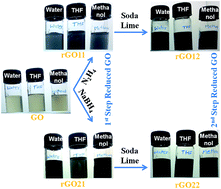
Here we report a two-step programmable reduction of graphene oxide (GO) which was synthesized by oxidation of graphite. X-Ray photoelectron spectroscopic (XPS) analysis confirmed the synthesis of exfoliated graphene oxide (GO) by introduction of oxygen as carboxylic (–COOH), epoxy (C–O–C) and hydroxyl (–OH) groups. The first step of GO reduction was achieved separately by (i) hydrazine (rGO11) and (ii) sodium borohydride (rGO21). Soda lime was used in the second-stage reduction of (a) hydrazine reduced GO (rGO12) and (b) sodium borohydride reduced GO (rGO22) to remove most of the remaining carboxylic functionalities from the rGO11 and rGO21 surface. XPS spectra of rGO21 showed a decrease (38 to 30%) in the oxygen whereas the further reduction of rGO21 with soda lime can further reduce the oxygen content. Quantitative analysis of C([double bond, length as m-dash]O)OX in GO shows about 43% of carbon atoms (C[thin space (1/6-em)]1s signal) as carboxylic functionalities whereas the reduction of the GO with sodium borohydride reduced this signal to about 10%. The use of soda lime for both rGO11 and rGO21 further reduced the amount of carboxylic functionalities. An increase in the proportion of carbon atoms as sp2 and decrease in the oxygen functionalities were controlled in the two-step reduction. A good correlation in the conductivity of reduced GO with the percentage proportion of sp2 carbon was observed.


1. Introduction

Since the discovery of the graphene, a two-dimensional carbonic material, there has been huge interest because of its distinct properties and potential applications.1–3 Several researchers had reported the potential use of graphene in ballistic transport at room temperature,4 its high electron and hole mobility,5 in supercapacitors,6 and in thin film transistors.7 Functional groups at the surface of chemically synthesized graphene and tunable optical properties have advantages in biosensing and optoelectronics applications.8–10 Several methods have been used to synthesize graphene such as (i) zip removal from carbon nanotubes,11 (ii) micromechanical method to exfoliate graphite12,13 and (iii) chemical vapor deposition.14–18 The method used for production of graphene by zip removal from carbon nanotubes can provide high purity samples but there are significant challenges with regard to commercial production. The unzipping of single-walled carbon nanotubes can provide higher purity samples but again it is not a commercially viable method. The use of micromechanical methods to exfoliate graphite leads to graphene crystallite plane sizes of the order of 1 mm but such samples are only on the research scale at the moment.12 Despite the several advantages of graphene, the large scale chemical synthesis of graphene with high purity is still a challenging task.

Graphene samples synthesized by chemical vapor deposition methods have been used in several applications such as photonics, nanoelectronics, transparent conductive layers, sensors, biomedical applications. Similarly chemically reduced graphene oxide samples with crystallite plane size of 100 μm are also used for coatings, paint/ink, composites, transparent conductive layers, energy storage devices and biological applications. Extensive studies haver been carried out for graphene synthesis by chemical routes.19–24 Studies showed that the reduction of GO via chemical methods removes most of the oxygen functionalities from the surface of graphene oxide.25–28

Graphene production at the large scale is possible by the chemical exfoliation of graphite. This includes, oxidation of graphite powder followed by reduction by strong reducing reagents such as hydrazine,29,30 hydroquinone,31 sodium borohydride and its derivatives,32,33 lithium aluminium hydride,34 ascorbic acid,35,36 saccharides,37 norepinephrine,38 KOH,39 ethylenediamine,40 polyelectrolyte,41 protein,42 sodium citrate,43 plant extracts,44–46 metal/acid,47–54 melatonin,55 amino acids,56–59 bacterial respiration,60–65 thermal treatment,66 photocatalytic,67–71 sonochemical,72 laser,73–76 plasmas,77 lysozyme,78 electrochemical79–81 electric current.82 However, the complete removal of oxygen functionalities at the surface of graphene oxide has not achieved via such reduction methods of GO. There have been reports assessing the potential of two-step reduction for removal of selective functional groups, but still these process are poorly understood.83,84 The graphene synthesized via chemical routes contains several impurities and is substantially disordered. Thus, the synthesis of large surface area sheets of graphene with high purity via a chemical route is also very challenging.

The oxidation of graphite via chemical method introduces mainly carboxylic, aldehyde and ketonic functional groups at the edges and epoxide and hydroxyl groups at the basal planes of graphene oxide.85–90 N2H4 and NaBH4 are the most commonly used reducing agents for reduction of graphene oxide. NaBH4 has ability to reduce aldehyde, ketone and carboxylic groups to hydroxyl groups whereas N2H4 can reduce epoxide and hydroxyl groups.91,92 The above reported reducing reagents in the literature can remove most of the oxygen from the surface of GO but still a large proportion of carboxylic and hydroxyl groups may remain at the surface of graphene oxide.

Here we report a two-step programmable reduction of graphene oxide which was synthesized by oxidation of graphite. The main objective of the study was to target the removal of carboxylic acids from the surface of reduced graphene oxide. The uniqueness of this study was the use of soda lime for removing carboxylic functional groups from the surface of reduced GO by decarboxylation.93 The first stage of reduction of GO was obtained via a chemical route by the separate use of (i) hydrazine and (ii) sodium borohydride. The physico-chemical nature of the synthesized graphene oxide and hydrazine and sodium borohydride reduced GO were characterized by different spectroscopic techniques. Subsequent effects of the soda lime on removal of carboxylic acid from the (i) hydrazine and (ii) sodium borohydride reduced GO were evaluated by X-ray photoelectron spectroscopy. In this study, we further quantified the proportion of carbon as sp2 and sp3 in reduced GO and GO by XPS.

2. Materials and methods

2.1 Materials

NaBH4, hydrazine hydrate and soda lime were purchased from Sigma Aldrich. Graphite flakes were obtained from CDH. H2SO4 and hydrochloric acid (HCl) were obtained from RANKEM. KMnO4 and H2O2 were purchased from MERCK and S D Fine-Chem Limited (SDFCL), respectively. Milli-Q water (18 M ohm) was used as a solvent for all the experiments. All other chemicals were of analytical grade and purchased from local suppliers.

2.2 Synthesis of exfoliated graphite oxide sheets

Here, we have modified Hummer’s method for the synthesis of graphene oxide.20,94 H2SO4 (46 ml) was added to a mixture of graphite flakes (2 g) and NaNO3 (1 g) and stirred at 0–4 °C using an ice-bath until the solution became homogeneous. Gradually 6 g of KMnO4 was added to the homogeneous graphite solution in 7 h at ∼20 °C by carrying out the reaction in an ice-bath during the reaction period. Further to this mixture, 6 g of KMnO4 was added to the graphite homogeneous solution in 4 h at 35–40 °C and stirred for another 8 h. This reaction mixture was allowed to cool to room temperature (25 °C) and poured onto ice prepared from ∼260 ml of Milli-Q water. Finally 6 ml of 30% H2O2 was added to complete the reaction. Then the mixture was filtered with Whatman paper and the filtrate was collected. The filtrate was washed with 10% HCl and ethanol. Finally the product was thoroughly washed with Milli-Q water. The wet graphite oxide was dried by vacuum at room temperature for 5 days.

2.3 Reduction of GO

2.3.1 Reduction of GO with NaBH4 and hydrazine hydrate. Synthesized graphite oxide (200 mg) was added to 200 ml water and ultrasonicated for 3 h while maintaining the pH at 8–9. NaBH4 (1.600 g, 62.2 mM) was added to well dispersed graphene oxide solution and stirred at 70–80 °C for 2 h. The reaction mixture was allowed to cool down to room temperature and was filtered by Whatman paper. The filtrate was washed with methanol and MQ water and dried in vacuum at room temperature for 3 days. For reduction of GO by hydrazine, 200 mg of GO was dispersed in 200 ml water by sonicating for 3 h and 2 ml of 64.2 mM of hydrazine was added and the solution was continuously stirred at 95 °C for 24 h. The reaction mixture was allowed to cool down to room temperature and was filtered by Whatman paper. The filtrate was washed with methanol and MQ water and dried in vacuum at room temperature for 3 days.
2.3.2 Reduction of hydrazine and NaBH4 reduced GO by soda lime. The reaction of soda lime with rGO can be described in terms of eqn (1):
 
R–COOH + 2(NaOH/CaO) → RH + Na2CO3 + H2O (1)
where R–COOH represents rGO with remaining COOH functionalities.93 The decarboxylation of rGO by soda lime removes carboxylic functionalities and generates reduced species “RH”.. 100 mg of NaBH4 reduced graphene oxide were dissolved in 100 ml water by ultrasonication for 2 h. 60 mg of 6.2 mM soda lime at pH (7 to 8) was added and stirred at 45–50 °C for 1 h. To adjust the pH of the solution, 1 M NaOH and 1 M HCl was used. The solution was filtered by Whatman filter papers and the filtrate was subsequently washed with 1 M HCl, methanol and MQ water. The material was dried by vacuum at room temperature for 3 days. Alternatively, 100 mg of hydrazine reduced graphene oxide were dissolved in 100 ml water by ultrasonication for 2 h. 30 mg of 3.1 mM soda lime at pH (7 to 8) was added and stirred at 45–50 °C for 1 h. To adjust the pH of the solution, 1 M NaOH and 1 HCl was used in the same way discussed above. The solution was filtered by Whatman filter papers and the filtrate was subsequently washed with 1 M HCl, methanol and MQ water. The material was dried by vacuum at room temperature for 3 days.

Using the above methods for synthesis of graphene oxide and reduced graphene oxides, we have synthesized five different types of carbonic materials as: (i) graphene oxide (GO), (ii) N2H4 reduced GO (rGO11), (iii) NaBH4 reduced GO (rGO21), (iv) soda lime reduced rGO11 (rGO12) and (v) soda lime reduced rGO21 (rGO22). The above materials were dissolved in three different solvents (water, tetrahydrofuran (THF) and methanol) and photographs of these are shown in Fig. 1. After sonication of 10 min, hydrazine reduced graphene oxide showed relatively less solubility as compared to the NaBH4 reduced GO. For both the soda lime reduced GO samples very good solubility in all three solvents was observed. The GO and reduced GO dissolved in water were further characterized by Raman spectroscopy, UV-visible spectroscopy, ATR-FTIR spectroscopy, TGA, XPS and XRD.


image file: c5ra18880f-f1.tif
Fig. 1 Optical images of graphene oxide (GO), N2H4 reduced GO (rGO11), NaBH4 reduced GO (rGO21), soda lime reduced rGO11 (rGO12) and soda lime reduced rGO21 (rGO22) in three different solvents (water, THF and methanol).

2.4 Characterization

The extent of disorder in the crystal structures of the carbonic materials (graphite, graphene oxide and reduced graphene oxide) were characterized by Raman spectroscopy. Raman spectra of GO and reduced GO were measured by a RENISHAW System at 532 nm laser. Absorption spectra of GO and reduced GO were measured by a UV-2600 SHIMADZU spectrophotometer. ATR-FTIR spectroscope (Alpha-e Bruker System) was used to obtain information about the surface functionalities of GO.

The crystallite structure of GO and reduced GO was characterized from the XRD patterns. The XRD spectra were obtained by using a XRD-6000 (Japan) X-ray diffractometer in the diffraction angle range 5–80° with Cu-Kα radiation (λ = 1.54060 Å). Electrical characteristics of GO and reduced GO was characterized by taking the same amount of the materials and solution casting between gold electrodes. The current was measured at different voltages and the current–voltage relationship plotted. X-Ray photoelectron spectra of GO and reduced GO were obtained by MultiLab200 with standard Mg-Kα radiation to quantify elemental composition, surface carbon and oxygen functionalities. All spectra were taken at a working pressure of ∼10−9 mbar. Wide scan XPS survey was used for elemental proportion quantification and high-resolution spectra of C[thin space (1/6-em)]1s was used for characterization of surface functionalities. The different surface states were obtained in the high resolution C[thin space (1/6-em)]1s spectra by specifying a line shape, relative sensitivity factor, peak position, full width at half maxima, and area constraints.

3. Results and discussion

Raman spectra of graphite, synthesized GO and reduced GO (rGO11, rGO12, rGO21 and rGO22) are shown in Fig. 2. The Raman spectra of graphite shows a sharp peak at 1576 cm−1 as shown in Fig. 2. Normally two distinct peaks in Raman spectra of graphite materials are observed due to (i) breathing of sp2 carbon atoms (known as D band at ∼1360 cm−1) and (ii) graphitic carbonic sp2 carbon atoms (known as G band at ∼1580 cm−1).94 The peak at 1576 cm−1 corresponds to the G-band which represents stretching of the C–C bond. The conversion of graphite into graphene oxide induces several disorders in sp2-hybridized carbon sheets; therefore an increase in D-band peak intensity of Raman spectra in GO occurs.29,95 The Raman spectra of GO shows a wide peak at 1597 cm−1 due to stretching of the C–C bond present in aromatic rings of GO containing sp2 carbons. The peak at 1358 cm−1 is mainly associated with disorder introduced by addition of oxygen atoms at the surface of graphite by oxidation process in GO. The observed ratio of the peak intensities of the D-band (ID) with the G-band (IG) peaks were 0.30 and 0.85 for graphite and GO, respectively. The relative peak intensity of the D-band at 1358 cm−1 was increased as compared to the G-band at 1597 cm−1 in GO in relation with graphite.
image file: c5ra18880f-f2.tif
Fig. 2 Raman spectra of graphite, graphene oxide (GO), N2H4 reduced GO (rGO11), NaBH4 reduced GO (rGO21), soda lime reduced rGO11 (rGO12) and soda lime reduced rGO21 (rGO22).

The Raman peaks for the D- and G-bands are at 1349 and 1581 cm−1, respectively, for rGO11. The measured intensity ratio of ID/IG for rGO11 was 1.17. The peak intensity of the D-band as compared to the G-band in the Raman spectrum of rGO11 was relatively higher which is similar to previous findings.29 A further reduction of rGO11 by soda lime which additionally deoxygenates the surface of rGO11 led to a decrease (ID/IG ∼ 1.08) in the intensity of the D-band (at 1333 cm−1) as compared to the G band (at 1596 cm−1) in the Raman spectra of rGO12. The Raman spectrum of NaBH4 reduced GO (rGO21) had a similar pattern to GO, with D- and G-band peaks at 1355 and 1589 cm−1, respectively. The intensity ratio of ID/IG (∼0.93) for rGO21 was slightly decreased as compared to rGO11. The Raman peak position for D band and G bands are at 1331 and 1599 cm−1, respectively, for rGO22 and the peak intensity ratio ID/IG was ∼1.14.

The Raman spectra of GO showed a large red shift in the G-band position after oxidation of graphite into GO and results are shown in ESI (SFig. 1). A previously similar red shift was observed by Bo et al.96 Gupta et al.97 had explained the red shift of the G band in the Raman spectra due to an increase in the number of layers of graphene. The change in Raman peak position and shape were used to estimate the number of layers of graphene.98 Thus, the observed red shift in the G-band position of GO Raman spectra in our finding indicated an increase in the thickness of the layered structures of graphene oxide sheets. Reduction of GO by either of the reducing agents (i) NaBH4 and (ii) N2H4 led to a decrease in the red shift of the D-band. The oxidation of graphite had showed a red-shift in the D band whereas the subsequent reduction of GO caused a blue shift in D band of the Raman spectra. This change may be due to change in sp2 hybridized cluster size by addition/removal of oxygen functional groups from the surface in the oxidation and reduction process. A further reduction with soda lime causes a large red shift in the spectra and the D-band positions are at 1596 and 1599 cm−1 for rGO12 and rGO22. We do not understand the mechanism, but it could be due to multiple folding of highly reduced graphene oxide. A previous Raman peak at 1582–1600 cm−1 corresponded to glassy carbon in carbonic materials.99

XRD spectra of synthesized GO and reduced GO (rGO11, rGO12, rGO21 and rGO22) are shown in Fig. 3. A sharp peak at 2θ ∼ 10°, corresponding to the reflection from the (002) plane, was observed in XRD spectra of GO.100 A peak at 2θ ∼ 43° may correspond to the turbostratic band of disordered carbon materials. XRD spectra of N2H4 reduced GO showed a broad peak at 2θ ∼ 43° and the peak at 2θ ∼ 10° completely disappeared. Reduced graphene oxide has a peak around 2θ ∼ 23°. The broad diffraction peak of rGO indicates poor ordering of the sheets along the stacking direction. rGO21 XRD had a broad peak at 2θ ∼ 10° with increased full width half maximum (FWHM). Further reduction of rGO21 with soda lime shows a peak shift towards higher 2θ and an increase in the peak broadening. This change in peak position and FWHM of the peak could be due to the exfoliation of GO sheets after removal of the intercalated carboxylic groups.101,102 These XRD results are closely related to the exfoliation and reduction processes of GO.


image file: c5ra18880f-f3.tif
Fig. 3 XRD spectra of graphene oxide (GO), N2H4 reduced GO (rGO11), NaBH4 reduced GO (rGO21), soda lime reduced rGO11 (rGO12) and soda lime reduced rGO21 (rGO22).

Fig. 4A shows UV-vis spectra of synthesized GO and reduced GO (rGO11, rGO12, rGO21 and rGO22) between the spectral range 200–700 nm. The absorbance peak of graphene oxide present at 225 nm corresponds to the π–π* transition and a peak at 303 nm is due to C[double bond, length as m-dash]O.103 GO reduced by NaBH4 and further by soda lime shows a peak shift to 262.5 and 263.5 nm, respectively. Reduction of GO by hydrazine and further by soda lime leads to peaks at 256 and 266.5 nm, respectively. Previous studies also showed a red shift in chemically reduced graphene oxide64,68,71 and our experimental results are also consistent with previous observations.


image file: c5ra18880f-f4.tif
Fig. 4 (A) UV-vis spectra and (B) TGA of graphene oxide (GO), N2H4 reduced GO (rGO11), NaBH4 reduced GO (rGO21), soda lime reduced rGO11 (rGO12) and soda lime reduced rGO21 (rGO22).

Thermal stability of synthesized GO and reduced GO (rGO11, rGO12, rGO21 and rGO22) was measured between 10 and 1000 °C by thermogravimetric analysis and results are shown in Fig. 4B. The thermal stability data of GO shows maximum wt% loss due to the higher number of oxygen functionalities and the pyrolysis of the labile oxygen functional groups at 150 °C. Further increase in the temperature shows a very small decrease in the remaining mass which may be due to release of CO and CO2 gases. The comparative data for rGO11 and rGO12 show about 50% weight loss at 430 and 520 °C, respectively. These results suggest a lower number of oxygen groups present in rGO12 as compared to rGO11. Similar observations were recorded for rGO21 and rGO22.

Fig. 5 shows ATR-FTIR spectra of graphite, synthesized GO and reduced GO (rGO11, rGO12, rGO21 and rGO22). The absence of any functional groups in the ATR-FTIR spectrum of graphite was observed. Oxidation of graphite show peaks at 1035, 1390, 1635 and 1751 cm−1 in the ATR-FTIR spectrum. The peak at 1751 cm−1 corresponds to saturated carboxylic acids and the peak at 1635 cm−1 corresponds to C[double bond, length as m-dash]C bonds. Peaks at 1035 and 1390 cm−1 may be due to the presence of C–O and C[double bond, length as m-dash]O. The presence of different oxygen functional groups and an increase in D-band of Raman spectra confirms the conversion of graphite into graphene oxide by the oxidation process used in this study. The ATR-FTIR spectra of rGO12 obtained after reduction of GO with N2H4 showed a very wide peak between 1300–1700 cm−1. The disappearance of several functional peaks and the weak peak intensity between 1300–1700 cm−1 confirms the removal of oxygen from the surface of GO by the reduction process. A small peak at 1255 cm−1 is due to the presence of C–O. The FTIR result suggests that a large proportion of oxygen functionalities were removed from the surface of GO after reduction. The use of soda lime led to further removal of carboxylic functional groups from the surface of reduced GO (rGO12).


image file: c5ra18880f-f5.tif
Fig. 5 ATR-FTIR spectra of graphite, graphene oxide (GO), N2H4 reduced GO (rGO11), NaBH4 reduced GO (rGO21), soda lime reduced rGO11 (rGO12) and soda lime reduced rGO21 (rGO22).

The strong appearance of the peak at 1645 cm−1 in NaBH4 reduced GO (rGO21) ATR-FTIR spectrum indicates that the reduced GO contains a large number of C[double bond, length as m-dash]C bonds. A peak at 905 in the spectrum was assigned to the alkenic functionalities in the reduced GO sample. Two small peaks at 1373 and 1108 cm−1 could be associated with the C–O and –OH which indicate the presence of small proportion of oxygen groups at NaBH4 reduced GO surface. ATR-FTIR spectra of soda lime deoxygenated NaBH4 reduced GO (rGO22) samples were very similar to the NaBH4 reduced GO ATR-FTIR spectra as shown in Fig. 5. The peak intensity at 1373 cm−1 was slightly reduced whereas a small increase in the peak intensity at 1645 cm−1 was observed. XPS analysis was carried out for quantitative analysis of carbon functionalities of GO and rGO.

Wide scan XPS spectra of synthesized GO and reduced GO (rGO11, rGO12, rGO21 and rGO22) were obtained to further quantify the chemical nature of GO and reduced GO. Wide scan XPS spectra of these samples are shown in ESI (SFig. 2). Oxygen/carbon elemental percentage proportions in synthesized GO were 38 and 62 atom%, respectively. XPS wide scan spectra of GO reduced with NaBH4 showed a significant decrease (30 atom%) in the oxygen content at the surface. An even smaller value of 11 atom% oxygen was observed in N2H4 treated GO. The further reduction of rGO11 and rGO21 with soda lime, to produce rGO12 and rGO22, respectively, further reduces the oxygen content (to 4 and 28 atom%, respectively) at the surface of rGO.

High-resolution XPS spectra of C[thin space (1/6-em)]1s of synthesized GO and reduced GO were obtained. The peak fitting for surface state quantification from C[thin space (1/6-em)]1s was done as described in previous studies104 and results are shown in Fig. 6. C[thin space (1/6-em)]1s peak mainly fitted as hydrocarbon (CC), hydroxyl (COX), C[double bond, length as m-dash]O/O–C–O and carboxylic functionality peaks.105–107 In our analysis and peak fitting, we have separately fitted two peaks of hydrocarbons as C[thin space (1/6-em)]1s (sp2) and C[thin space (1/6-em)]1s (sp3) for a better representation of the XPS observations.108 An additional peak at the tail of the spectra towards higher binding energy, which is the shake-up peak associated with carbon in aromatic rings, was also separately assigned during the peak fitting.109 Thus, the higher resolution C[thin space (1/6-em)]1s XPS spectra of GO was fitted with six peaks of different carbon environments as: hydrocarbon (C[double bond, length as m-dash]C) at 283.5 eV, (C–C/C–H) at 285.7 eV, (C–OX) at 287.4 eV, (C[double bond, length as m-dash]O/O–C–O) at 288.9 eV, (C([double bond, length as m-dash]O)OX) at 290.8 eV and a satellite peak at 293.5 eV due to π–π interactions. The positions of each peak associated with C–OX, (C[double bond, length as m-dash]O/O–C–O) and (C([double bond, length as m-dash]O)OX) were fixed by assigning 1.5 ± 0.3 eV shift in the binding energy, respectively.110 Previously Chu et al.108 had characterized amorphous and nanocrystalline carbon films and observed about ∼1.7 eV difference in the binding energy associated with C[thin space (1/6-em)]1s (sp2) and C[thin space (1/6-em)]1s (sp3) peaks of carbon. During peak fitting we have observed about ∼1.7 ± 0.3 eV difference in the binding energy for C[thin space (1/6-em)]1s (sp2) and C[thin space (1/6-em)]1s (sp3). The percentage proportion of different carbon environments in C[thin space (1/6-em)]1s was 8.3, 17.7, 13.2, 17.6 and 42.1 corresponding to the C[double bond, length as m-dash]C, C–C/C–H, C–OX, C[double bond, length as m-dash]O/O–C–O and C([double bond, length as m-dash]O)OX, respectively.


image file: c5ra18880f-f6.tif
Fig. 6 Peak fitted C[thin space (1/6-em)]1s XPS spectra of graphene oxide (GO), N2H4 reduced GO (rGO11), NaBH4 reduced GO (rGO21), soda lime reduced rGO11 (rGO12) and soda lime reduced rGO21 (rGO22).

The higher resolution C[thin space (1/6-em)]1s XPS spectra of NaBH4 reduced GO was fitted with six peaks of different carbon environments and the values for hydrocarbon (C[double bond, length as m-dash]C) at 284.2 eV, (C–C/C–H) at 286.1 eV, (C–OX) at 287.6 eV, (C[double bond, length as m-dash]O/O–C–O) at 289.1 eV, (C([double bond, length as m-dash]O)OX) at 290.6 eV and shake-up peak was at 293.2 eV. The percentage proportion of different carbon environments in C[thin space (1/6-em)]1s was 22.9, 30.1, 24.2, 12.4 and 9.7, corresponding to C[double bond, length as m-dash]C, C–C/C–H, C–OX, C[double bond, length as m-dash]O/O–C–O and C([double bond, length as m-dash]O)OX, respectively. The higher resolution C[thin space (1/6-em)]1s XPS spectra of soda lime reduced rGO21 was also fitted with similar six peaks as: hydrocarbon (C[double bond, length as m-dash]C) at 284.2 eV, (C–C/C–H) at 286 eV, (C–OX) at 287.4 eV, (C[double bond, length as m-dash]O/O–C–O) at 288.9 eV, (C([double bond, length as m-dash]O)OX) at 290.5 eV and a shake-up peak at 293.1 eV due to aromatic carbon atoms. The percentage proportion of different carbon environments in C[thin space (1/6-em)]1s was 43.9, 34.1, 12.6, 4.9 and 3.8 corresponding to C[double bond, length as m-dash]C, C–C/C–H, C–OX, C[double bond, length as m-dash]O/O–C–O and C([double bond, length as m-dash]O)OX, respectively.

The higher resolution C[thin space (1/6-em)]1s XPS spectra of N2H4 reduced GO was fitted as: hydrocarbon (C[double bond, length as m-dash]C) at 284.2 eV, (C–C/C–H) at 285.7 eV, (C–OX) at 287.1 eV, (C[double bond, length as m-dash]O/O–C–O) at 288.6 eV, (C([double bond, length as m-dash]O)OX) at 290.3 eV and shake-up peak at 292.8 eV. The percentage proportion of different carbon environments in C[thin space (1/6-em)]1s was 64.8, 17.2, 8.4, 3.2 and 5.6, corresponding to C[double bond, length as m-dash]C, C–C/C–H, C–OX, C[double bond, length as m-dash]O/O–C–O and C([double bond, length as m-dash]O)OX, respectively. The C[thin space (1/6-em)]1s XPS spectra of soda lime reduced rGO11 was fitted as: hydrocarbon (C[double bond, length as m-dash]C) at 284.2 eV, (C–C/C–H) at 285.8 eV, (C–OX) at 287.2 eV, (C[double bond, length as m-dash]O/O–C–O) at 288.3 eV, (C([double bond, length as m-dash]O)OX) at 290.5 eV and shake-up peak at 292.9 eV. The percentage proportion of different carbon environments in C[thin space (1/6-em)]1s was 56.4, 26.9, 11.1, 1.9 and 3.7, corresponding to C[double bond, length as m-dash]C, C–C/C–H, C–OX, C[double bond, length as m-dash]O/O–C–O and C([double bond, length as m-dash]O)OX, respectively. The XPS spectra of GO showed a spectra shift towards lower binding energy which may be due to insulating nature of the sample.111 However, there was no spectral shift observed in reduced GO, which indicates the conducting nature of reduced GO.

Fig. 7 shows quantitative analysis of C([double bond, length as m-dash]O)OX in synthesized GO and reduced GO (rGO11, rGO12, rGO21 and rGO22). The synthesized GO has about 43% proportion of carbon atoms in C[thin space (1/6-em)]1s as carboxylic functionalities whereas the reduction with NaBH4 reduced this level to about 10%. The use of soda lime further reduced the percentage proportion of carboxylic functionalities. The rGO11 showed a very low percentage of carboxylic functionality as compared to NaBH4 reduced GO. The remaining carboxylic functionalities of rGO11 and rGO21 were further reduced by soda lime and a low level of carboxylic functionalities were achieved. Soda lime reduced the carboxylic group amount significantly in both NaBH4 and N2H4 reduced GO. An increase in the proportion of carbon atoms as sp2 and decrease in the oxygen functionality was controlled in the two-step process in a much more precise way. Table 1 shows comparison of D and G band shifts in Raman spectra, peak intensity ratio of D to G Raman band and oxygen functionalities for different rGO samples.


image file: c5ra18880f-f7.tif
Fig. 7 Percentage proportion of carboxylic functionalists in C[thin space (1/6-em)]1s XPS spectra of N2H4 reduced GO (rGO11), NaBH4 reduced GO (rGO21), soda lime reduced rGO11 (rGO12) and soda lime reduced rGO21 (rGO22).
Table 1 D and G Raman band shifts and oxygen functionalities for different rGO
Sample Raman D-band (cm−1) Raman G-band (cm−1) ID/IGa XPS COOXb (%)
a Experimentally observed peak intensity ratio of D-band (ID) and G-band (IG) from Raman spectra.b Percentage of carboxylic functionalities in C[thin space (1/6-em)]1s XPS spectra.
rGO11 1349 1581 1.17 5.6
rGO12 1333 1596 1.09 2.7
rGO21 1355 1589 0.94 9.7
rGO22 1331 1599 1.14 3.8


Fig. 8 shows IV characteristics of GO and reduced GO (rGO11, rGO12, rGO21 and rGO22). As expected, the GO did not show current conduction whereas the IV response of hydrazine reduced GO (rGO11) showed a linear response. Two-step reduction led to a change in the current response, with the response of rGO21 being very distinct from that of rGO11. Initially a low current conduction was observed up to the 2 V, but a further increase in the bias voltage showed a rapid increase in the current and this reached saturation at 4 V bias voltage.


image file: c5ra18880f-f8.tif
Fig. 8 IV response of GO (black squares) and reduced GO samples (green squares). (A) rGO11, (B) rGO12, (C) rGO21 and (D) rGO22.

Change in the relative conductivity of different types of reduced GO can be assessed by comparison of current at contact bias voltage in the IV response curve. The conductivity of different types of reduced GO was observed according to the chemical and structural nature of the reduced GO. It is seen that the conductivity of the reduced GO decreases with the percentage of sp2 carbon obtained from XPS analysis (ESI SFig. 3). The two-step process of reduction described here may provide an improvement and better structural, functional and electrical properties control in the reduction of GO. The two-step process of reduction described here may also provide a better conversion of graphite into graphene.

4. Conclusions

Here we have described a method for a synthesis of chemically reduced graphene oxide which has a higher proportion of graphene. First we have prepared highly exfoliated graphene oxide from graphite which was reduced by hydrazine and sodium borohydride. The Raman spectroscopic and XPS analysis confirm the synthesis of exfoliated graphene oxide by chemically introduced oxygen as carboxylic groups (–COOH), hydroxyl (–OH) and epoxy groups (C–O–C). Two distinct peaks of graphene oxide and reduced graphene in Raman spectra are present due to the breathing mode of sp2 atoms (graphitic carbonic sp2 of carbon atoms). Raman spectra of hydrazine reduced GO showed relatively higher intensity of the D-band as compared to the G-band in the spectra. A strong red shift in the G-band position was observed after oxidation of graphite into GO due to an increase in the number of layers of graphene. The reduced GO by either reducing agent NaBH4 and N2H4 showed a decrease in the red shift of the D-band due to decrease in the thickness of the reduced GO sheets. The synthesized GO has very high percentage of carbon atoms as carboxylic functionalities whereas reduction with NaBH4 and particularly hydrazine reduces this level. Soda lime reduced the carboxylic group significantly further in both NaBH4 and N2H4 reduced GO. The two step process of reduction described here provides an improvement in the reduction of GO, and therefore better conversion of graphite into graphene.

Acknowledgements

This work was supported by network project (NanoSHE) of Council for Scientific and Industrial Research (CSIR), Ministry of Science and Technology, Govt. of India. KD is a Project Student at CCMB from Centre for Converging Technologies, University of Rajasthan, Jaipur 302004, India.

Notes and references

  1. A. K. Geim, Science, 2009, 324, 1530 CrossRef CAS PubMed .
  2. Y. Zhu, S. Murali, W. Cai, X. Li, J. W. Suk, J. R. Potts and R. S. Ruoff, Adv. Mater., 2010, 22, 3906 CrossRef CAS PubMed .
  3. H. Raza, NanoScience and Technology, Springer, 2012 Search PubMed .
  4. J. Baringhaus, M. Ruan, F. Edler, A. Tejeda, M. Sicot, A. Taleb-Ibrahimi, A. P. Li, Z. Jiang, E. H. Conrad, C. Berger, C. Tegenkamp and W. A. Heer, Nature, 2014, 506(7488), 349–354 CrossRef CAS PubMed .
  5. Y. Zhang, Y. W. Tan, H. Stormer and P. Kim, Nature, 2005, 438, 201–204 CrossRef CAS PubMed .
  6. B. T. Yu and J. M. Lee, J. Mater. Chem. A, 2013, 1, 14814–14843 Search PubMed .
  7. G. Eda, G. Fanchini and M. Chhowalla, Nat. Nanotechnol., 2008, 3, 270–274 CrossRef CAS PubMed .
  8. Y. Liu, D. Yu, C. Zeng, Z. Miao and L. Dai, Langmuir, 2010, 26(9), 6158–6160 CrossRef CAS PubMed .
  9. S. W. Crowder, D. Prasai, R. Rath, D. A. Balikov, H. Bae, K. Bolotin and H. Sung, Nanoscale, 2013, 5(10), 4171–4176 RSC .
  10. M. Zhang, B. Yin, X. Wang and B. Ye, Chem. Commun., 2011, 47, 2399–2401 RSC .
  11. D. V. Kosynkin, A. L. Higginbotham, A. Sinitskii, J. R. Lomeda, A. Dimiev, B. K. Price and J. M. Tour, Nature, 2009, 458, 872–876 CrossRef CAS PubMed .
  12. K. S. Novoselov, A. K. Geim, S. V. Morozov, D. Jiang, M. I. Katsnelson, I. V. Grigorieva, S. V. Dubonos and A. A. Firsov, Nature, 2005, 438, 197–200 CrossRef CAS PubMed .
  13. Y. Zhang, J. P. Small, M. E. S. Amori and P. Kim, Phys. Rev. Lett., 2005, 94, 176803 CrossRef PubMed .
  14. A. Reina, X. Jia, J. Ho, D. Nezich, H. Son, V. Bulovic, M. S. Dresselhaus and J. Kong, Nano Lett., 2009, 9, 30–35 CrossRef CAS PubMed .
  15. J. C. Delgado, J. M. R. Herrera, X. Jia, D. A. Cullen, H. Muramatsu, Y. A. Kim, T. Hayashi, Z. Ren, D. J. Smith, Y. Okuno, T. Ohba, H. Kanoh, K. Kaneko, M. Endo, H. Terrones, M. S. Dresselhaus and M. Terrones, Nano Lett., 2008, 8, 2773–2778 CrossRef PubMed .
  16. N. G. Shang, P. Papakonstantinou, M. McMullan, M. Chu, A. Stamboulis, A. Potenza, S. S. Dhesi and H. Marchetto, Adv. Funct. Mater., 2008, 18, 3506–3514 CrossRef CAS .
  17. K. S. Kim, Y. Zhao, H. Jang, S. Y. Lee, J. M. Kim, K. S. Kim, J. H. Ahn, P. Kim, J. Y. Choi and B. H. Hong, Nature, 2009, 457, 706–710 CrossRef CAS PubMed .
  18. W. Cai, R. D Piner, F. J. Stadermann, S. Park, M. A. Shaibat, Y. Ishii, D. Yang, A. Velamakanni, S. J. An, M. Stoller, J. An, D. Chen and R. S. Ruoff, Science, 2008, 321, 1815–1817 CrossRef CAS PubMed .
  19. B. C. Brodie, Ann. Chim. Phys., 1860, 59, 466–472 Search PubMed .
  20. W. Hummers and R. Offeman, J. Am. Chem. Soc., 1958, 80, 1339 CrossRef CAS .
  21. L. Staudenmaier, Ber. Dtsch. Chem. Ges., 1898, 31, 1481–1499 CrossRef CAS .
  22. A. B. Bourlinos, D. Gournis, D. Petridis, T. Szabo, A. Szeri and I. Dekany, Langmuir, 2003, 19(15), 6050–6055 CrossRef CAS .
  23. U. Hofmann and A. Frenzel, Kolloid-Z., 1934, 68, 149–151 CrossRef CAS .
  24. P. Xiao, M. Xiao, P. Liu and K. Gong, Carbon, 2000, 38(4), 626–628 CrossRef CAS .
  25. N. A. Kotov, I. Dekany and J. H. Fendler, Adv. Mater., 1996, 8(8), 637–641 CrossRef CAS .
  26. A. Celzard, Carbon, 2002, 40, 2801–2815 CrossRef CAS .
  27. D. S. McLachlan, J. Phys. C: Solid State Phys., 1986, 19(9), 1339–1354 CrossRef .
  28. D. S. McLachlan, J. Phys. C: Solid State Phys., 1987, 20(7), 865–877 CrossRef .
  29. S. Stankovich, D. A. Dikin, R. D. Piner, K. A. Kohlhaas, A. Kleinhammes, Y. Jia, Y. Wu, S. T. Nguyen and R. S. Ruoff, Carbon, 2007, 45, 1558–1565 CrossRef CAS .
  30. S. Gilje, S. Han, M. Wang, K. L. Wang and R. B. Kaner, Nano Lett., 2007, 7, 3394–3398 CrossRef CAS PubMed .
  31. G. Wang, J. Yang, J. Park, X. Gou, B. Wang, H. Liu and Y. J. Facile, J. Phys. Chem. C, 2008, 112, 8192–8195 CAS .
  32. J. H. Shin, K. K. Kim, A. Benayad, S. M. Yoon, H. K. Park, I. S. Jung, M. H. Jin, H. K. Jeong, J. M. Kim, J. Y. Choi and Y. H. Lee, Adv. Funct. Mater., 2009, 19, 1987–1992 CrossRef .
  33. C. K. Chua and M. Pumera, J. Mater. Chem. A, 2013, 1, 1892 CAS .
  34. A. Ambrosi, C. K. Chua, A. Bonanni and M. Pumera, Chem. Mater., 2012, 24, 2292–2298 CrossRef CAS .
  35. M. J. F. Merino, L. Guardia, J. I. Paredes, S. V. Rodil, P. S. Fernández, A. M. Alonso and J. M. D. Tascón, J. Phys. Chem. C, 2010, 114, 6426–6432 Search PubMed .
  36. J. Gao, F. Liu, Y. Liu, N. Ma, Z. Wang and X. Zhang, Chem. Mater., 2010, 22, 2213–2218 CrossRef CAS .
  37. C. Zhu, S. Guo, Y. Fang and S. Dong, ACS Nano, 2010, 4, 2429–2437 CrossRef CAS PubMed .
  38. S. M. Kang, S. Park, D. Kim, S. Y. Park, R. S. Ruoff and H. Lee, Adv. Funct. Mater., 2011, 21, 108–112 CrossRef CAS .
  39. X. Fan, W. Peng, Y. Li, X. Li, S. Wang, G. Zhang and F. Zhang, Adv. Mater., 2008, 20, 4490–4493 CrossRef CAS .
  40. J. F. Che, L. Y. Shen and Y. H. Xiao, J. Mater. Chem., 2010, 20, 1722–1727 RSC .
  41. S. Zhang, Y. Shao, H. Liao, M. H. Engelhard, G. Yin and Y. Lin, ACS Nano, 2011, 5, 1785–1791 CrossRef CAS PubMed .
  42. J. Liu, S. Fu, B. Yuan, Y. Li and Z. Deng, J. Am. Chem. Soc., 2010, 132, 7279–7281 CrossRef CAS PubMed .
  43. W. Wan, Z. Zhao, H. Han, Y. Gogotsi and J. Qiu, Mater. Res. Bull., 2013, 48, 4797–4803 CrossRef CAS .
  44. Y. Wang, Z. Shi and J. Yin, ACS Appl. Mater. Interfaces, 2011, 3, 1127–1133 CAS .
  45. S. Thakur and N. Karak, Carbon, 2012, 50, 5331–5339 CrossRef CAS .
  46. B. Haghighi and M. A. Tabrizi, RSC Adv., 2013, 3, 13365–13371 RSC .
  47. Z. Fan, K. Wang, T. Wei, J. Yan, L. Song and B. Shao, Carbon, 2010, 48, 1686–1689 CrossRef CAS .
  48. Z. J. Fan, W. Kai, J. Yan, T. Wei, L. J. Zhi, J. Feng, Y. M. Ren, L. P. Song and F. Wei, ACS Nano, 2011, 5, 191–198 CrossRef CAS PubMed .
  49. X. Mei and J. Ouyang, Carbon, 2011, 49, 5389–5397 CrossRef CAS .
  50. P. B. Liu, Y. Huang and L. Wang, Mater. Lett., 2013, 91, 125–128 CrossRef CAS .
  51. R. S. Dey, S. Hajra, R. K. Sahu, C. R. Raj and M. K. Panigrahi, Chem. Commun., 2012, 48, 1787–1789 RSC .
  52. N. A. Kumar, S. Gambarelli, F. Duclairoir, G. Bidan and L. Dubois, J. Mater. Chem. A, 2013, 1, 2789–2794 CAS .
  53. V. H. Pham, H. D. Pham, T. T. Dang, S. H. Hur, E. J. Kim, B. S. Kong, S. Kim and J. S. Chung, J. Mater. Chem., 2012, 22, 10530–10536 RSC .
  54. B. K. Barman, P. Mahanandia and K. K. Nanda, RSC Adv., 2013, 3, 12621–12624 RSC .
  55. A. Esfandiar, O. Akhavan and A. Irajizad, J. Mater. Chem., 2011, 21, 10907–10914 RSC .
  56. D. Chen, L. Li and L. Guo, Nanotechnology, 2011, 22, 325601 CrossRef PubMed .
  57. S. Bose, T. Kuila, A. K. Mishra, N. H. Kim and J. H. Lee, J. Mater. Chem., 2012, 22, 9696–9703 RSC .
  58. J. K. Ma, X. R. Wang, Y. Liu, T. Wu, Y. Liu, Y. Q. Guo, R. Q. Li, X. Y. Sun, F. Wu, C. B. Li and J. P. Gao, J. Mater. Chem. A, 2013, 1, 2192–2201 CAS .
  59. T. A. Pham, J. S. Kim, J. S. Kim and Y. T. Jeong, Colloids Surf., A, 2011, 384, 543–548 CrossRef CAS .
  60. E. C. Salas, Z. Sun, A. Luttge and J. M. Tour, ACS Nano, 2010, 4, 4852–4856 CrossRef CAS PubMed .
  61. G. Wang, F. Qian, C. Saltikov, Y. Jiao and Y. Li, Nano Res., 2011, 4, 563–570 CrossRef CAS .
  62. O. Akhavan and E. Ghaderi, Carbon, 2012, 50, 1853–1860 CrossRef CAS .
  63. S. Gurunathan, J. W. Han, V. Eppakayala and J. H. Kim, Colloids Surf., B, 2013, 102, 772–777 CrossRef CAS PubMed .
  64. P. Khanra, T. Kuila, N. H. Kim, S. H. Bae, D. S. Yu and J. H. Lee, Chem. Eng. J., 2012, 183, 526–533 CrossRef CAS .
  65. T. Kuila, S. Bose, P. Khanra, A. K. Mishra, N. H. Kim and J. H. Lee, Carbon, 2012, 50, 914–921 CrossRef CAS .
  66. H. C. Schniepp, J. L. Li, M. J. McAllister, H. Sai, M. H. Alonso and D. H. Adamson, J. Phys. Chem. B, 2006, 110(17), 8535–8539 CrossRef CAS PubMed .
  67. G. Williams, B. Seger and P. V. Kamat, ACS Nano, 2008, 2, 1487–1491 CrossRef CAS PubMed .
  68. X. Huang, X. Zhou, S. Wu, Y. Wei, X. Qi, J. Zhang, F. Boey and H. Zhang, Small, 2010, 6, 513–516 CrossRef CAS PubMed .
  69. O. Akhavan and E. Ghaderi, J. Phys. Chem. C, 2009, 113, 20214–20220 CAS .
  70. S. R. Kim, M. K. Parvez and M. Chhowalla, Chem. Phys. Lett., 2009, 483, 124–127 CrossRef CAS .
  71. H. B. Yao, L. H. Wu, C. H. Cui, H. Y. Fang and S. H. Yu, J. Mater. Chem., 2010, 20, 5190–5195 RSC .
  72. K. Vinodgopal, B. Neppolian, I. V. Lightcap, F. Grieser, M. A. Kumar and P. V. Kamat, J. Phys. Chem. Lett., 2010, 1, 1987–1993 CrossRef CAS .
  73. D. A. Sokolov, K. R. Shepperd and T. M. Orlando, J. Phys. Chem. Lett., 2010, 1, 2633–2636 CrossRef CAS .
  74. Y. Zhou, Q. Bao, B. Varghese, L. A. L. Tang, C. K. Tan, C. H. Sow and K. P. Loh, Adv. Mater., 2010, 22, 67–71 CrossRef CAS PubMed .
  75. Y. Zhang, L. Guo, S. Wei, Y. He, H. Xia, Q. Chen, H. B. Sun and F. S. Xiao, Nano Today, 2010, 5, 15–20 CrossRef CAS .
  76. V. Abdelsayed, S. Moussa, H. M. Hassan, H. S. Aluri, M. M. Collinson and M. S. E. Shall, Chem. Lett., 2010, 1, 2804–2809 CAS .
  77. M. Baraket, S. G. Walton, Z. Wei, E. H. Lock, J. T. Robinson and P. Sheehan, Carbon, 2010, 48, 3382–3390 CrossRef CAS .
  78. F. Yang, Y. Liu, L. Gao and J. Sun, J. Phys. Chem. C, 2010, 114, 22085–22091 CAS .
  79. C. Fu, Y. Kuang, Z. Huang, X. Wang, N. Du, J. Chen and S. Zhou, Chem. Phys. Lett., 2010, 499, 250–253 CrossRef CAS .
  80. M. Zhou, Y. Wang, Y. Zhai, J. Zhai, W. Ren, F. Wang and S. Dong, Chem.–Eur. J., 2009, 15, 6116–6120 CrossRef CAS PubMed .
  81. L. Chen, Y. Tang, K. Wang, C. Liu and C. Luo, Electrochem. Commun., 2011, 13, 133–137 CrossRef CAS .
  82. P. Yao, P. Chen, L. Jiang, H. Zhao, H. Zhu, D. Zhou, D. Hu, B. H. Han and M. Liu, Adv. Mater., 2010, 22, 5008–5012 CrossRef CAS PubMed .
  83. W. Gao, L. B. Alemany, L. Ci and P. M. Ajayan, Nat. Chem., 2009, 1, 403–408 CrossRef CAS PubMed .
  84. J. Geng, L. Liu, S. B. Yang, S. C. Youn, D. W. Kim, D. W. Lee, J. K. Choi and H. T. Jung, J. Phys. Chem. C, 2010, 114, 14433–14440 CAS .
  85. H. He, T. Riedl, A. Lerf and J. Klinowski, J. Phys. Chem., 1996, 100(51), 19954–19958 CrossRef CAS .
  86. H. He, J. Klinowski, M. Forster and A. Lerf, Chem. Phys. Lett., 1998, 287(1–2), 53–56 CrossRef CAS .
  87. A. Lerf, H. He, T. Riedl, M. Forster and J. Klinowski, Solid State Ionics, 1997, 101–103, 857–862 CrossRef CAS .
  88. A. Lerf, H. He, M. Forster and J. Klinowski, J. Phys. Chem. B, 1998, 102(23), 4477–4482 CrossRef CAS .
  89. T. Szabo, O. Berkesi and I. Dekany, Carbon, 2005, 43(15), 3186–3189 CrossRef CAS .
  90. C. H. Lucas, A. J. L. Peinado, J. D. L. Gonzalez, M. L. R. Cervantes and R. M. M. Aranda, Carbon, 1995, 33(11), 1585–1592 CrossRef .
  91. H. C. Brown and S. Krishnamurthy, Tetrahedron, 1979, 35, 567–607 CrossRef CAS .
  92. M. B. Smith and J. March, March’s Advanced Organic Chemistry, Mechanisms and Structure, John Wiley & Sons, Inc., New Jersey, 6th edn, 2007,  DOI:10.1002/0470084960.
  93. C. G. Lyons, S. McLintock and N. H. Lumb, A Concise Text-Book of Organic Chemistry, Pergamon Press Ltd, 1965 Search PubMed .
  94. D. C. Marcano, D. V. Kosynkin, J. M. Berlin, A. Sinitskii, Z. Sun, A. Slesarev, L. B. Alemany, W. Lu and J. M. Tour, ACS Nano, 2010, 4, 4806–4814 CrossRef CAS PubMed .
  95. A. C. Ferrari and J. Robertson, Phys. Rev. B: Condens. Matter, 2000, 61(20), 14095–140107 CrossRef CAS .
  96. Z. Bo, X. Shuai, S. Mao, H. Yang, J. Qian, J. Chen, J. Yan and K. Cen, Sci. Rep., 2014, 4, 4684 Search PubMed .
  97. A. Gupta, G. Chen, P. Joshi, S. Tadigadapa and P. C. Eklund, Nano Lett., 2006, 6, 2667–2673 CrossRef CAS PubMed .
  98. A. C. Ferrari, J. C. Meyer, V. Scardaci, C. Casiraghi, M. Lazzeri, F. Mauri, S. Piscanec, D. Jiang, K. S. Novoselov, S. Roth and A. K. Geim, Phys. Rev. Lett., 2006, 97, 187401 CrossRef CAS PubMed .
  99. P. Lespade, A. Marchand, M. Couzi and F. Cruege, Carbon, 1984, 22, 375 CrossRef CAS .
  100. W. Gao, L. B. Alemany, L. Ci and P. M. Ajayan, Nat. Chem., 2009, 1, 403 CrossRef CAS PubMed .
  101. A. K. Geim and K. S. Novoselov, Nat. Mater., 2007, 6, 183–191 CrossRef CAS PubMed .
  102. H. M. Ju, S. H. Choi and S. H. Huh, J. Korean Phys. Soc., 2010, 57(6), 1649–1652 CAS .
  103. H. Dong, W. Gao, F. Yan, H. Ji and H. Ju, Anal. Chem., 2010, 82, 5511–5517 CrossRef CAS PubMed .
  104. D. Yang, A. Velamakanni, G. Bozoklu, S. J. Park, M. Stoller, R. D. Piner, S. Stankovich, I. H. Jung, D. A. Field, C. A. J. Ventrice and R. S. Ruoff, Carbon, 2009, 47, 145–152 CrossRef CAS .
  105. M. Dhayal, R. Kapoor, P. G. Sistla, R. R. Pandey, S. Kar, K. K. Saini and G. Pande, Mater. Sci. Eng., C, 2014, 37, 99–107 CrossRef CAS PubMed .
  106. M. Dhayal, M. R. Alexander and J. W. Bradley, Appl. Surf. Sci., 2006, 252(22), 7957–7963 CrossRef CAS .
  107. Z. Bo, X. Shuai, S. Mao, H. Yang, J. Qian, J. Chen, J. Yan and K. Cen, Sci. Rep., 2014, 4, 4684 Search PubMed .
  108. P. K. Chu and L. Li, Mater. Chem. Phys., 2006, 96, 253–277 CrossRef CAS .
  109. Y. S. Yuji, Y. Iijima, D. Asakawa and K. Hiraoka, Surf. Interface Anal., 2010, 42(6–7), 658–661 Search PubMed .
  110. J. F. Moulder, W. F. Stickle, P. E. Sobol and K. Bomben, Handbook of X-ray Photoelectron Spectroscopy, ed. J. Chastain, Perkin-Elmer Corporation, Physical Electronics, 1992 Search PubMed .
  111. K. S. Kim and N. Winograd, Chem. Phys. Lett., 1975, 31(2), 312–317 CrossRef CAS .

Footnote

Electronic supplementary information (ESI) available. See DOI: 10.1039/c5ra18880f

This journal is © The Royal Society of Chemistry 2015
Click here to see how this site uses Cookies. View our privacy policy here.