O. Erolab and
H. I. Unal*a
aGazi University, Chemistry Department, Smart Materials Research Lab., Ankara 06500, Turkey. E-mail: hiunal@gazi.edu.tr
bNational Nanotechnology Research Center-UNAM, Bilkent University, Ankara 06800, Turkey
First published on 18th November 2015
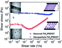
As a new electrorheological (ER) material, core/shell nanorods composed of a titania core and conducting polymer poly(3,4-ethylenedioxythiophene) (PEDOT) shell were prepared via covalent bonding to achieve a thin polymer shell and make the interfacial interactions between the two components more impressive. The successful coating of PEDOT on the nanorod-TiO2 particles was confirmed by TEM analysis. The antisedimentation stability of the core/shell nanorod-TiO2/PEDOT particles was determined to be 100%. The ER properties of the materials were studied under controlled shear, oscillatory shear and creep tests. The dielectric spectra of the dispersions were obtained to further understand their ER responses and fitted with the Cole–Cole equation. The ER behavior of the dispersions was also observed using an optical microscope. The flow curves of these ER fluids were determined under various electric field strengths and their flow characteristics examined via a rheological equation using the Cho–Choi–Jhon (CCJ) model. In addition, the results were also compared with nanoparticle-TiO2/PEDOT. It was concluded that the conducting thin polymer shell and elongated structure of the hybrid material introduced a synergistic effect on the electric field induced polarizability and colloidal stability against sedimentation, which resulted in stronger ER activity, storage modulus and higher recovery after stress loadings when compared to nanoparticle-TiO2/PEDOT.
Researchers have made many efforts to explore various promising materials based on ER fluids, which include consideration of inorganic and polymeric materials such as metal oxides,15,16 porous particles17,18 conducting polymers and their composites,19,20 carbonaceous particles,21 and core–shell nanoparticles.14,22 Among these materials, titania has been frequently studied as a potential ER active material because of its stability against high electric field strengths and suitable dielectric constant.23,24 The synthesis of titania with various morphologies, including elongated structures, such as nanotubes, nanowires and nanofibers, is also possible. In particular, one-dimensional anisotropic titania nanostructures have become interesting due their combination of high aspect ratio and specific surface area. However, similar to the other inorganic nanoparticles, titania tends to agglomerate in an insulating liquid because of its highly hydrophilic character, which may decrease the effective volume fraction of the ER fluid. For this reason, the surface of titania nanoparticles can be modified with convenient molecules or polymers.25,26 On the other hand, a good ER material should not only exhibit a large dielectric constant and appropriate dielectric loss tangent but also an appropriate conductivity.27 Core/shell-structured nanocomposites give an opportunity to combine dielectric inorganic materials and conducting materials in a unique structure with controlled size, shape and composition. Among the methods used to prepare core–shell nanocomposites, modification and functionalization of the nanoparticle surfaces are required for their intended applications to enhance the adhesive interactions between the inner core and outer shell. To obtain functional groups on the surface of the nanoparticles, a covalent strategy can be used to tailor a strong and stable binding between the surface of the nanoparticles and the functional linker groups,28,29 which was the goal of this current study.
Especially, using conducting polymers as either the core or shell species of core/shell-structured ER particles has become a hot topic.30 In particular, PEDOT as a derivative of polythiophene has received a considerable amount of attention due to its various advantages among conducting polymers such as processability in an aqueous solution, good thermal and structural stability, low band gap and higher charge carrier mobility due to the electron donating effect of the alkoxy-substituted group.31 In the literature, the ER properties of PEDOT and its composites have been reported in a limited number of studies. Hong and Jang developed silica/conducting polymer nanospheres31 and reported that the ER efficiency was correlated with the charge transport behaviour of the conductive polymer shell and PEDOT was found to be slightly more favoured when compared to both polypyrrole and polythiophene. In another study, a physical adsorption route32 and Pickering emulsion polymerization33 was used to fabricate PEDOT/poly(styrene sulfonic acid) coated polystyrene microspheres with PEDOT used as an electroactive material. Moreover, PEDOT was used as a conductive filler in various poly(dimethylsiloxane)/PEDOT/poly(styrene sulfonic acid)/ethylene glycol blends as potential actuator materials.34 None of these PEDOT containing hybrid materials were covalently bonded to their substrates and neither of the studies were focused on the effects of anisotropy on the polarizability, dielectric and ER performances of the ER dispersions.
In this study, as novel ER materials, core/shell-structured nanocomposites comprised of nanorods and particulate titania cores and conducting polymer PEDOT shells were prepared via a bottom-up surface engineering strategy to achieve covalently bonded thin polymer shells and make the interfacial interactions between the two components more impressive. The as-obtained nanocomposites were characterized in terms of their structural, surface, morphological, thermal, and electrical properties. The antisedimentation stability, dielectric properties and ER performance of the core/shell-structured TiO2/PEDOT nanocomposites in SO at various volume fractions were evaluated. The microstructural alterations of the ER fluids under E were also revealed using an optical microscope (OM).
To make a comparison, nanoparticle-TiO2/PEDOT was also synthesized by following the same synthesis procedure described above.
Further evidence of the surface functionalization and PEDOT bonding on the TiO2 surface were provided by XPS analyses. Fig. 3 shows the XPS survey-scan spectra of the nanorod-TiO2, nanorod-TiO2-APTS, nanorod-TiO2-3TA and nanorod-TiO2/PEDOT. The survey-scan XPS spectrum of nanorod-TiO2 showed that the bare particles mainly contained the elements of Ti, O and C. The C 1s peak at a binding energy of 285.0 eV was probably due to hydrocarbon contamination of the nanorod-TiO2 during XPS operation.41 After modification with APTS, the intensities of the C 1s, Si 2s and Si 2p peaks at 285, 154 and 102 eV, respectively, were increased and the N 1s peak appeared at 399 eV. Apart from these peaks, the thiophene ended surface showed new signals at about 152 eV and 164 eV, which were assigned to S 2s and S 2p, respectively.37 This result was consistent with a reaction between the –NH2 group of silane and the –COOH group of 3TA. Similar distinctive characteristic peaks were also observed from XPS survey-scan of nanoparticle-TiO2, surface modified-nanoparticle-TiO2 and nanoparticle-TiO2/PEDOT (ESI, Fig. S2†).
For further conclusions on the bond formation and molecule configuration on nanorod-TiO2 surface, core-level XPS spectra of the materials are given in Fig. S3† (for C 1s, O 1s, N 1s, Si 2p, and S 2p). Before APTS attachment, the C 1s spectrum (curve b in Fig. S3†) can be fitted into two components with binding energies at about 285.0 and 286.8 eV, which were attributed to –C/C–H and
–O bonds, respectively, probably due to hydrocarbon contamination typically obtained when air exposed samples are introduced into the XPS chamber.41 After APTS grafting, the C 1s spectrum was fitted into two components at 285.0 eV and 286.2 eV, which can be attributed to
–C/
–H and
–N bonds, respectively. The C 1s fitted spectrum of nanorod-TiO2-3TA contained peaks corresponding to
–C/
–H (285.0 eV) and
–N/
–S (286.3 eV) bonds beside N–
O (288.2 eV) bonds, which were attributed to amide bond formation (curve c in Fig. S3†). Exclusive of these peaks, after PEDOT grafting on the nanorod-TiO2 surface, the intensity of the C 1s signal increased due to the contribution of the DBSA dopant molecules at a binding energy of 285.0 eV in addition to the presence of
–O–C and N–
O bonds at 286.4 and 288.0 eV, respectively (curve d in Fig. S3†). In the O 1s spectrum (curve b in Fig. S3†), after APTS grafting, a clear decrease in the O 1s peak at 529.8 eV (assigned to Ti–
bond) was obtained, whereas a new peak at binding energy of 532.2 eV occurred, which can be attributed to the surface species of
–Si–R.
The O 1s spectrum of nanorod-TiO2-3TA was resolved into three components centred at 530.0 eV, 531.5 eV and 532.4 eV, corresponding to –Ti,
–Si and N–C
species (curve c in Fig. S3†). For nanorod-TiO2/PEDOT, the intensity of the peak at 530.3 eV, corresponding to the
–Ti bond, was decreased due to the thickness of the PEDOT shell. The other two peaks at 531.7 eV and 532.6 eV were also attributed to the –SO3− group in DBSA42 and the
–Si and C–
–C bonds in PEDOT.43 The core-level N 1s spectrum exhibited two clear peaks at 399.2 eV and 400.9 eV after APTS-grafting, which can be assigned to free amine termination (desired silane coupling) and protonated amine (reverse attachment), respectively (curve b in Fig. S3†). After PEDOT grafting on the nanorod-TiO2 surfaces, the intensity of the N 1s peak at 400.6 eV was decreased and broadened as expected (curve d in Fig. S3†). APTS-grafting can also be proved by the sharp Si 2p peak at 102.5 eV (curve b in Fig. S3†) and after PEDOT grafting, the intensity of the Si 2p peak was clearly decreased due to the thickness of the PEDOT shell on the nanorod-TiO2 surface (curve d in Fig. S3†). Nanorod-TiO2 and nanorod-TiO2-APTS showed no S 2p signals, whereas the high-resolution S 2p spectrum of nanorod-TiO2-3TA (curve c in Fig. S3†) consisted of a spin-split doublet for S 2p3/2 and S 2p1/2 at 164.5 and 165.8 eV, respectively, indicating the presence of the C–
bond in the thiophene residue.44 The binding energies of S 2p for nanorod-TiO2/PEDOT at 163.9 eV/165.2 eV and 168.3 eV/168.7 eV were attributed to the presence of the S atoms in the thiophene ring found in PEDOT and the sulfonate in DBSA (curve d in Fig. S3†), respectively.45 The signal intensities of N 1s, Ti 2p and Si 2p in the nanorod-TiO2/PEDOT were reduced and the signal intensities of C 1s, S 2s and S 2p were increased when nanorod-TiO2 was covered with PEDOT. In addition, similar results were obtained for nanoparticle-TiO2 (ESI, Fig. S4†). Both the FTIR and XPS results indicated that the functionalizations of the nanorod and nanoparticle-TiO2 surfaces and PEDOT graftings onto these surfaces were successfully carried out using the bottom-up surface engineering approach.
ζ-potential and water CA measurements also confirmed the surface functionalization and PEDOT grafting on the TiO2 surface. The isoelectric point (IEP) obtained from the ζ-potential-pH curve for nanorod-TiO2 was 4.3, which was lower than that found for nanoparticle-TiO2 (IEP = 6.1) and attributed to its larger surface area and good adsorption performance46 (ESI, Fig. S5†). The IEPs of the nanorod-TiO2 and nanoparticle-TiO2 after APTS functionalization were determined to be 8 and 8.5, respectively, indicating the presence of amino groups on the TiO2 surface.47 Surface functionalization of TiO2-APTS with 3TA decreased the IEPs of both the geometries to 7.3, which indicated a decrease in the number of amino groups on the particles surface after the coupling reaction between the amino and carboxylic acid groups. On the other hand, after PEDOT coating on TiO2-APTS-3TA, the IEPs shifted to 6.6 and 5.7 for the nanorod-TiO2/PEDOT and nanoparticle-TiO2/PEDOT, respectively.
The contact angle (θ) between the water droplet on the sample and the surface gives information on the ratio between the interfacial tension (water/air, water/solid, and solid/air).48 For this reason, CA is a useful tool to monitor the alterations that occurred on the surface of the materials. The hydrophilic character of the samples was determined to decrease in the following order: nanorod-TiO2 < nanorod-TiO2-APTS < nanorod-TiO2-3TA < PEDOT < nanorod-TiO2/PEDOT and nanoparticle-TiO2 < nanoparticle-TiO2-APTS < nanoparticle-TiO2-3TA < PEDOT < nanoparticle-TiO2/PEDOT (Fig. S6†). It can be concluded that the functionalizations and PEDOT coatings of the both TiO2 surfaces were successfully performed. Moreover, the water CAs obtained for both nanocomposites were significantly higher than PEDOT and indicated that their hydrophobic characteristics were increased after the grafting process. It is well known that the wettability of a solid surface is governed by both the chemical compositions and physical properties of the materials used. In addition, the surface roughness can enhance both the hydrophilicity of the hydrophilic surfaces and the hydrophobicity of hydrophobic surfaces.49 It can be said that the obtained nanocomposites possessed rougher surfaces with loose porous structures when compared to PEDOT.50
According to the XRD patterns (Fig. 4), nanoparticle-TiO2 showed peaks at 25.1°, 37.8°, 48.1°, 54.1°, 55.1°, 62.8°, 69.0° and 70.0°, which were in good agreement with the anatase phase of TiO2.51 These anatase peaks were observed to disappear after the alkaline hydrothermal treatment of the nanoparticle-TiO2 for nanorod-TiO2 fabrication. The peak at 2θ = 11.5° corresponded to an interlayer spacing of 0.77 nm between the titanate sheets, which can be assigned to the hydrogen titanate structure of nanorod-TiO2.52 It has been reported that during nanorod-TiO2 formation, the Ti–O–Ti bonds are broken and replaced with Ti–O–Na bonds to form TiO6 octahedra frameworks comprised of Na+ ions between the interlayers of the three dimensional frameworks in highly concentrated NaOH(aq) and at elevated temperature.53 After the HCl(aq) treatment, hydrogen-titanate was formed by exchanging Ti–O–Na to Ti–O–H. According to the XRD pattern of PEDOT, a broad peak at 2θ = 23.4° was observed, which was attributed to the inter-chain planar ring-stacking formed due to the amorphous nature of the homopolymer.54,55 For nanorod-TiO2/PEDOT and nanoparticle-TiO2/PEDOT nanocomposites, the peaks obtained for nanoparticle-TiO2 and nanorod-TiO2 were still observed but became slightly broader after coating with PEDOT.
SEM (Fig. 5a) and TEM (Fig. 6a) images of nanoparticle-TiO2 proved that after the alkaline hydrothermal treatment all the nanoparticle-TiO2 was transformed into rod-like structures (Fig. 5b and 6b) with smooth surfaces, diameters of 30–100 nm and length on the micrometre scale.
![]() | ||
Fig. 5 SEM images of the nanoparticle-TiO2 (a), nanorod-TiO2 (b), nanoparticle-TiO2/PEDOT (c), and nanorod-TiO2/PEDOT (d). |
![]() | ||
Fig. 6 TEM images of the nanoparticle-TiO2 (a), nanorod-TiO2 (b), nanoparticle-TiO2/PEDOT (c), and nanorod-TiO2/PEDOT (d). |
It is clear that after PEDOT coating, the particle size of the nanoparticle-TiO2/PEDOT was increased due to the core-nanoparticles tendency to easily form clusters (Fig. 5c and 6c). On the other hand, it was revealed that uniformly distributed nanorod-TiO2 particles were successfully coated with PEDOT shells with a thickness of 6–7 nm (Fig. 6d).
Fig. 7 shows the TGA curves of the materials. The slight weight losses below 110 °C were assigned to the physically adsorbed solvent and/or moisture in the samples. PEDOT remained thermally stable up to 300 °C and major decomposition occurred between 323 and 404 °C, which may be attributed to the decomposition of the polymer skeleton. A 48.5% total weight loss was determined at 700 °C. For nanorod-TiO2, continuous degradation occurred until 326 °C, which corresponded to the dehydration and further oxidation with a 6.5% total weight loss at 700 °C.56 After surface modification with APTS, continuous weight loss appeared between 240 and 529 °C for nanorod-TiO2-APTS, which was associated with the thermal decomposition of 3-aminopropyl groups and an 11% total weight loss occurred at 700 °C.57 On the basis of these results, the grafting ratio of APTS was calculated to be 4.5%. Nanorod-TiO2-3TA exhibited continuous weight loss between 233 and 453 °C similar to that observed for nanorod-TiO2-APTS and 13% total weight loss was obtained at 700 °C; the grafting ratio of 3TA was determined to be 2%. Finally, the initial weight loss of ∼9% up to ∼340 °C for nanorod-TiO2/PEDOT was due to the removal of volatile components, small molecules and dopant anions. The subsequent weight loss between 345 and 452 °C corresponded to the degradation of the PEDOT chains and the total weight loss was determined to be 32%. Based on the data, the mass ratio of PEDOT in the nanorod-TiO2/PEDOT was estimated to be ∼19%. Similar results were determined for the nanoparticle-TiO2, surface functionalized nanoparticle-TiO2 and nanoparticle-TiO2/PEDOT (ESI, Fig. S7†). For the nanoparticle-TiO2, 1.4% weight loss was obtained. Grafting ratios for the nanoparticle-TiO2 were determined to be 7.4% and 3.2% after modification with APTS and 3TA, respectively. The grafting ratio of PEDOT was obtained as 17.4% in the nanoparticle-TiO2/PEDOT nanocomposite. According to the TGA results, both nanocomposites have approximately the same amount of PEDOT grafted in their structures as targeted.
![]() | ||
Fig. 7 TGA curves for the nanorod-TiO2 (a), nanorod-TiO2-APTS (b), nanorod-TiO2-3TA (c), nanorod-TiO2/PEDOT (d), and PEDOT (e). |
A summary of the density and conductivity values of the materials are tabulated in Table 1.
Sample | Density (g mL−1) | Conductivity (S cm−1) | Conductivity after washing with NH4OH(aq) (S cm−1) |
---|---|---|---|
Nanoparticle-TiO2 | 3.655 | 7.5 × 10⁻⁷ | — |
Nanorod-TiO2 | 3.575 | 6.2 × 10⁻⁷ | — |
Nanoparticle-TiO2/PEDOT | 2.558 | 2.63 | 6.8 × 10⁻² |
Nanorod-TiO2/PEDOT | 2.253 | 2.69 | 7.8 × 10⁻² |
PEDOT | 1.792 | 3.5 × 10² | 16.4 |
It was observed that densities of the nanocomposites were lower than the core-TiO2 particles due to the coating with low density PEDOT shells as targeted for enhanced colloidal stability. It was also noted that when PEDOT was grafted onto both types of TiO2 particles, higher conductivity values were obtained for the hybrid nanocomposites. The conductivity originated primarily from the surface coated conducting PEDOT layer. However, these conductivity values were very high for ER applications and to avoid electrical breakdown under various electric field strengths, the nanocomposites were further treated with 0.1 M NH4OH(aq) overnight in a dedoping process58 and then washed with deionized water until at pH ≅ 7. As a result, the conductivities of the nanocomposites were reduced ∼100×. On the other hand, PEDOT could not be used for ER purposes because of its high conductivity even after the dedoping process.
Volume fraction (%) | Antisedimentation ratio (%) | |||
---|---|---|---|---|
Nanoparticle-TiO2 | Nanorod-TiO2 | Nanoparticle-TiO2/PEDOT | Nanorod-TiO2/PEDOT | |
0.625 | 29 | 79 | 20 | 85 |
1.25 | 59 | 85 | 21 | 97 |
2.5 | 88 | 92 | 35 | 98 |
5 | 91 | 97 | 63 | 100 |
One-dimensional nano-sized anisotropic rod structures with high surface area, enhanced rod to rod interactions and interparticle entanglements formed due to their limited rotational motions have led to the improved dispersion stability and hindered settling observed for the nanorod-TiO2/PEDOT. On the other hand, nanoparticle-TiO2/PEDOT consisted of clusters showed the lowest antisedimentation stability against gravitational forces with a 63% antisedimentation ratio. It was concluded that the particle morphology, size and surface properties are very efficient parameters for colloidal stability and the nanorod-TiO2/PEDOT/SO system was a perfect candidate for potential industrial applications.
![]() | (1) |
Sample (φ = 1.25%) | ε0 | ε∞ | Δε | λ (s) | fmax (Hz) | α |
---|---|---|---|---|---|---|
Nanoparticle-TiO2 | 3.07 | 2.60 | 0.47 | 5 × 10⁻³ | 32 | 0.69 |
Nanoparticle-TiO2/PEDOT | 3.20 | 2.60 | 0.60 | 3 × 10⁻³ | 53 | 0.63 |
Nanorod-TiO2 | 3.97 | 2.67 | 1.30 | 1.9 × 10⁻⁵ | 8377 | 0.69 |
Nanorod-TiO2/PEDOT | 4.20 | 2.52 | 1.68 | 1.3 × 10⁻⁴ | 1224 | 0.72 |
In the equation, ε* is the complex dielectric constant; ε′ and ε′′ are the dielectric constant and the dielectric loss, respectively; ε0 and ε∞ are the static and infinite frequency dielectric constants, ω is angular frequency, λ is dielectric relaxation time denoted by λ = 1/2fmax (where fmax is the relaxation frequency defined by a local maximum of the dielectric loss factor, ε′′), α is the scattering degree of relaxation time and Δε (Δε = ε0 − ε∞) shows the difference between the dielectric constant at 0 and infinite frequency. Δε and λ are related with the magnitude and the rate of the interfacial polarization of the ER fluid, respectively and are considered to be important for the observed particle polarizations and strength of the ER fluid.
An appropriate dielectric loss peak position (f = 102 to 105 Hz) and large Δε do not only result in increased interactions between the dispersed particles but also maintain the stable chain structure formed by particles under applied electric and shear fields.63
No distinct dielectric loss peak was observed for nanoparticle-TiO2 and nanoparticle-TiO2/PEDOT (Fig. 8a), whereas the nanorod-TiO2 and nanorod-TiO2/PEDOT dispersions gave dielectric loss peaks within 102 to 105 Hz (Fig. 8b). The achievable polarizability values were obtained as follows: Δεnanorod-TiO2/PEDOT > Δεnanorod-TiO2/PEDOT > Δεnanoparticle-TiO2/PEDOT > Δεnanoparticle-TiO2. It can be concluded that the nanorod-TiO2/PEDOT has stronger interfacial polarization, which leads to stiffer chain structures formed by the particles under an applied E and shows the highest ER activity when compared to the particulate nanocomposite. Wang and Zhao reported the relationship between ER performance and the dielectric characteristics for core/shell kaolinite/TiO2 particles in which a larger dielectric constant enhancement increased the interfacial polarizability of particles and induced a higher ER effect.64 Among the ER fluids examined in this study, nanorod-TiO2/PEDOT have the highest Δε value and will lead to stronger particle–particle attractions, higher performance in the yield stress and high modulus under E. However, on the basis of relaxation time (λ), core nanorod-TiO2 may show faster interfacial polarization than that of nanorod-TiO2/PEDOT under E in terms of dielectric loss model. Therefore, considering both λ and Δε, it can be concluded that the Δε value was more dominant for ER performance because it is directly related to the strength of fibrillar structures in dispersions.65 It has been known that the high aspect ratio of the dispersed particles plays a dominant role in enhancing the performance of ER fluids.66 In this study, the geometrical effect originating from the aspect ratio and obtained larger magnitude of the polarizability gave enhanced ER performance. Furthermore, interfacial polarization was more dominant than conductivity when we considered the dielectric properties and conductivity values of the materials.67
![]() | ||
Fig. 9 OM images of the nanoparticle-TiO2 (a), nanoparticle-TiO2/PEDOT (b), nanorod-TiO2 (c), and nanorod-TiO2/PEDOT (d) dispersions at various particle volume fractions. |
For nanoparticle-TiO2 and nanoparticle-TiO2/PEDOT, much denser fibrillar structures were observed after a certain volume fraction. As the volume fraction increased, thicker columnar structures were formed for nanoparticle-TiO2/PEDOT, whereas denser, closer and well aligned fibrillar structures were turned into a network structure for nanoparticle-TiO2. After the network structure was formed, the applied electric field did not change its shape and only made the network structure become significantly stronger, which was also in good agreement with the ER results. On the other hand, nanorod-TiO2 and nanorod-TiO2/PEDOT dispersions formed denser chain structures even for φ = 0.0625% and formed a network structure at φ = 2.5%. For this reason, φ = 5% was not conducted by OM for rod-like dispersions. Due to their high wetted surface area and rod–rod interactions, one-dimensional structured nanorod-TiO2 and nanorod-TiO2/PEDOT particles aligned along the field direction and linked with neighbouring ones with the side by side interactions, which led to the formation of more complex structures. According to the previous studies, it was reported that one-dimensional elongated particles tend to form complicated dendrite-like network structures under an electric field rather than chain-like structures formed by the granular particles.68–70 Moreover, this particle overlap can contribute to the solid friction between the neighbouring particles and enhance the yield stress.13 The electric field induced structures remain stable as long as the electrical field was applied. It was clearly observed that dense fibrillar structures attached at both sides of the electrodes for nanorod-TiO2/PEDOT were formed, which exhibited a higher shear stress when compared to the nanoparticle-TiO2/PEDOT dispersion under the same electric field strength.
Fig. 10 shows the flow curves for shear stress and shear viscosity as a function of shear rate for the dispersions (φ = 5%). In the absence of an electric field, the dispersions showed non-Newtonian shear thinning flow behaviour having low yield stresses even with Newtonian dispersant medium due to the formation of a particle network caused by the interparticle interactions at high particle volume fractions. The off-field viscosities of the dispersions were about 1.2–1.6 Pa s in the high shear rate region. When the electric field was applied, the viscosities and shear stresses of the dispersions increased abruptly and showed pseudo-plastic behaviours with yield stresses due to the formation of chain-like/columnar or network structures. In addition, the shear stresses increased stepwise over the entire shear rate range with rising electric field strengths.
Yield stress is one of the critical design parameters for ER fluids. The widely accepted rheological model for ER fluids, i.e. the Bingham fluid model, (τ = τy + η, where τ is shear stress, τy is yield stress, η is viscosity, and
is shear rate) did not fit well to the flow curves of our ER systems except for nanoparticle-TiO2, especially in the low shear rate region. This deviation in flow behaviour reflects that the dispersed particles possess a different ER response under electric and shear fields caused by the differences in shape and surface chemistry of the particles. On the other hand, a suggested equation, the CCJ model, provided more effective fitting for many ER systems71 and for all the dispersions examined in this study. The CCJ model was applied to describe the flow curves of the dispersions, as shown in eqn (2):
![]() | (2) |
In the low shear rate region, the electrostatic interactions induced by the electric field strength among the particles were dominant when compared to the hydrodynamic interactions induced by the shear flow. The chain-like structures of the particles started to break down with a further increase in shear rate. The destruction rate of the columnar structures became higher than the reforming rate of columns induced by the electric field above the critical shear rate; thus, the flow curves behaved much like those without an electric field. At elevated shear rates, the fibrillar structures were broken into particles or particle clusters by shearing due to the domination of hydrodynamic interactions. For both the nanoparticle-TiO2/PEDOT and nanorod-TiO2/PEDOT nanocomposite ER systems, the shear stress tended to decrease as a function of shear rate to a minimum value and then increased again. The decrease in shear stress at low shear deformations means that the reformation of the fibrillar structures was slower than the destruction of field induced fibrillar structures and also the reformed structures were not completely similar to those before applying shear deformation.
It was observed that the nanorod-TiO2/PEDOT dispersions have a higher critical shear rate and sustained its fibrillar or network structure over a wide range of shear deformations. Interestingly, although the surface properties were similar for both the nanocomposites, the ER effect increased after the nanorod-TiO2 was coated with PEDOT layers with fine distributions, whereas it decreased after the nanoparticle-TiO2 was coated with PEDOT with clusters. This can be attributed to their final structures when compared with their uncoated core nanoparticles.
The τy values determined according to the CCJ model and their dependence on φ and E are given in Fig. 11. It was observed that τy increased with increasing φ and E, which suggested that the particle–particle interactions and electrostatic forces became strong enough to resist against the hydrodynamic forces.
![]() | ||
Fig. 11 Changes in the τy values determined according to the CCJ model with E. Inset figure indicates the effect of φ on τy. |
Fig. 12 shows the frequency dependence of storage (G′) and loss (G′′) moduli for various electric field strengths for the dispersions. The results agree with those of steady state rheological measurements. Without an electric field, G′′ was smaller than G′ for nanoparticle-TiO2 and nanorod-TiO2 dispersions and showed an increase with increasing frequency. This indicated that the formation of weak 3D structures resulted in elastic interactions, which may be attributed to the higher particle volume fraction and larger probability of particle–particle interactions. However, for the nanocomposites, the G′ and G′′ values were closer to each other and increased with increasing frequency and therefore G′′ became dominant at higher frequencies, indicating viscous-like structures without E.
![]() | ||
Fig. 12 The frequency dependence of G′ and G′′ at various electric field strengths for the dispersions (G′: full symbols, G′′: open symbols, φ = 5%). |
With applied E, the G′ values significantly increased and began to dominate over the G′′ values and stable plateau regions were observed over a wide frequency range, indicating that the dispersions showed solid-like elastic behaviour. These increases were significant for the nanorod-TiO2/PEDOT dispersions when compared to the particulate form. It can be said that the rod-like structure will be more suitable for vibration damping applications. When compared to others, the nanorod-TiO2/PEDOT dispersion possessed higher storage moduli. Under constant conditions (f = 1 Hz and E = 3 kV mm−1), G′ values of the dispersions were determined as follows: G′nanorod-TiO2/PEDOT = 206 MPa > G′nanorod-TiO2 = 170 MPa > G′nanoparticle-TiO2 = 37 MPa > G′nanoparticle-TiO2/PEDOT = 10.5 MPa. This revealed that the nanorod-TiO2/PEDOT showed higher rigidity under an electric field, which was also in accordance with its higher yield stress.
Therefore, both the steady shear viscosity and dynamic viscoelastic results indicate that the nanorod-TiO2/PEDOT dispersion exhibited an enhanced ER effect when compared to the nanorod-TiO2 and nanoparticle-TiO2/PEDOT dispersions. The ER enhancement may be attributed to several reasons, including increased polarizability, interparticle friction and viscous drag force that stem from the elongated structure and increased colloidal stability.73
For the nanoparticle-TiO2/PEDOT dispersion (Fig. 13a) under all the electric field strengths and for the nanorod-TiO2/PEDOT dispersion (Fig. 13b) under no electric field strength, the strain values were increased linearly under applied stress with time and no recovery occurred after removing the applied stress. This means that they behaved like viscous materials under these conditions. This applied stress value was higher than the yield stresses of the nanoparticle/PEDOT dispersion under all the E cases. For this reason, the results were in accordance with the steady-state flow measurements.
On the other hand, the nanorod-TiO2/PEDOT dispersion represented time-dependent non-linear viscoelastic deformation under an applied stress and subsequent time-dependent reformation after setting the applied stress to τ = 0 Pa. An instantaneous decrease in strain corresponded to the elastic recovery and reversible viscoelastic recoveries were subsequently obtained under E in the recovery process. This viscoelastic response of the nanorod-TiO2/PEDOT dispersion arose from the fibrillar aggregates of the dispersed particles, which was an indication of solid-like behaviour under E. For the case of the nanoparticle-TiO2/PEDOT, above the yield point, the fibrillar structures were repeatedly broken down and did not reform, which resulted in viscous flow. The strain values obtained during the creep-recovery process were decreased with increasing E indicating that the nanorod-TiO2/PEDOT dispersion formed stronger solid-like structure.
The recovery ratio (χ) was defined to evaluate the elasticity of the ER fluid and calculated using the following equation75
![]() | (3) |
Footnote |
† Electronic supplementary information (ESI) available: ATR-FTIR spectra, XPS survey-scan, core-level spectra, TGA curves, ζ-potentials and contact angle images of the samples. See DOI: 10.1039/c5ra20284a |
This journal is © The Royal Society of Chemistry 2015 |