DOI:
10.1039/C5RA19381H
(Paper)
RSC Adv., 2015,
5, 104275-104283
Hydrogen titanate constructed by ultrafine nanobelts as advanced anode materials with high-rate and ultra-long life for lithium-ion batteries†
Received
20th September 2015
, Accepted 1st December 2015
First published on 3rd December 2015
Abstract
Hydrogen titanate (H2Ti3O7) anode materials with desirable electrochemical performance, such as long cycling life and high capacity, have rarely been reported so far. In the present work, as-prepared hydrogen titanate with a particularly hierarchical nanostructure exhibits a breakthrough in electrochemical performance as an anode material for lithium-ion batteries by morphology engineering and heat treatment: 168 mA h g−1 at 16.8 A g−1, meaning that a full charge only needs 40 seconds; 184.8, 176.4 and 156.8 mA h g−1 at 0.168, 1.68 and 10.08 A g−1 after 1000 cycles, respectively; 120 mA h g−1 at 10.08 A g−1 even after 4000 cycles. It is believed that such excellent performance makes hydrogen titanate a promising anode material for advanced lithium-ion batteries with ultra-long life and high power density.
1 Introduction
With intrinsic advantages of low cost, superior cycling stability and improved safety over graphite, titanium dioxide (TiO2) has attracted great interest as a promising anode material for next generation lithium-ion batteries (LIBs).1 But the practical application of TiO2 is severely hindered by the limited capacity and poor rate capability due to the low ionic conductivity and intrinsic poor electronic conductivity of the semiconductor.2 Currently, great efforts have been made to focus on improving the capacity and rate capability of TiO2 anode materials for LIBs. It is commonly accepted that these limitations in rate capability of LIBs are mainly ascribed to slow solid-state diffusion of lithium ion within the electrode materials.3 So, considerable works have been devoted to tailoring TiO2 materials towards the nanoscale, because the large specific surface area and confined material dimension characteristics of the nanostructured materials can endow TiO2 electrodes with a shortened diffusion distance and large contact area for enhanced lithium-ion intercalation kinetics.4 As precursor for preparing TiO2 with peculiar nanostructures such as nanobelts, nanowires and nanotubes, one-dimensional hydrogen titanate (H2Ti3O7) has received increasing attention due to the simple preparation method.5 Besides as precursor for preparing peculiarly nanostructured TiO2, the H2Ti3O7 with layered structure is also potential lithium intercalation hosts at a voltage window of 1 to 3 V.6 Particularly, the layered structure can offer H2Ti3O7 electrodes a very large accessible surface area for lithium storage and a more open tunnel than general intercalation materials.7 But, the work about the H2Ti3O7 anode materials with desirable electrochemical performance (such as long cycling life and high capacity) has rarely been reported so far.6–10
Herein, we focus on the improvement of the lithium intercalation properties of nanostructured hydrogen titanate electrodes. Our approach is based on the alkaline-hydrothermal treatment and subsequent acid washing, and then heating treatment. The resultant material, denoted as HTO-HT, has less adsorbed water than H2Ti3O7 precursor and a peculiar morphology of micro/nanoparticles constructed by ultrafine nanobelts with 1–3 nm thickness, 3–6 nm width and several micrometer (μm) length, with larger surface area of 333 m2 g−1, pore volume of 1.134 cm3 g−1 and average pore size of 6.8 nm. As a result, the HTO-HT exhibits remarkable rate capability and excellent cycling stability (168 mA h g−1 at 16.8 A g−1, meanings full charge only needs 40 second; 184.8, 176.4 and 156.8 mA h g−1 at 0.168, 1.68 and 10.08 A g−1 after 1000 cycles, respectively; 120 mA h g−1 at 10.08 A g−1 even after 4000 cycles). The breakthrough in electrochemical properties can be attributed to the hierarchical mesoporous micro/nanoparticles constructed by ultrafine nanobelts and appropriate heating treatment, as illustrated in Scheme 1. On one hand, the interspaces and mesoporous nature, and ultrafine nanobelt units of the HTO-HT provide a very large accessible surface area for contact of the electrode with electrolyte and promoted transportation of lithium ions and electrons, which can effectively improve the cycling performance, lithium storage and rate capability. On the other hand, the reduction of adsorbed water in HTO-HT makes the layered structure very open which can provide a very large interspaces for lithium storage besides reducing the adverse effect to the electrochemical performance.
 |
| | Scheme 1 Illustration of lithium insertion mechanism in the HTO-HT with shortened diffusion distance of lithium-ions and electrons. | |
2 Experimental section
Preparation of samples
All chemicals were analytical grade and used without further purification. Typically, 1 mL of tetrabutyl titanate (TBOT) was slowly dropped into 20 mL of 7.5 M NaOH aqueous solution (NaOH concentration-dependent experiments: 6.25 and 10 M NaOH aqueous solution were also investigated). After stirred for 30 min, this white suspension was transferred into a Teflon-lined stainless steel autoclave, and then placed in an oven at 180 °C for 48 h (temperature-dependent experiments: 150 and 200 °C were also investigated). After cooled down to room temperature naturally, the white product was collected by centrifugation, washed with deionized water and ethanol thoroughly, followed by dispersed in 50 mL of 0.1 M HCl and stirred for 24 h to exchange Na+ completely by H+. After that, the H2Ti3O7 was collected by centrifugation, washed with deionized water and ethanol thoroughly, and dried in an oven at 60 °C under vacuum overnight. Finally, the HTO-HT was obtained from H2Ti3O7 calcination at 300 °C for 3 h with a heat rate of 1 °C min−1 under air. Other samples were also prepared at the same conditions with HTO-HT but calcination temperature changed. Another control sample, denoted as HTO-HTB, was prepared by using monodispersed broad H2Ti3O7 nanobelts (Fig. S2(b1 and b2)†) as precursor under the same conditions with HTO-HT.
Materials characterization
The morphologies and microstructures of the products were investigated by using field emission scanning electron microscopy (SEM, JEOL JSM-7401F) and transmission electron microscopy (TEM, JEOL JEM-2010) with an energy dispersive X-ray spectrometer (EDX) and a selected area electron diffraction pattern (SAED). The crystal structures and composition were characterized by X-ray diffraction measurement (XRD, Rigaku, D/max-Rbusing Cu K radiation) and X-ray photoelectron spectroscopy (XPS, AXIS ULTRA DLD instrument, using aluminum Kα X-ray radiation). The mesoporous structure and surface area were studied by nitrogen (N2) adsorption/desorption isotherms (Micromeritics ASAP 2010 instrument). The chemical bonding was investigated by the fourier transform infrared (FTIR; Varian Carry) spectrum. Thermogravimetric analysis (TGA) was carried out with a thermogravimetric analysis instrument (TGA, SDT Q600 V8.2 Build 100) at a heating rate of 10 °C under N2 (the temperature firstly keeps at 80 °C for 2 h to removal the adsorbed water from air, then increases to 800 °C at a heating rate of 10 °C).
Electrochemical characterization
Electrochemical measurements were conducted using 2016-type coin cells assembled in argon-filled glove box (German, M. Braun Co., [O2] < 1 ppm, [H2O] < 1 ppm). The working electrodes were composed of active material (HTO-HT), conductive material (acetylene black, AB), and binder (sodium carboxymethyl cellulose, CMC) in a weight ratio of 7![[thin space (1/6-em)]](https://www.rsc.org/images/entities/char_2009.gif) :2:1 and pasted on a Cu foil, followed by dried in a vacuum oven at 110 °C for overnight. The mass of active material loaded on electrode was ca. 1.12 mg. The pure lithium foil and glass fiber (GF/A) from Whatman were used as the counter electrode and separator, respectively. The electrolyte consisted of a solution of 1 M LiPF6 in ethylene carbonate and dimethyl carbonate (EC + DMC) (1:1 in volume). The galvanostatic discharge/charge cycles were performed on a CT2001a cell test instrument (LAND Electronic Co.) over a voltage range of 1.0 to 3.0 V at room temperature (25 °C). Cyclic voltammetry (CV) was implemented on a CHI660D electrochemical workstation at a scan rate of 0.5 mV s−1 between 1 and 3.0 V. Electrochemical impedance spectrum (EIS) measurements were also conducted using a CHI660D electrochemical workstation in the frequency range from 100 kHz to 0.01 Hz with an ac perturbation of 5 mV s−1. The specific capacities reported and current densities used here were based on the mass of HTO-HT.
:2:1 and pasted on a Cu foil, followed by dried in a vacuum oven at 110 °C for overnight. The mass of active material loaded on electrode was ca. 1.12 mg. The pure lithium foil and glass fiber (GF/A) from Whatman were used as the counter electrode and separator, respectively. The electrolyte consisted of a solution of 1 M LiPF6 in ethylene carbonate and dimethyl carbonate (EC + DMC) (1:1 in volume). The galvanostatic discharge/charge cycles were performed on a CT2001a cell test instrument (LAND Electronic Co.) over a voltage range of 1.0 to 3.0 V at room temperature (25 °C). Cyclic voltammetry (CV) was implemented on a CHI660D electrochemical workstation at a scan rate of 0.5 mV s−1 between 1 and 3.0 V. Electrochemical impedance spectrum (EIS) measurements were also conducted using a CHI660D electrochemical workstation in the frequency range from 100 kHz to 0.01 Hz with an ac perturbation of 5 mV s−1. The specific capacities reported and current densities used here were based on the mass of HTO-HT.
3 Results and discussion
The preparation process of HTO-HT is detailed in Experimental section and briefly comprises three steps: alkaline-hydrothermal treatment (preparation of sodium titanate (Na2Ti3O7) precursor), acid washing (preparation of H2Ti3O7 precursor by exchange between Na+ and H+), and heating treatment (removing excess adsorbed water of H2Ti3O7). Sodium hydroxide (NaOH) concentration-dependent (Fig. S1†) and hydrothermal temperature-dependent (Fig. S2†) experiments indicated that the NaOH concentration and hydrothermal temperature played important roles in control of H2Ti3O7 morphologies. Here, we focus on the investigation of as-prepared H2Ti3O7 (7.5 M of NaOH and hydrothermal treatment at 180 °C) with a unique morphology of micro/nanoparticles constructed by intertwined seeming nanowires (unless otherwise specified, the H2Ti3O7 mentioned in the following is this kind of nanostructured H2Ti3O7), because the rich interspaces and ultrafine seeming nanowires may be beneficial to improve the electrochemical performance of H2Ti3O7. So, to get more distinct observation of the unique morphology of H2Ti3O7, the TEM measurement was conducted on it (Fig. S3†). It can be seen from Fig. S3(a and b)† that the as-prepared H2Ti3O7 micro/nanoparticles are indeed constructed by ultrafine one-dimensional substance units. More importantly, the one-dimensional substance units are not nanowires but nanobelts, and further determined by magnified TEM images (Fig. S3c†). Thus, it can be considered that the ultrafine nanobelts grew into broader nanobelts after increasing concentration of NaOH or elevating temperature during NaOH concentration-dependent and hydrothermal temperature-dependent experiments. Then, to reduce the adsorbed water which would be adverse to the electrochemical performance of H2Ti3O7 electrodes, as well as get more open tunnel between layered structures, the heating treatment was conducted on H2Ti3O7 precursor. For confirming the appropriate calcination temperature for H2Ti3O7, the TEM and XRD characterizations were used to observe the influence of calcination temperature on the structures of calcined samples. Fig. S4 and S5† give the TEM images of samples obtained from the H2Ti3O7 heating treatment at different temperatures from 200 to 600 °C. When the calcination is performed below 400 °C, the samples can remain the morphology of H2Ti3O7 precursor perfectly (Fig. S4†). But, the calcination temperatures selected at or over 400 °C, the obtained samples (Fig. S5(a1 and a2)†) show a changed morphology compared to H2Ti3O7 precursor, some nanobelt units collapse into nanorods or particles, and the micro/nanoparticles become compact. Fig. 1 shows the XRD patterns of H2Ti3O7 precursor and calcined samples. It is found that the patterns of the samples obtained from H2Ti3O7 calcination below 400 °C with a relatively low intensity can be assigned to layered protonated titanate (JCPDS, card 47-0124) with poor crystallinity (Fig. S6a†).6 When the calcination temperatures increase to or over 400 °C, the layered protonated titanate transforms to anatase phase (JCPDS card no. 21-1272) (tiny TiO2-B phase observed at 400 °C, JCPDS card no. 74-1940), which is attributed to the fully dehydration of layered titanate.9 The dehydration process of H2Ti3O7 was also monitored by thermogravimetric analysis (TGA), as shown in Fig. S6.† The steep weight loss occurred before 200 °C is generally attributed to the adsorbed water and interlayer water.10 The weight loss above 200 °C is thought to be the dehydration of structural water. Such a dehydration process did not finish until 500 °C, where the transition of crystal from titanate to anatase could occur as demonstrated previously by differential scanning calorimetry (DSC).11 The XRD and TEM analysis suggest that the H2Ti3O7 loses some structural water at the first step, then reconstructs slowly to anatase phase from about 400 °C. Comparing the TGA of HTO-HT, H2Ti3O7 (precursor) and H2Ti3O7 calcinated at 200 °C, it can be found that the HTO-HT has lowest weight loss, which indicates HTO-HT has less water than other two samples. N2 adsorption/desorption isotherms were used to investigate the specific surface areas and pore structures of samples (Fig. S7†). The Brunauer–Emmett–Teller (BET) specific surface areas of HTO-HT, H2Ti3O7, H2Ti3O7 calcined at 200 °C, TiO2-400 (obtained from H2Ti3O7 calcination at 400 °C) and TiO2-500 (obtained from H2Ti3O7 calcination at 500 °C) are about 353.2, 333, 339.7, 201.3 and 112 m2 g−1, respectively, as shown in Fig. S7c.† Obviously, the samples obtained from H2Ti3O7 calcined at below 400 °C possess much higher surface areas than those of H2Ti3O7 calcined at above 300 °C. The higher surface area of the samples (calcined at below 400 °C) compared with those of samples (calcined at above 300 °C) is attributed to hierarchical micro/nanoparticles constructed by ultrafine nanobelts. In fact, we can find that a sharp drop of BET specific surface area and pore size (Fig. S7b†) between HTO-HT and TiO2-400, which could be attributed to the morphologies changed from hierarchical micro/nanoparticles constructed by ultrafine nanobelts with rich interspaces to hierarchical compact micro/nanoparticles composed of nanorods or particles. The result is in good agreement with observation of TEM measurement (Fig. S4 and S5†). Thus, the appropriate calciantion temperature of H2Ti3O7 should be selected at 300 °C since the obtained HTO-HT not only remain the same morphology and crystal nature with H2Ti3O7, but also has less adsorbed water which would be adverse to the electrochemical performance. Importantly, the HTO-HT expectedly exhibited excellent electrochemical performances (presented in the following), so the HTO-HT should be a promising anode material for LIBs. The necessarily detailed investigation of microstructure and electrochemical properties is performed on HTO-HT in the following.
 |
| | Fig. 1 XRD patterns of samples obtained from H2Ti3O7 calciantion at different temperatures under air: (a) the calcination temperature selected lower than 400 °C and (b) higher than 400 °C. | |
The chemical components of HTO-HT were firstly examined using FTIR spectroscopy in the range between 4000 and 400 cm−1. As displayed in Fig. S8a,† a stretching vibration of O–H around 3392 cm−1 and a bending vibration of H–O–H around 1625 cm−1 are clearly seen, implying that bonded H exists within the HTO-HT.12 The bands around 520 and around 916 cm−1 can be assigned to the characteristic peaks of Ti compound.13 Then, the EDS spectrum taken from HTO-HT is illustrated in Fig. S8b.† As can be seen from EDS spectrum that the strong peaks for Ti and O elements are clearly observed. But peaks for sodium ion are not observed, indicating the sodium ion is completely exchanged by proton during acid washing process. Therefore, it can be concluded that the HTO-HT are composed of Ti, O and H elements.
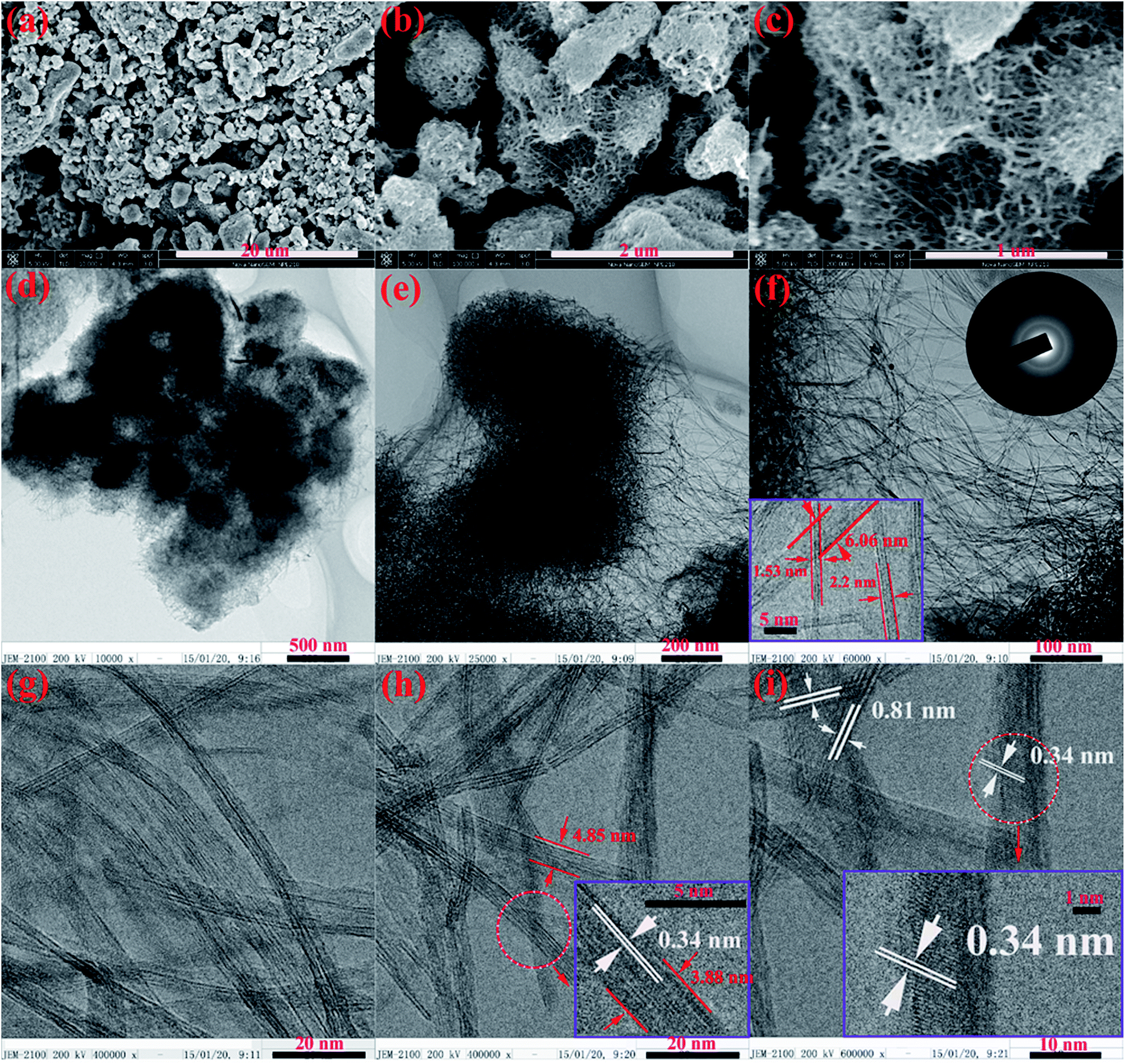
Fig. 2 shows the SEM and TEM images of HTO-HT. As presented in Fig. 2a–c, the HTO-HT preserves the morphology of H2Ti3O7 precursor perfectly, and consists of micro/nanoparticles constructed by ultrafine nanobelt units. This unique nanostructure of HTO-HT was further confirmed by TEM characterization as shown in Fig. 2d–i. The intertwined ultrafine nanobelt units are clearly revealed in Fig. 2f, and their length can reach micrometer scale. The SAED patterns indicate the polycrystalline and low crystallinity nature of HTO-HT, which is in good agreement with XRD patterns (Fig. 1a). The HRTEM images (Fig. 2f–g) and magnified inset in Fig. 2f depict the ultrafine nanobelt units have a thickness of about 1–3 nm and a width of about 3–6 nm. The crystallized layered structure of HTO-HT is clearly showed in HRTEM (Fig. 2i), and the observed interlayer spacing is about 0.81 nm, which is consistent with the reported value.7 The visible lattice fringes with a spacing of 0.34 nm are also observed, which corresponds to the d101-spacing in the XRD patterns.14 After that, SEM with electron dispersive X-ray spectroscopy (EDS) was used to map the elemental composition at the HTO-HT (Fig. 3a–d). It can be seen that the edge of Ti and O EDS maps showed in Fig. 3c and d well match the observation in the SEM image (Fig. 3a), further indicating that the HTO-HT contains Ti and O elements. Then, the X-ray photoelectron spectroscopy (XPS) was used to further insight into the chemical states of Ti and O. Fig. 3e is the high resolution XPS spectrum of Ti 2p, showing two peaks located at 458.9 (Ti 2p3/2) and 464.6 eV (Ti 2p1/2), respectively, which are assigned to Ti4+.15 Fig. 3f shows the typical high-resolution XPS spectra of O 1s, it can be deconvoluted into two individual peaks located at 530.4 and 531.2 eV, respectively, which can be attributed to O2− in TiO2 and H2O, respectively.15,16 The XPS result is consistent with characterization of FTIR spectroscopy.
 |
| | Fig. 2 The SEM and TEM images of HTO-HT: (a–c) SEM images with different magnifications; (d–f) TEM images with different magnifications, and the insets in (f) showing the SAED (top right) and magnified TEM image (bottom left), respectively; (g–i) HRTEM, the insets in (h) and (i) showing the enlarged pictures of the design areas indicated by red circles in (h) and (i), respectively. | |
 |
| | Fig. 3 (a) SEM image of HTO-HT; (b) EDS; (c, d) SEM EDS mappings of HTO-HT; (e) and (f) the typical high-resolution XPS spectra of Ti 2p and O 1s, respectively. | |
The electrochemical properties of the HTO-HT were evaluated as anode materials for LIBs. Fig. 4a shows cyclic voltammograms (CV) of HTO-HT and other samples electrodes between 1 and 3 V at a scan rate of 0.5 mV s−1. Except the TiO2-400 (it displays a typical behavior of mixed phase of anatase TiO2 and TiO2-B),9a all other samples show the typical behavior of hydrogen titanate.6–10 For HTO-HT, H2Ti3O7 and H2Ti3O7 calcined at 200 °C (H2Ti3O7-200), only broad redox peaks can be observed during the entire voltage range, which is well match with the sloping voltage profile during the discharge/charge cycles. The increase in intensities of current peaks may be due to larger surface area and less adsorbed water of HTO-HT, which is beneficial to improve the lithium-ion intercalation kinetics.17 Fig. 4b depicts the discharge/charge profiles of different samples at 168 mA g−1. Except the TiO2-400, other three samples exhibit the similar behaviors, indicating they are all hydrogen titanate only with different adsorbed water. The smooth sloping nature of the voltage profiles implies that lithium intercalation into the HTO-HT ultrafine nanobelts remains a single-phase process after cycling, which means that no two phases equilibrium happen during the lithium intercalation process. This electrochemical behavior may be ascribed to the common open layered and tunnel-like structure of hydrogen titanate.7 The first discharge and charge capacities of HTO-HT, H2Ti3O7, H2Ti3O7-200 and TiO2-400 are 538.1 and 268.4, 532.5 and 214.7, 564 and 284, and 341.8 and 213.7 mA h g−1, respectively, with a coulombic efficiency of 50, 40.3, 50 and 62.5%, respectively. The large initial capacity loss may be mainly ascribed to the nanostructured characteristic and trace water adsorbed on the materials surface because of the large specific surface area of HTO-HT, H2Ti3O7 and H2Ti3O7-200.18 Therefore, it is suggested that the relatively low initial coulombic efficiency of HTO-HT should be attributed to its relatively large specific surface area which would provide a larger contact interface between electrode and electrolyte for formation of more SEI film and abundant irreversible surface defect lithium storage. Fig. 4c shows the cycling performances of HTO-HT at different current densities, delivering a high capacity of 184.8, 176.4 and 156.8 mA h g−1 after 1000 cycles at 0.168, 1.68 and 10.08 A g−1, respectively. In Fig. 4c, the coulombic efficiencies for current density of 0.168 A g−1 are also given and stable at 99% after 30 cycles, indicating the excellent cycling stability of HTO-HT. In contrast, the control samples of H2Ti3O7, H2Ti3O7-200 and TiO2-400 only delivers a discharge capacity of 115.2, 151.2 and 154.4 mA h g−1 after 1000 cycles at 1.68 A g−1, respectively, as displayed in Fig. 4d. For better confirming that the excellent performance of HTO-HT is related to its unique morphology, another control sample, denoted as HTO-HTB, has also been prepared by monodispersed broad H2Ti3O7 nanobelts (Fig. S2b2†) calcination at the same conditions with HTO-HT. The obtained HTO-HTB shows an ill-shaped broad nanobelts morphology (lost the morphology of monodispersed broad H2Ti3O7 nanobelt precursor after calcination), and has a protonated titanate phase (JCPDS, card 47-0124), which are determined by characterizations of TEM and XRD measurements, as shown Fig. S9 and S10,† respectively. Compared with HTO-HT, the HTO-HTB shows an inferior performance, only delivering 127.4 mA h g−1 after 1000 cycles at 1.68 A g−1 (Fig. 4d). So, it is suggested that the excellent performance of HTO-HT should be benefited from the unique morphology and rational heating treatment. More over, the ultra-long cycling performance of HTO-HT is also investigated as shown in Fig. 4e. Even after 4000 cycles, HTO-HT can deliver a capacity of 120 mA h g−1 at 10.08 A g−1. Surprisingly, the HTO-HT yet delivers a capacity of 168 mA h g−1 after 150 cycles when the current density increases to 16.8 A g−1 (full charge only needs about 40 second), as shown in Fig. 4f. The rate capability of HTO-HT is further tested between 67 and 5040 mA g−1 as shown in Fig. S11.† The fifth reversible capacity of 282.4, 230.2, 215.8, 202, 183 and 176.2 mA h g−1 is delivered at a rate of 67, 336, 672, 1344, 3360 and 4200 mA g−1, respectively. When the current density backs to 67 mA g−1 after high rate tests, a capacity of 204.7 mA h g−1 can be recovered at 425th cycle, indicating good rate performance and stability of HTO-HT electrode. The excellent performance of HTO-HT should be attributed to the unique nanostructure and appropriate heating treatment: on one hand, the ultrafine belts can greatly reduce the diffusion distance of lithium ion and electron, and the hierarchical structure assembled by ultrafine nanobelts (rich interspaces formed among ultrafine nanobelt units) can effectively and partly prevent from aggregation of nanobelts and provide large specific surface area for electrode to ensure high contact area between the electrode and electrolyte, shortened lithium-ion and electron diffusion distance, easy penetration of HTO-HT by liquid electrolyte and effective relaxation of strain induced by repeated lithiation/delithiation process; on the other hand, the reduction of adsorbed water in HTO-HT makes the layered structure very open which can provide a very large interspaces for lithium storage besides reducing the adverse effect to the electrochemical performance. The electrochemical impedance spectroscopy (EIS) measurements were used to better demonstrate the effect of peculiar architecture on the electron transfer resistance of the HTO-HT. The measurements were carried out after initial several charge/discharge cycle in order to effectively eliminate the irreversible reaction stage. Generally, the impedance response of LIBs can be regarded as the sum of following processes: (i) conduction of Li-ions and electrons, (ii) Li-ion migration through the SEI layer, (iii) charge transfer-involved electrochemical reaction of active materials, and (iv) Li-ion diffusion into the lattice of active materials.19 And the impedance response of LIBs can be finally reflected by EIS plots, in which the diameter of the depressed semicircle is correlated with the electron transfer resistance on the electrode interface, and the angled line is related to a diffusion controlled process. Fig. 4g displays the Nyquist plots of the three samples after 5 discharge/charge cycles. Obviously, all electrodes show Nyquist plots composing of a depressed semicircle at high frequency range and an angled line in the low frequency range. And the HTO-HT exhibits smallest diameter of the high frequency semicircles among three samples, indicating the peculiarly hierarchical structure constructed by ultrafine nanobelts may play a key role in improving electron and lithium-ion transport ability.
 |
| | Fig. 4 (a) Cyclic voltammograms and (b) discharge/charge profiles of different samples; cycling performances of (c) HTO-HT and (d) control samples; (e) ultra-long cycling performance of HTO-HT; (f) cycling performance of HTO-HT at a high current density of 16.8 A g−1; (g) Nyquist plots of different samples after five charge/discharge cycles. | |
Furthermore, cyclic voltammogram at various scan rates were carried out to insight into the superior kinetic behavior of lithium storage in HTO-HT, as shown in Fig. 5. Usually, the change in the cathodic and anodic peaks with sweeping rates reflects the kinetics of lithium insertion/extraction at the electrode/electrolyte interface. To gain a deeper investigation on the nature of both anodic and cathodic currents, the peak current density (jp) is plotted against the scan rate (ν) in the format of log(jp) − log(ν), by assuming that jp follows a power-law relationship with ν (eqn (1))
where the power
z implies the nature of the current: a value of 1 would indicate that the current is surface-controlled.
20 In
Fig. 5b and c, the
z value for the anodic peak (
za) and cathodic peak (
zc) is about 1.03 and 1.1, respectively, indicating the both delithiation and lithiation process are governed by surface-controlled, which can be considered as faradic pseudocapacitance.
6,21 The pseudocapacitive behavior is readily explained in terms of the interaction taking place on the nanosheet surface.
22 It was accepted that the pseudocapacitance becomes the dominant reversible charge storage mechanism for nanoscale electrode materials with large specific surface area at high charge/discharge current densities, for example, nanostructured TiO
2-based electrodes.
9c And there are three plausible reasons for the novel kinetic performance of HTO-HT: firstly, the high specific surface area (333 m
2 g
−1) and unique hierarchical structure ensure a perfect contact between the electrode and electrolyte, which will significantly improve the interfacial characteristic; secondly, the ultrafine belts with 1–3 nm thickness, 3–6 nm width and several μm length will greatly shorten the diffusion distance of lithium-ion and electron in the solid state compared to a normal lithium-ion battery electrode derived from a high-temperature solid-state reaction; finally, the interlayer spacing in HTO-HT is much larger than that in normal transitional metal oxide intercalation compounds, which will also facilitate the lithium-ion diffusion in this open and loose structure. For getting the information of structure stability, we further studied the structure of HTO-HT electrode by XRD and TEM characterizations after cycled 17 cycles, as shown in
Fig. 6. It can be clearly seen from
Fig. 6a and b that the HTO-HT relatively well preserved the protonated titanate phase after cycled 17 cycles. More importantly, the HTO-HT exhibited outstanding structure stability as the peculiar hierarchical nanostructure was perfectly maintained after cycled 17 cycles as shown in
Fig. 6c and d. Thus, the ultra-long life of HTO-HT electrode should be attributed to the excellent structure stability.
 |
| | Fig. 5 (a) Cyclic voltammograms of HTO-HT at six various scan rates; (b) and (c) plots of log(current density) − log(scan rate), that is, log(jp, mA) − log(ν, mV s−1), based on the cyclic voltammograms in (a). | |
 |
| | Fig. 6 (a) XRD patterns and (c, d) TEM images with different magnifications of HTO-HT electrode after 17 cycles; (b) XRD patterns of different samples without cycle. | |
4 Conclusions
In summary, the electrochemical performance of hydrogen titanate as anode materials for lithium-ion batteries has been successfully and significantly improved by morphology-engineering and heating treatment. The as-prepared HTO-HT possesses a peculiar morphology of micro/nanoparticles constructed by ultrafine nanobelts, and less adsorbed water. Especially, as anode materials for lithium-ion batteries, the HTO-HT exhibits a remarkable rate capability and excellent cycling stability. Such excellent performance makes HTO-HT a promising anode material for advanced lithium-ion batteries with ultra-long life and high power density.
Acknowledgements
The authors are grateful for financial support from the National Natural Science Foundation of China (grant No. 21103108 and 21173148) and materials characterization from Instrumental Analysis Center of Shanghai Jiao Tong University.
References
- G. N. Zhu, Y. G. Wang and Y. Y. Xia, Energy Environ. Sci., 2012, 5, 6652 CAS.
- G. Q. Zhang, H. B. Wu, T. S. Song, U. Y. Paik and X. W. Lou, Angew. Chem., Int. Ed., 2014, 53, 12590 CAS.
- P. G. Bruce, Chem. Commun., 1997, 19, 1817 RSC.
- L. J. Fu, T. Zhang, Q. Cao, H. P. Zhang and Y. P. Wu, Electrochem. Commun., 2007, 9, 2140 CrossRef CAS.
-
(a) A. R. Armstrong, G. Armstrong, J. Canales and P. G. Bruce, Angew. Chem., Int. Ed., 2004, 43, 2286 CrossRef CAS PubMed;
(b) H. W. Shim, D. K. Lee, I. S. Cho, K. S. Hong and D. W. Kim, J. Nanotechnol., 2010, 21, 255706 CrossRef;
(c) J. M. Li, W. Wan, H. H. Zhou, J. J. Li and D. S. Xu, Chem. Commun., 2011, 47, 3439 RSC;
(d) Q. H. Tian, Z. X. Zhang, L. Yang and S.-I. Hirano, J. Mater. Chem. A, 2015, 3, 14721 RSC.
- J. R. Li, Z. L. Tang and Z. T. Zhang, Chem. Phys. Lett., 2006, 418, 506 CrossRef CAS.
- J. R. Li, Z. L. Tang and Z. T. Zhang, Chem. Mater., 2005, 17, 5848 CrossRef CAS.
-
(a) M. D. Wei, K. W. Wei, M. Ichihara and H. S. Zhou, Electrochem. Commun., 2008, 10, 1164 CrossRef CAS;
(b) F. X. Wu, Z. X. Wang, X. H. Li and H. J. Guo, J. Mater. Chem., 2011, 21, 12675 RSC.
-
(a) S. H. Liu, H. P. Jia, L. Han, J. L. Wang, P. F. Gao, D. D. Xu, J. Yang and S. A. Che, Adv. Mater., 2012, 24, 3201 CrossRef CAS PubMed;
(b) G. Armstrong, A. R. Armstrong, J. Canales, R. Garcia and P. G. Bruce, Adv. Mater., 2005, 17, 862 CrossRef;
(c) J. Y. Shin, D. Samuelis and J. Maier, Adv. Funct. Mater., 2011, 21, 3464 CrossRef CAS.
- Y. Lan, X. P. Gao, H. Y. Zhu, Z. F. Zheng, T. Y. Yan, F. Wu, S. P. Ringer and D. Y. Song, Adv. Funct. Mater., 2005, 15, 1310 CrossRef CAS.
- M. Zhang, Z. S. Jin, J. W. Zhang, X. Y. Guo, J. J. Yang, W. Li, X. D. Wang and Z. J. Zhang, J. Mol. Catal. A: Chem., 2004, 217, 203 CrossRef CAS.
- P. Y. Dong, B. Liu, Y. H. Wang, L. N. Guo, Y. J. Huang and S. Yin, J. Electochem. Soc., 2011, 158, k183 CrossRef CAS.
- G. Li, Surf. Rev. Lett., 2009, 16, 149 CrossRef CAS.
-
(a) P. G. Bruce, Chem. Commun., 1997, 19, 1817 RSC;
(b) J. R. Li, Z. L. Tang and Z. T. Zhang, Chem. Mater., 2005, 17, 5848 CrossRef CAS.
- Y. Qiao, X. L. Hu, Y. Liu, C. J. Chen, H. H. Xu, D. F. Hou, P. Hu and Y. H. Huang, J. Mater. Chem. A, 2013, 1, 10375 CAS.
- Y. M. Li, X. J. Lv, J. Lu and J. H. Li, J. Phys. Chem. C, 2010, 114, 21771 Search PubMed.
- C. Wessel, L. A. Zhao, S. Urban, R. Ostermann, I. Djerdj, B. M. Smarsly, L. Q. Chen, Y. G. Hu and S. Sallard, Chem. - Eur. J., 2011, 17, 775 CrossRef CAS.
- J. Li, Z. Tang and Z. Zhang, Electrochem. Solid-State Lett., 2005, 8, A316 CrossRef CAS.
-
(a) C. J. Chae, K. W. Kim, S. J. Kim, D. Lee, Y. Jo, Y. J. Yun, J. Moon, Y. Choi, S. S. Lee, S. Choi and S. Jeong, Nanoscale, 2015, 7, 10368 RSC;
(b) J. X. Song, S. R. Chen, M. J. Zhou, T. Xu, D. P. Lv, M. L. Gordin, T. J. Long, M. Melnyk and D. H. Wang, J. Mater. Chem. A, 2014, 2, 1257 RSC;
(c) J. X. Song, M. J. Zhou, R. Yi, T. Xu, M. L. Gordin, D. H. Tang, Z. X. Yu, M. Regula and D. H. Wang, Adv. Funct. Mater., 2014, 24, 5904 CrossRef CAS.
- V. Augustyn, J. Come, M. A. Lowe, J. W. Kim, P. L. Taberna, S. H. Tolbert, H. D. Abruna, P. Simon and B. Dunn, Nat. Mater., 2013, 12, 518 CrossRef CAS.
- G. Nuspl, K. Yoshizawa and T. Yamabe, J. Mater. Chem., 1997, 7, 2529 RSC.
- L. Kavan, M. Kalbac, M. Zukalova, I. Exnar, V. Lorenzen, R. Nesper and M. Gratzel, Chem. Mater., 2004, 16, 47 Search PubMed.
Footnote |
| † Electronic supplementary information (ESI) available. See DOI: 10.1039/c5ra19381h |
|
| This journal is © The Royal Society of Chemistry 2015 |
Click here to see how this site uses Cookies. View our privacy policy here.