Formulation characteristics of triacylglycerol oil-in-water emulsions loaded with ergocalciferol using microchannel emulsification

Nauman Khalidabc, Isao Kobayashi*a, Zheng Wangac, Marcos A. Nevesac, Kunihiko Uemuraa, Mitsutoshi Nakajimaac and Hiroshi Nabetaniab
aFood Engineering Division, National Food Research Institute, NARO, 2-1-12 Kannondai, Tsukuba, Ibaraki 305-8642, Japan. E-mail: isaok@affrc.go.jp; Fax: +81-29-838-8122; Tel: +81-29-838-8025
bGraduate School of Agricultural and Life Sciences, The University of Tokyo, 1-1-1 Yayoi, Bunkyo-ku, Tokyo 113-8657, Japan
cFaculty of Life and Environmental Sciences, University of Tsukuba, 1-1-1 Tennoudai, Tsukuba, Ibaraki 305-8572, Japan

Received 8th September 2015 , Accepted 23rd October 2015

First published on 12th November 2015


Abstract

Ergocalciferol is one important form of vitamin D that is needed for proper functioning of the human metabolic system. The study formulates monodisperse food grade ergocalciferol loaded oil-in-water (O/W) emulsions by microchannel emulsification (MCE). The primary characterization was performed with grooved MCE, while the storage stability and encapsulating efficiency (EE) were investigated with straight-through MCE. The grooved microchannel (MC) array plate has 5 × 18 μm MCs, while the asymmetric straight-through MC array plate consists of numerous 10 × 80 μm microslots each connected to a 10 μm diameter circular MC. Ergocalciferol at a concentration of 0.2–1.0% (w/w) was added to various oils and served as the dispersed phase, while the continuous phase constituted either of 1% (w/w) Tween 20, decaglycerol monolaurate (Sunsoft A-12) or β-lactoglobulin. The primary characterization indicated successful emulsification in the presence of 1% (w/w) Tween 20 or Sunsoft A-12. The average droplet diameter increased slowly with the increasing concentration of ergocalciferol and ranged from 28.3 to 30.0 μm with a coefficient of variation below 6.0%. Straight-through MCE was conducted with 0.5% (w/w) ergocalciferol in soybean oil together with 1% (w/w) Tween 20 in Milli-Q water as the optimum dispersed and continuous phases. Monodisperse O/W emulsions with a Sauter mean diameter (d3,2) of 34 μm with a relative span factor of less than 0.2 were successfully obtained from straight-through MCE. The resultant oil droplets were physically stable for 15 days (d) at 4 °C without any significant increase in d3,2. The monodisperse O/W emulsions exhibited an ergocalciferol EE of more than 75% during the storage period.

Introduction

Vitamin D plays a vital role in maintaining and developing a healthy human skeletal system, since it maintains calcium levels in the body. Deficiency of vitamin D results in an increased risk of diabetes, hypertension, cancer and autoimmune diseases.1–4 Broad spectrum deficiencies of vitamin D include rickets in children, osteomalacia in adults and osteoporosis in women; all of these lead to softening and weakening of bones.5–7 Nutritional and cultural factors leading to vitamin D deficiency include insufficient fortified food consumption, sun-block usage, limited body exposure to sun and fear for excessive intake of vitamin D.7,8 Vitamin D is synthesized in the skin and involves the phytochemical conversion of pro-vitamin D by the action of ultraviolet (UV-B) rays. This process takes place if the UV-B rays fall between 290 and 315 nm of the spectrum. These rays are only emitted in the regions that lie below 35° latitude.9,10

The terminology and classification related to vitamin D is confusing and can be classified into 5 different forms and metabolites. Among these, vitamin D2 (ergocalciferol) and D3 (cholecalciferol) are important (Fig. 1).11,12 Vitamin D2 is naturally present in some plants and is produced commercially by UV irradiation of yeast, while vitamin D3 is naturally produced in human and animal bodies. Vitamin D2 is substantially used for fortification and supplementation in the food and pharmaceutical industries.12 Several researchers pointed out the rapid metabolism of vitamin D2 in comparison to vitamin D3, while the action becomes bio-equivalent if taken daily.13,14 Both forms of vitamin D are converted to 25-hydroxy vitamin D [25-(OH)D] in the liver. The quantification of 25-(OH)D in blood gives the quantitation of the vitamin D status. A cutoff value of 30 ng mL−1 is sometimes used for an optimal vitamin D status.12 Ergocalciferol was produced in the 1920s through UV-B exposure of foods, leading to the formation of the first medicinal preparation called viosterol.15 Ergocalciferol has limited natural sources and the most significant source is wild mushrooms.16 Ergocalciferol is prone to oxidation and is also isomerized to isotachysterol in the presence of sunlight and mostly under acidic conditions.16,17 Ergocalciferol is mostly supplemented and fortified in fat-based products due to its hydrophobic nature.


image file: c5ra18354e-f1.tif
Fig. 1 Chemical structures of important forms of vitamin D: (a) ergocalciferol and (b) cholecalciferol.

Emulsification technologies play an important role in the production of encapsulated foods, pharmaceuticals, cosmetics and chemicals.18,19 The emulsions are usually either single (O/W and W/O) emulsions or double emulsions (W/O/W and O/W/O).19 These different emulsion systems are produced by either conventional devices including colloid mills, high pressure homogenizers and rotor-stator homogenizers or modern devices such as microfluidic devices (lab-on-a-chip), membrane emulsification and microchannel emulsification (MCE) devices.19,20 Conventional devices produce polydisperse emulsions with broader size distributions, which in turn reduce the emulsion stability and functionality. Microfabricated emulsification devices have the potential to produce monodisperse emulsions with the smallest coefficient of variation (CV) of less than 10% for membrane emulsification and around 5% for microfluidic devices and MCE.20

MCE is a progressive technique that enables production of monodisperse emulsions by spontaneous transformation of the oil–water interface specifically driven by interfacial tension dominant on a micron scale.21 MCE studies were comprehensively reviewed by Vladisavljević et al.20,22 Similarly, this emulsification technique allows integration of hundreds of thousands of droplet generation units on a single plate.23,24 MCE has been used for producing O/W, W/O and W/O/W emulsions with diameters ranging from 1 μm to 550 μm.22 Based on the microfabrication design, the MCE devices can either be categorized into grooved microchannel (MC) arrays each consisting of uniform microgrooves and a slit-like terrace or straight-through MC arrays each having uniform asymmetric microholes together with microslots.22 Grooved MC array plates are further classified into dead-end and cross-flow types. Grooved MCE plates of the cross-flow type are particularly useful to observe the entire droplet generation process together with droplet collection at low dispersed phase flow rates.25,26 On the other hand, straight-through MC arrays are designed to improve the throughput capacity of MCE. Straight-through MC arrays have the ability to increase the production capacity of monodisperse emulsion droplets over 2000 L m−2 h−1.22

MCE has also been used to produce monodisperse microdispersions (e.g. solid lipid microspheres,26 gel microbeads,27 and giant vesicles28). MCE has promising potential for producing uniformly sized oil droplets containing functional lipids such as β-carotene,29 γ-oryzanol,30 L-ascorbic acid,31 ascorbic acid derivatives,32 oleuropein,33 and vitamin D.34 Different food grade materials (e.g. refined vegetable oils, a medium-chain triglyceride oil, hydrophobic and hydrophilic emulsifiers, proteins, and hydrocolloids) were utilized to produce monodisperse O/W, W/O, and W/O/W emulsions and microparticles by MCE.35,36

We previously encapsulated vitamin D (both vitamin D2 and D3) in O/W emulsions using MCE and reported long-term stability studies and encapsulation efficiencies of O/W emulsions encapsulating vitamin D2 and D3.34 Ergocalciferol is a plant-based vitamin D type, and previous studies seldom report its encapsulation in different formulations. Keeping its rapid metabolism rate and importance in the human diet, the present study was conducted to encapsulate only ergocalciferol in triacylglycerol oil-in-water emulsions using MCE. The basic characterization and optimization of these emulsions were performed using a grooved MC array plate. Straight-through MCE was carried out to investigate the effect of the dispersed phase flux on the production characteristics as well as the physical and chemical stability of the ergocalciferol-loaded O/W emulsions. The effects of different triacylglycerol oils and emulsifiers on the preparation characteristics of O/W emulsions by MCE were also evaluated. The results of this research are expected to help formulate new aqueous-based functional foods.

Experimental

Chemicals

Ergocalciferol, polyoxyethylene (20) sorbitan monolaurate (Tween 20), olive oil, and soybean oil were purchased from Wako Pure Chemical Industries Ltd. (Osaka, Japan). Medium-chain triacylglycerol (MCT, Sunsoft MCT-7) with a fatty acid residue composition of 75% caprylic acid and 25% capric acid and decaglycerol monolaurate (Sunsoft A-12) were procured from Taiyo Kagaku Co. Ltd (Yokkaichi, Japan). Safflower oil was purchased from MP biomedicals (lllkirch, France). β-Lactoglobulin (β-lg) from bovine milk (>90% purity) was purchased from Sigma-Aldrich Co. LLC (St. Louis, USA). All other chemicals used in this study were of analytical grade and used as received.

Preparation of solutions

The continuous phase was prepared by dissolving either 1% (w/w) Tween 20, 1% (w/w) Sunsoft A-12 or 1% (w/w) β-lg in Milli-Q water with a resistivity of 18 MΩ cm. The dispersed phase was prepared by dissolving 0.2–1.0% (w/w) ergocalciferol in MCT, soybean, olive or safflower oil at 85 °C ± 3 °C for 20 min and afterwards cooling at room temperature for 2 h before storing at 4 ± 1 °C. During storage, dissolved ergocalciferol molecules could form nuclei whose growth eventually makes crystals large enough to sediment. Before initiating the experiments, the samples were therefore shaken slightly to avoid formation of any ergocalciferol crystals.

Silicon plates for MCE

The experiments have been carried out using silicon MC array plates (model CMS 6-2 and WMS 11-1, EP. Tech Co., Ltd., Hitachi, Japan). Fig. 2a is a schematic representation of a CMS 6-2 plate with 540 parallel channels on 10 consecutive MC arrays. Each MC array contains 54 parallel MCs with a depth of 5 μm, a width of 18 μm and a length of 140 μm and a terrace with a depth of 5 μm and a length of 60 μm. Each continuous phase channel outside the terrace outlet has a depth of 100 μm. Fig. 2b is a schematic representation of a WMS 11-1 plate with 27[thin space (1/6-em)]400 MCs compactly arranged within a 10 × 10 mm square region in the plate center. Each MC consists of a cylindrical 10 μm diameter straight microhole with a depth of 200 μm and a 10 × 80 μm microslot with a depth of 40 μm. The slot aspect ratio of 8 was above the threshold value of 3 for successfully generating monodisperse emulsion droplets.37 The distance between the two adjacent MCs vertically was 105 μm, and the distance between the centers of the MCs in adjacent rows was 70 μm. The MC array plates were subjected to plasma oxidation in a plasma reactor (PR41, Yamato Science Co. Ltd., Tokyo, Japan) to activate a silicon dioxide layer on their surfaces. The activated silicon dioxide layer is capable of maintaining their hydrophilicity during MCE.
image file: c5ra18354e-f2.tif
Fig. 2 (a) Schematic drawings of the grooved MC array plate (CMS 6-2) and part of an MC array together with different dimensions. (b) Schematic drawings of the straight-through MC array plate (WMS 11-1) and MC dimensions.

Experimental procedure for MCE

For grooved MCE, the setup consists of an MC module, a 10 mL liquid chamber that contains the disperse phase and a syringe pump (Model 11, Harvard Apparatus Inc., Holliston, USA) that feeds the continuous phase with a 50 mL glass syringe (Fig. 3a). MCE was carried out for approximately 3 h and monitored through an inverted metallographic microscope equipped with an objective lens of 2.5× to 20× and a CCD camera (MS-511B, Seiwa Kougaku Sesakusho Ltd., Tokyo, Japan). The whole process was recorded with a video recorder (RDR-HX67, Sony Co., Tokyo, Japan). Droplet generation experiments were performed with the grooved MCE setup depicted in Fig. 3b. The module was initially filled with the continuous phase before mounting the CMS 6-2 plate. The pressurized dispersed phase was introduced into the module. The pressure applied to the dispersed phase (ΔPd) was gradually increased. ΔPd can be given by:
 
ΔPd = ρdΔhdg (1)
where ρd is the dispersed phase density, Δhd is the difference in the hydraulic heads between the chamber containing the dispersed phase and the channels of the module, and g is the acceleration due to gravity. To generate droplets, the dispersed phase was forced through the MCs onto the terrace and into the continuous phase channel.

image file: c5ra18354e-f3.tif
Fig. 3 (a) Schematic drawing of a grooved MCE setup. (b) Schematic drawing of droplet generation via part of a MC array having a 5 μm depth.

For straight-through MCE, the MC array plate was degassed in a continuous aqueous phase by an ultrasonic bath for 20 min and the setup consists of a MC module (comprising of six steel parts and two glass plates of different dimensions and rubber seals) and syringe pumps (Model 11, Harvard Apparatus Inc.) that feed the continuous and dispersed phases (Fig. 4a). MCE was carried out for approximately 1 h and monitored through a FASTCAM-1024 PCI high speed video system at 250 to 500 fps (Photron Ltd., Tokyo, Japan) attached to the inverted metallographic microscope. The droplet generation process is depicted in Fig. 4b. Droplet generation started with injecting the dispersed phase through the syringe pump at a dispersed phase flow rate (Qd) ranging from 0.5 to 2.0 mL h−1 (5 to 20 L m−2 h−1 in a dispersed phase flux (Jd)). The generated droplets were removed by varying the continuous phase flow rate (Qc) from 100 to 500 mL h−1 through the gap between the MC array plate and the glass plate. The shear stress (τ) in the module surrounding the WMS 11-1 plate is given by:

 
image file: c5ra18354e-t1.tif(2)
where h = 1 mm is the gap height and W = 12 mm is the gap width, and ηc is the continuous phase viscosity. τ had a negligible value of 0.002 to 0.02 Pa at the Qc range applied in this study. After each experiment the MC array plates were cleaned in three steps. In the first step the MC array plates were washed with neutral detergent together with Milli-Q water in an ultrasonic bath (VS-100 III, As One Co., Osaka, Japan) for 20 min, followed by treatment with 50% Milli-Q water and 50% ethanol in an ultrasonic bath, and lastly cleaned in an ultrasonic bath with Milli-Q water and stored in 50 mL of Milli-Q water prior to reuse for MCE.


image file: c5ra18354e-f4.tif
Fig. 4 (a) Schematic representation of a straight-through MCE setup. (b) Droplet generation representation through asymmetric MCs.

Determination of droplet size and droplet size distribution

The size and size distribution of the resultant O/W emulsion droplets from the grooved MCE were determined as follows. The average droplet diameter (dav) was defined by:
 
image file: c5ra18354e-t2.tif(3)
where di is the diameter of the ith droplet measured using WinRoof software (Mitani Co., Ltd., Fukui, Japan) and n is the number of droplets measured (n = 250). The droplet size distribution was expressed as CV, and is defined as:
 
image file: c5ra18354e-t3.tif(4)
where σ is the standard deviation and dav is the average droplet diameter.

The droplet size distribution of the O/W emulsions obtained from straight-through MCE was measured using a laser scattering instrument that works on the principle of Polarization Intensity Differential Scattering Technology (LS 13 320, Beckman Coulter, Inc., Brea, USA). This instrument has the ability to measure the size ranging from 0.04 to 2000 μm with a resolution of 116 particle size channels. The mean droplet size was expressed as the Sauter mean diameter (d3,2), defined as the diameter of a droplet having the same area per unit volume as that of the total collection of droplets in emulsions. The width of the droplet size distribution was expressed as relative span factor (RSF), defined as:

 
image file: c5ra18354e-t4.tif(5)
where dv0.9 and dv0.1 are the representative diameters where 90% and 10% of the total volume of the liquid is made up of droplets with diameters smaller than or equal to the stated value, and dv0.5 is the representative diameter where 50% of the total volume of the liquid is made up of droplets with diameters larger than the stated value and 50% is made up of droplets with diameters smaller than the stated value.

Measurement of fluid properties

The densities of the dispersed and continuous phases were measured using a density meter (DA-130 N, Kyoto Electronics Manufacturing Co., Ltd., Kyoto, Japan) at 25 ± 2 °C. Their viscosities were measured with a vibro viscometer (SV-10, A&D Co., Ltd., Tokyo, Japan) at 25 ± 2 °C by taking either 10 or 35 mL of samples in a measuring vessel followed by immersion of sensor plates in that vessel. Viscosity was measured by detecting the electric current needed to resonate the sensor plates. The static interfacial tension between the preceding two phases was measured with a fully automatic interfacial tensiometer (PD-W, Kyowa Interface Sciences Co., Ltd., Saitama, Japan) using a pendant drop method. The key physical properties of the dispersed and continuous phases used in this study are presented in Table 1.
Table 1 Fluid properties of the systems containing ergocalciferol together with different oils used for preparing O/W emulsions
  Dispersed phase Continuous phase
ηd (mPa s) ρd (kg m−3) γd (mN m−1) ζb Emulsifiers in Milli-Q water ηc (mPa s) ρc (kg m−3) γc (mN m−1)
a Dispersed phase contains 0.5% (w/w) ergocalciferol and interfacial tension was measured in the presence of 1% (w/w) Tween 20 in Milli-Q water.b Viscosity ratio (ζ) was defined as the ratio of dispersed phase viscosity over continuous phase viscosity.
MCTa 22.5 ± 0.3 946.9 ± 0.2 5.3 ± 0.4 24.7 0.5% Tween 20 0.89 ± 0.1 997.3 ± 0.6 5.1 ± 0.2
Soybean oila 53.0 ± 0.1 921.9 ± 0.4 5.6 ± 0.1 58.2 1.0% Tween 20 0.91 ± 0.1 998.4 ± 0.6 5.2 ± 0.1
Olive oila 68.2 ± 0.1 911.9 ± 0.2 6.2 ± 0.2 75.0 1.5% Tween 20 0.96 ± 0.1 999.1 ± 0.8 5.2 ± 0.2
Safflower oila 53.2 ± 0.1 918.9 ± 0.1 5.3 ± 0.2 58.5 2.0% Tween 20 0.99 ± 0.1 1000.1 ± 0.1 5.4 ± 0.3
  1.0% β-lg 0.95 ± 0.1 999.9 ± 0.2 12.6 ± 0.9
1.0% Sunsoft A-12 0.97 ± 0.1 998.5 ± 0.6 4.8 ± 0.2


Physical and chemical stability of O/W emulsions

The physical stability of the O/W emulsion droplets loaded with ergocalciferol was evaluated according to the method described in an earlier section. The d3,2, RSF, consistency and coalescence during 15 d of storage at 4 ± 1 °C under dark conditions were observed.

The amount of ergocalciferol encapsulated in the O/W emulsions was determined spectrophotometrically. All spectral measurements of the ethanolic extracts of these O/W emulsions were carried out using a UV/VIS/NIR spectrophotometer (UV-1700, Shimadzu Co., Kyoto, Japan). First, 1 mL of the emulsion was mixed with 9 mL of ethanol, followed by ultrasonication for 20 min. The ethanolic extracts were then centrifuged (Avanti HP-25, Beckman Coulter, Inc.) at 20[thin space (1/6-em)]000g for 15 min. A 1 mL aliquot of the subnatants was diluted ten times with ethanol and then injected into a quartz cell with a 10 mm pass length. The absorbance of ergocalciferol in the emulsion extract was measured at 310 nm using an appropriate blank. A representative standard curve of absorbance versus concentration gave the linear least-squares regression with a coefficient of determination (r2) of 0.9996. All experiments were repeated in triplicate and mean values were calculated. Beer’s law was obeyed in the concentration range of 0.1–0.5 mg mL−1 and the sensitivity of measurement has a relative standard deviation of 0.85% (n = 15). The molar absorptivity (ε) for ergocalciferol during this study was 2.52 mM−1 cm−1. The encapsulation efficiency (EE) of ergocalciferol in the samples were calculated with the equation:

 
image file: c5ra18354e-t5.tif(6)
where Wt is the total amount of ergocalciferol in the O/W emulsions at a specific time (t) and W0 is the total amount of ergocalciferol initially quantified at day 1.

Results and discussion

Basic droplet generation characteristics through grooved MCE

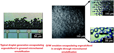
Effect of dispersed phase composition. Fig. 5a illustrates the effect of different oils on the dav and CV of the O/W emulsions prepared using the CMS 6-2 plate. MCE was carried out using 0.5% (w/w) ergocalciferol in MCT, soybean, olive or safflower oil as the dispersed phase and 1% (w/w) Tween 20 in Milli-Q water as the continuous phase. A gradual increase in ΔPd caused the dispersed phase to enter the terrace in front of the MC inlets. When ΔPd reached the break-through pressure of about 3.0 kPa, the dispersed phase started to pass through the MCs, leading to the periodic generation of oil droplets. The MCE was performed at a ΔPd of 3.2 kPa slightly higher than the break-through pressure. Qc was fixed at 2 mL h−1 throughout the experiment. Successful generation of monodisperse O/W emulsion droplets took place, regardless of the oil type used. The dav of the resultant O/W emulsions loaded with ergocalciferol ranged from 28.3 to 30.0 μm with a CV between 3.6 and 6.1%. Fig. 5b(i–iv) shows the droplet detachment process with different dispersed phases. The different dispersed phase solutions exhibited successful emulsification with smooth detachment of droplets from the terrace outlets, and narrower droplet size distributions were seen in the emulsions prepared with olive oil, safflower oil and soybean oil in comparison to MCT (Fig. 5c). The O/W emulsions prepared with MCT had a somewhat greater CV of about 6%. Uniformly sized droplets are stably generated in MCE, if the inflow of the continuous phase toward the terrace is sufficiently fast compared to the outflow of the dispersed phase from the MC outlets.38 The viscosity ratio (ζ = ηd/ηc) is also a key factor determining the monodispersity of emulsions.38 The viscosity ratio of all the oils used in this study was sufficiently high (Table 1), leading to successful preparation of monodisperse O/W emulsions encapsulating ergocalciferol. The lower monodispersity with MCT might be attributed to some attractive interaction of MCT with the MCs and terrace surfaces. Such an interaction causes a slight increase in the CV of the O/W emulsion droplets in comparison to other viscous oils. Tan et al.39 pointed out that the hydrophobicity of the dispersed phase is the critical parameter affecting the generation of oil droplets in MCE.
image file: c5ra18354e-f5.tif
Fig. 5 (a) Effect of different dispersed phase compositions on the dav and CV of O/W emulsions. (image file: c5ra18354e-u1.tif) denotes the dav of different dispersed phases, while (image file: c5ra18354e-u2.tif) denotes the CV of different dispersed phases. (b) Typical generation behaviors of O/W emulsions droplets encapsulating ergocalciferol using different dispersed phase oils. (c) Micrographic images of droplets encapsulating ergocalciferol using different dispersed phases.
Effect of different emulsifiers. The effect of the emulsifier type on oil droplet generation by grooved MCE was also investigated. Food grade hydrophilic emulsifiers (Tween 20, Sunsoft A-12 and β-lg) with a noticeable ability to prepare O/W emulsions were used at a concentration of 1% (w/w) in Milli-Q water. These aqueous emulsifier solutions were used as the continuous phase. 0.5% (w/w) ergocalciferol in soybean oil was used as the dispersed phase. The important physical properties of the different emulsifiers are presented in Table 1. There was not a prominent difference in the viscosity and density values of the emulsifier solutions, while β-lg had a higher interfacial tension (12.6 mN m−1) in comparison to Tween 20 and Sunsoft A-12. All of these emulsifier solutions exhibited successful emulsification with smooth detachment of droplets from the terrace outlets (Fig. 6a). Uniformly sized emulsion droplets were stably generated from the MCs especially in the presence of Tween 20 and Sunsoft A-12. There was neither the generation of bigger droplets nor a continuous outflow of the dispersed phase. Fig. 6b illustrates the effect of the emulsifier type on the dav and CV of the O/W emulsions. The dav and CV of the resultant O/W emulsions were 28.5 μm and 5.9% for Tween 20 and 28.1 μm and 6.6% for Sunsoft A-12. Uniformly sized droplets stabilized by Tween 20 or Sunsoft A-12 were successfully generated due to high interfacial activity, as these emulsifiers have low interfacial tension values of about 5.0 mN m−1. The droplet generation and detachment processes for β-lg at pH 7.3 were initially stable with the smallest dav of 26.6 μm. However, after 5 min a few droplets started sticking in the well (Fig. 6a(iii) and c) and coalesced with the passage of time, resulting in the increase of the CV value to 7.2%. The result suggests that β-lg did not adsorb strongly at the newly created interface, presumably because the weak electrostatic interactions between ergocalciferol and β-lg clearly caused a high interfacial tension value.
image file: c5ra18354e-f6.tif
Fig. 6 Effect of different emulsifiers on droplet generation behavior in grooved MCE. (a) Droplet generation with (i) Tween 20, (ii) Sunsoft A-12 and (iii) β-lg. (b) Effect of different emulsifiers on the dav and CV of O/W emulsions, (image file: c5ra18354e-u3.tif) denotes the dav of different emulsifiers, while (image file: c5ra18354e-u4.tif) denotes the CV of different emulsifiers. (c) Typical droplet generation characteristics with β-lg as the emulsifier.

Kobayashi and Nakajima40 investigated the effect of the emulsifier type on the droplet generation characteristics using a straight-through extrusion filter. They reported Tween 20 and decaglyerol monolaurate as suitable, hydrophilic food grade emulsifiers in MCE without ergocalciferol. Patel and San Martin-Gonzalez17 also demonstrated the successful preparation of solid lipid nanoparticles loaded with ergocalciferol stabilized by Tween 20. The preceding results demonstrate that Tween 20 and Sunsoft A-12 are potential emulsifiers for generating ergocalciferol-loaded O/W emulsions, either with conventional homogenization techniques (data not shown) or MCE.

Effect of ergocalciferol concentration. Fig. 7 illustrates the effect of concentration of ergocalciferol on the dav and CV of the O/W emulsions prepared using two different emulsifiers and a CMS 6-2 plate. The concentration of ergocalciferol varied from 0.2% to 1.0% (w/w) in soybean oil. According to US Pharmacopeia, ergocalciferol is sparingly soluble in different oils but has good solubility in organic solvents, except hexane. In our study, we noticed a maximum solubility of 1% (w/w) ergocalciferol in different oils at 85 ± 2 °C with no solubility at room temperature. Successful MCE was conducted with different concentrations of ergocalciferol by keeping ΔPd at 3.2 kPa and Qc around 2 mL h−1. The dav of the ergocalciferol-loaded O/W emulsions increased slowly with the increasing concentration of ergocalciferol when emulsified with 1% (w/w) Tween 20. Their dav ranged between 23.8 and 28.5 μm with the CV between 5.9 and 6.2%. Comparatively similar results were obtained with Sunsoft A-12 (Fig. 7). The ergocalciferol-loaded O/W emulsions stabilized with 1% (w/w) Sunsoft A-12 had a dav of 24.5 to 27.5 μm and a CV of 6.5 to 8.4%. A better droplet size distribution expressed as a smaller CV was seen in the emulsions stabilized with Tween 20 in comparison to those stabilized with Sunsoft A-12 (Fig. 7). A reason behind the increased dav with the increasing ergocalciferol concentration could be attributed to the weak attractive interaction between the oil encapsulating ergocalciferol and the MC and terrace surfaces during MCE. These types of interactions with terrace surfaces in MCE during L-ascorbic acid encapsulation were previously reported by Khalid et al.41 To remain in the optimum range and ease of the process we conducted stability and encapsulating efficiency experiments using 0.5% (w/w) ergocalciferol in soybean oil as an optimum dispersed phase. Moreover, this concentration has no effect on different parameters, since we evaluated the low and high concentration effect also. The other reason for choosing this concentration is to make the process more practical at an industrial scale; i.e., 0.5% (w/w) ergocalciferol in the dispersed phase is mostly desired in different industries.
image file: c5ra18354e-f7.tif
Fig. 7 Effect of ergocalciferol concentration in soybean oil on the dav and CV of O/W emulsions either stabilized by 1% (w/w) Tween 20 or Sunsoft A-12.
Effect of dispersed phase flow rate. The dispersed phase flow rate (Qd) is an important parameter in MCE that correlates with droplet productivity in the stable droplet generation regime. Fig. 8a depicts the effect of Qd on the dav and CV of the droplets generated using the CMS 6-2 plate. The dispersed phase constitutes 0.5% (w/w) ergocalciferol in soybean oil, while the continuous phase includes 1% (w/w) Tween 20 in Milli-Q water.
image file: c5ra18354e-f8.tif
Fig. 8 (a) Effect of Qd on the dav and CV of the O/W emulsions encapsulating soybean oil loaded-ergocalciferol produced using the CMS 6-2 plate. (b) Effect of Qd on the droplet generation frequency per hour.

At the lowest Qd of 2 × 10⁻³ mL h−1, the resultant droplets with a monomodal and very narrow size distribution had a dav of 23.5 μm and a CV of 5.4%. When Qd was increased stepwise, monodisperse emulsions with a CV of 4 to 10% were produced at a Qd of 8 × 10⁻² mL h−1 or less. In this Qd range, the dav of the resultant droplets ranged from 23.7 μm to 26.7 μm. The microscopic observations during MCE confirmed that the resultant droplet size hardly changed at Qc between 0 mL h−1 and 5.0 mL h−1. The generation of droplets even without external flow of the continuous phase depicts the unique spontaneous transformation of the interface in MCE. In contrast, at a Qd of 8 × 10⁻² mL h−1 or more, the dav and CV of the O/W emulsions dramatically increased to >30 μm with CV values of more than 10% (Fig. 8a). Moreover the droplet size distribution became wider and shifted towards a large droplet size area. The CMS 6-2 plate used here enabled the production of O/W emulsions with uniformly sized droplets at a critical Qd of 8 × 10⁻² mL h−1, which was higher than a maximum Qd (5 × 10⁻³ mL h−1) for the previously reported studies from grooved MCE.42

After reaching the critical Qd, some of the dispersed phase that passed through the MCs expanded instead of generating droplets, suggesting that the flow state of the dispersed phase was affected by the dispersed phase velocity inside the MC. Sugiura et al.21 reported that the droplet generation behavior inside the MCs is related to the capillary number of the dispersed phase that flows inside the MCs. The capillary number (Ca), which indicates the balance between the viscous force and interfacial force, can be determined by:

 
Ca = ndUd/γ (7)
where ηd is the dynamic viscosity (Pa s) of the dispersed phase, Ud is the dispersed phase velocity inside a MC (m s−1) and γ is the interfacial tension between the two phases (N m−1). Ca at the critical Qd of 8 × 10⁻² mL h−1 was 0.017. This critical Ca value was similar to the previous findings with grooved MCE.21,43

The influence of Qd on the droplet generation frequency per MC array plate (f) (Fig. 8b) can be estimated by:

 
image file: c5ra18354e-t6.tif(8)
where Vav is the average droplet volume. f increased with the increasing Qd in the range of 8 × 10⁻² mL h−1 or less. A further increase in Qd lowered f, and uniform fine droplets were generated at a maximum f of 8.0 × 10⁶ h−1 (Fig. 8b).

Stability evaluation of ergocalciferol-loaded O/W emulsions prepared by MCE

The stability and EE of the emulsion system are directly dependant on the droplet size and droplet size distribution. The more monodisperse the system is, the better the efficiency of the process parameters. Grooved MCE provides useful information regarding the basic characterization of droplet generation, whereas its drawback lies in low droplet productivity (e.g. a maximum of 1.5 × 10⁻³ L h−1).25 In comparison, straight-through MCE can increase the throughput capacity of droplets and work even at a Qd of 0.27 L h−1 with uniform droplet productivity.44 Straight-through MC arrays are comprised of narrow microholes and microslots that can accommodate >104 asymmetric MCs per 1 cm2.45 Here we focus on the stability and encapsulation efficiency of soybean oil loaded-ergocalciferol O/W emulsions prepared by straight-through MCE.
Effect of dispersed phase flux on droplet size stability during storage. Fig. 9a shows the effect of Jd on the d3,2 and RSF of the oil droplets containing ergocalciferol prepared using a WMS 11-1 plate. The dispersed phase flux (Jd) is a useful indicator of droplet productivity via MCs as well as other microfabricated devices. Jd is defined as:
 
image file: c5ra18354e-t7.tif(9)
where AMCA is the total active area of the MC array (10 × 10 mm2). The maximum Qd used here was 2 mL h−1 which corresponds to a Jd of 20 L m−2 h−1, as it was the critical value in this study. After crossing this critical Jd there was continuous outflow of the dispersed phase via some MCs, resulting in unstable droplet production. There was little increase in the d3,2 of the resultant O/W emulsions with the increasing Jd of 20 L m−2 h−1 or less (Fig. 9a). Their d3,2 values ranged between 33.9 and 35.4 μm. Their RSF was less than 0.4 and slowly increased with the increasing Jd, demonstrating the monodispersity of the ergocalciferol-loaded O/W emulsions prepared here. The droplet production behavior with a varying Jd is presented in Fig. 9b. There was smooth detachment of oil droplets before reaching the critical Jd value.

image file: c5ra18354e-f9.tif
Fig. 9 (a) Effect of dispersed phase flux (Jd) on the Sauter mean diameter (d3,2) and relative span factor (RSF) of the O/W emulsions encapsulating ergocalciferol. (b) Typical droplet generation behavior at (i) a low flux of 5 L m−2 h−1 and (ii) a critical flux of 20 L m−2 h−1. (c) Effect of continuous phase flow rate (Qc) on the d3,2 and RSF of the O/W emulsions encapsulating ergocalciferol.

The results presented in Fig. 9 deviate from the previous findings of Vladisavljević et al.44 They reported the size stable zone of soybean oil-in-water emulsions which ranged between 0 and 50 L m−2 h−1. Moreover, they reported a critical Jd of 260 L m−2 h−1 for soybean oil loaded emulsions without loading any bioactive computed from CFD simulations. Our results are somewhat similar to the findings of Neves et al.30 They formulated soybean oil-in-water emulsions loaded with polyunsaturated fatty acid at a critical Jd of 80 L m−2 h−1. It should be noted that the flow rate of the continuous phase hardly affected the d3,2 and RSF of the O/W emulsions encapsulating ergocalciferol (Fig. 9c), which is another advantage for the stable preparation of emulsion droplets.

The monodisperse O/W emulsions loaded with ergocalciferol prepared using the WMS 11-1 plate were stored at 4 ± 1 °C for 15 d. MCE was performed by keeping Jd at 5 L m−2 h−1 and a Qc of 150 mL h−1. Immediately after collection, the O/W emulsion samples had a colorless turbid appearance with good flowability. Their appearance did not change with storage time. Fig. 10 depicts time changes in the d3,2 and RSF values of the resultant O/W emulsions loaded with ergocalciferol. There was hardly any increase in their d3,2 and RSF values during the evaluated storage time, indicating a high physical stability of the monodisperse O/W emulsions loaded with ergocalciferol.


image file: c5ra18354e-f10.tif
Fig. 10 Storage stability of the O/W emulsions encapsulating ergocalciferol stored at 4 ±1 °C. The data are presented in terms of the d3,2 and RSF.
Encapsulation efficiency of ergocalciferol in O/W emulsions. The freshly prepared O/W emulsions had an initial ergocalciferol retention of 0.06 mg mL−1 which was regarded as 100% encapsulated efficiency (EE), since in MCE it was difficult to maintain the volume fraction with the passage of time in comparison to conventional emulsification processes. Fig. 11 shows the EE and retention of ergocalciferol in the O/W emulsions prepared by MCE. The EE of ergocalciferol slightly decreased with storage time, reaching 76% after 15 d of storage at 4 °C. This result is comparable with the EE of previously encapsulated bioactives in MCE. For example, the EE of L-ascorbic acid in the W/O/W emulsions prepared through MCE was >80% after 10 d of storage.31 These high EE values can be ascribed to a very mild droplet generation process as well as a narrow droplet size distribution. It should be noted that droplet generation in MCE is based upon spontaneous transformation of the oil–water interface over the MC outlets rather than high energy homogenization processes.46 The EE with conventional devices like a high pressure homogenizer and rotor-stator homogenizer was significantly lower (about <50%, data not shown) than that of MCE (>75%). The energy efficiency in MCE was around 27%, while that in conventional devices like high pressure homogenizers was very low at around 0.1%.47,48 However, scale-up of MCE is still an ongoing process.
image file: c5ra18354e-f11.tif
Fig. 11 Encapsulating efficiency and retention profile of O/W emulsions with storage time. The emulsions were prepared at a Jd of 5 L m−2 h−1, and the data are presented over 15 days of storage time.

The potential reason for such a decrease is the conversion of ergocalciferol to its isomeric form, isotachysterol. This conversion is accelerated in the presence of light and under slightly acidic conditions. In a conventional emulsification system this process is highly accelerated due to heat and other stress conditions, and the EE is always lower at around 60% even within one week of storage. In comparison the MCE system improves the stability as well as the encapsulation efficiency up to two weeks of storage. In our research these emulsions were stored at 4 °C and most of the emulsion samples encapsulating ergocalciferol were subjected to this optimum temperature for storage. In our previous study we found no prominent different in the EE of O/W emulsions encapsulating both ergocalciferol and cholecalciferol at 4 and 25 °C.34

Semo et al.49 encapsulated ergocalciferol in casein micelles, having an initial EE of over 85%. Moreover, the concentration of ergocalciferol in casein micelles was about 5.5 times the concentration in serum surrounding the micelles. Ron et al.50 encapsulated ergocalciferol in β-lg stabilized nanoparticles. The concentration of ergocalciferol in nanoparticles was 55 times higher in comparison to unbounded ergocalciferol. Ergocalciferol encapsulated in the above-mentioned studies performed better against oxygen diffusion, interaction with oxidizing agents and harmful effects of UV radiation. The evaluation of these mechanisms is beyond the scope of the present study.

Conclusions

Monodisperse food grade O/W emulsions loaded with ergocalciferol were successfully formulated through grooved MCE and straight-through MCE. The key point of our findings is the stable generation of uniformly sized O/W droplets that encapsulate ergocalciferol via MC arrays of asymmetric microstructures, without any coalescence or wetting of the dispersed phase during MCE. Successful grooved MCE is achieved with different food grade ingredients as dispersed and continuous phases. The high throughput studies with straight-through MCE indicate successful operating conditions under a critical dispersed phase flux as well as under 1% (w/w) Tween 20 as an optimum emulsifier in the continuous phase. There was hardly any increase in the droplet size during 15 days of storage. The resultant O/W emulsions containing ergocalciferol have an encapsulating efficiency of more than 75% after 15 days of storage time. The improved physical and chemical stability correlate well with the monodispersity of the emulsion system. Our results indicate that MCE is a promising technique for encapsulating bioactive compounds, with superior control of the processing parameters and various other physical conditions. The forthcoming scaling up of MCE devices is expected to further improve the production capacity of emulsions, to make it practical on an industrial scale through much more throughput production capacities.

References

  1. M. T. Cantorna and B. D. Mahon, Exp. Biol. Med., 2004, 229, 1136–1142 CAS.
  2. E. Hypponen, E. Laara, A. Reunanen, M. R. Jarvelin and S. M. Virtanen, Lancet, 2001, 358, 1500–1503 CrossRef CAS.
  3. J. Kendrick, G. Targher, G. Smits and M. Chonchol, Atherosclerosis, 2009, 205, 255–260 CrossRef CAS.
  4. J. M. Lappe, D. Travers-Gustafson, K. M. Davies, R. R. Recker and R. P. Heaney, Am. J. Clin. Nutr., 2007, 85, 1586–1591 CAS.
  5. F. Bandeira, A. G. Costa, M. A. Soares Filho, L. Pimentel, L. Lima and J. P. Bilezikian, Arq. Bras. Endocrinol. Metabol., 2014, 58, 504–513 CrossRef PubMed.
  6. I. G. Song and C. J. Park, Blood Res., 2014, 49, 84 CrossRef PubMed.
  7. L. M. Ward, I. Gaboury, M. Ladhani and S. Zlotkin, Can. Med. Assoc. J., 2007, 177, 161–166 CrossRef PubMed.
  8. R. W. Chesney, Pediatr. Int., 2003, 45, 509–511 CrossRef PubMed.
  9. M. F. Holick, J. Cell. Biochem., 2003, 88, 296–307 CrossRef CAS PubMed.
  10. M. F. Holick, J. Invest. Med., 2011, 59, 872–880 CAS.
  11. R. B. Japelt and J. Jakobsen, Front. Plant Sci., 2013, 4, 136 CrossRef PubMed.
  12. T. D. Thacher and B. L. Clarke, Mayo Clin. Proc., 2011, 86, 50–60 CrossRef CAS PubMed.
  13. L. A. Armas, B. W. Hollis and R. P. Heaney, J. Clin. Endocrinol. Metab., 2004, 89, 5387–5391 CrossRef CAS PubMed.
  14. H. M. Trang, D. E. Cole, L. A. Rubin, A. Pierratos, S. Siu and R. Vieth, Am. J. Clin. Nutr., 1998, 68, 854–858 CAS.
  15. L. A. Houghton and R. Vieth, Am. J. Clin. Nutr., 2006, 84, 694–697 CAS.
  16. R. R. Eitenmiller, W. Landen Jr and L. Ye, Vitamin analysis for the health and food sciences, CRC Press, 2007 Search PubMed.
  17. M. R. Patel and M. F. San Martin-Gonzalez, J. Food Sci., 2012, 77, N8–N13 CrossRef CAS PubMed.
  18. L. L. Schramm, Microscience and Applications of Emulsions, Foams, Suspensions, and Aerosols, John Wiley & Sons, 2014 Search PubMed.
  19. D. J. McClements, Crit. Rev. Food Sci. Nutr., 2007, 47, 611–649 CrossRef CAS PubMed.
  20. G. T. Vladisavljević, N. Khalid, M. A. Neves, T. Kuroiwa, M. Nakajima, K. Uemura, S. Ichikawa and I. Kobayashi, Adv. Drug Delivery Rev., 2013, 65, 1626–1663 CrossRef PubMed.
  21. S. Sugiura, M. Nakajima, N. Kumazawa, S. Iwamoto and M. Seki, J. Phys. Chem. B, 2002, 106, 9405–9409 CrossRef CAS.
  22. G. T. Vladisavljević, I. Kobayashi and M. Nakajima, Microfluid. Nanofluid., 2012, 13, 151–178 CrossRef.
  23. I. Kobayashi, S. Mukataka and M. Nakajima, Ind. Eng. Chem. Res., 2005, 44, 5852–5856 CrossRef CAS.
  24. I. Kobayashi, Y. Wada, Y. Hori, M. A. Neves, K. Uemura and M. Nakajima, Chem. Eng. Technol., 2012, 35, 1865–1871 CrossRef CAS.
  25. I. Kobayashi, Y. Wada, K. Uemura and M. Nakajima, Microfluid. Nanofluid., 2010, 8, 255–262 CrossRef CAS.
  26. S. Sugiura, M. Nakajima, J. H. Tong, H. Nabetani and M. Seki, J. Colloid Interface Sci., 2000, 227, 95–103 CrossRef CAS PubMed.
  27. F. Ikkai, S. Iwamoto, E. Adachi and M. Nakajima, Colloid Polym. Sci., 2005, 283, 1149–1153 CAS.
  28. S. Sugiura, T. Kuroiwa, T. Kagota, M. Nakajima, S. Sato, S. Mukataka, P. Walde and S. Ichikawa, Langmuir, 2008, 24, 4581–4588 CrossRef CAS PubMed.
  29. M. A. Neves, H. S. Ribeiro, I. Kobayashi and M. Nakajima, Food Biophys., 2008, 3, 126–131 CrossRef.
  30. M. A. Neves, H. S. Ribeiro, K. B. Fujiu, I. Kobayashi and M. Nakajima, Ind. Eng. Chem. Res., 2008, 47, 6405–6411 CrossRef CAS.
  31. N. Khalid, I. Kobayashi, M. A. Neves, K. Uemura, M. Nakajima and H. Nabetani, Colloids Surf., A, 2014, 458, 69–77 CrossRef CAS.
  32. N. Khalid, I. Kobayashi, M. A. Neves, K. Uemura, M. Nakajima and H. Nabetani, Colloids Surf., A, 2014, 459, 247–253 CrossRef CAS.
  33. S. Souilem, I. Kobayashi, M. Neves, S. Sayadi, S. Ichikawa and M. Nakajima, Food Bioprocess Technol., 2014, 7, 2014–2027 CrossRef CAS.
  34. N. Khalid, I. Kobayashi, Z. Wang, M. A. Neves, K. Uemura, M. Nakajima and H. Nabetani, Int. J. Food Sci. Technol., 2015, 50, 1807–1814 CrossRef CAS.
  35. Z. Wang, M. A. Neves, I. Kobayashi and M. Nakajima, Controlling properties of micro-to nano-sized dispersions using emulsification devices, Wiley-Blackwell, Oxford, UK, 2015 Search PubMed.
  36. O. Brand, G. K. Fedder, C. Hierold, J. G. Korvink, O. Tabata and N. Kockmann, Micro process engineering: fundamentals, devices, fabrication, and applications, John Wiley & Sons, 2013 Search PubMed.
  37. I. Kobayashi, S. Mukataka and M. Nakajima, J. Colloid Interface Sci., 2004, 279, 277–280 CrossRef CAS PubMed.
  38. K. van Dijke, I. Kobayashi, K. Schroen, K. Uemura, M. Nakajima and R. Boom, Microfluid. Nanofluid., 2010, 9, 77–85 CrossRef CAS.
  39. C. Tan, I. Kobayashi and M. Nakajima, Preparation of monodispersed refined-bleached-deodorized (RBO) palm olein-in-water emulsions by microchannel emulsification, Proceedings of the International Chemical Congress of Pacific Basin Societies, 15-20, Honolulu, Hawaii, December 2005 Search PubMed.
  40. I. Kobayashi and M. Nakajima, Eur. J. Lipid Sci. Technol., 2002, 104, 720–727 CrossRef CAS.
  41. N. Khalid, I. Kobayashi, M. A. Neves, K. Uemura, M. Nakajima and H. Nabetani, Biosci., Biotechnol., Biochem., 2015, 79, 1852–1859 CrossRef CAS PubMed.
  42. T. Kawakatsu, G. Tragardh, Y. Kikuchi, M. Nakajima, H. Komori and T. Yonemoto, J. Surfactants Deterg., 2000, 3, 295–302 CrossRef CAS.
  43. I. Kobayashi, M. A. Neves, Y. Wada, K. Uemura and M. Nakajima, Procedia Food Sci., 2011, 1, 109–115 CrossRef CAS.
  44. G. T. Vladisavljević, I. Kobayashi and M. Nakajima, Microfluid. Nanofluid., 2011, 10, 1199–1209 CrossRef.
  45. I. Kobayashi, S. Mukataka and M. Nakajima, Langmuir, 2005, 21, 7629–7632 CrossRef CAS PubMed.
  46. S. Sugiura, M. Nakajima, S. Iwamoto and M. Seki, Langmuir, 2001, 17, 5562–5566 CrossRef CAS.
  47. I. Kobayashi, T. Takano, R. Maeda, Y. Wada, K. Uemura and M. Nakajima, Microfluid. Nanofluid., 2008, 4, 167–177 CrossRef.
  48. S. Schultz, G. Wagner, K. Urban and J. Ulrich, Chem. Eng. Technol., 2004, 27, 361–368 CrossRef CAS.
  49. E. Semo, E. Kesselman, D. Danino and Y. D. Livney, Food Hydrocolloids, 2007, 21, 936–942 CrossRef CAS.
  50. N. Ron, P. Zimet, J. Bargarum and Y. D. Livney, Int. Dairy J., 2010, 20, 686–693 CrossRef CAS.

This journal is © The Royal Society of Chemistry 2015
Click here to see how this site uses Cookies. View our privacy policy here.