
Nitrogen-doped carbon dots as fluorescent probe for detection of curcumin based on the inner filter effect†
Qingyan Zhang,
Caihong Zhang,
Zengbo Li,
Jinyin Ge,
Chenzhong Li,
Chuan Dong and
Shaomin Shuang*
Department of Chemistry and Chemical Engineering, Shanxi University, Taiyuan 030006, People's Republic of China. E-mail: smshuang@sxu.edu.cn; Fax: +86-351-7011688; Tel: +86-351-7018842
First published on 28th October 2015
Abstract
A facile, economical and green one-step hydrothermal method for N-doped CDs is presented by using citric acid as carbon source and urea as nitrogen source. The as-prepared N-doped CDs possess an average size of 5.23 nm and exhibit a high fluorescent quantum yield of 25.4%, good water solubility, and excellent photostability. The fluorescence of N-doped CDs may be quenched dramatically from curcumin via the inner filter effect. The sensitive detection method of curcumin in aqueous solution was developed with the linear range of 2.0 × 10⁻⁷ to 1.0 × 10⁻⁵ mol L−1 and detection limit of 8.48 × 10⁻⁸ mol L−1 (S/N = 3). The other common ions and related compounds could not interfere with the detection of curcumin enabling good selectivity. The proposed method was successfully applied to the determination of curcumin in urine samples and the recoveries were 95.71–103.81%.
1 Introduction
Curcumin (Fig. S1†), a polyphenolic pigment derived from the root of the plant Curcuma longa L., is cultivated in some tropical parts of Asia1 and used as a common ingredient in spices, cosmetics and traditional Chinese medicine.2–5 It attracts considerable interest due to its biological and pharmacological activities including anti-inflammatory, anti-oxidant, anti-carcinogenic, anti-HIV etc.6 However, recent studies have demonstrated that an excessive dose of curcumin treatment shows prooxidant activity on DNA7 as well as causing intracellular ATP levels to decrease and triggering necrosis processes.8 Therefore, investigation of the determination of curcumin with a reliable, rapid and simple assay is essential for clinical medicine and pharmacology.Numerous analytical assays for curcumin have been involved including spectrofluorometry,9–11 UV-Vis spectrophotometry,12 capillary electrophoresis,13 voltammetric,14 HPLC,15–17 and LC-MS18 and so on. Most of them were compromised by requiring expensive equipment and the time-consuming, complicated sample pretreatment. The fluorimetry took the fancy of the public because of its intrinsically high sensitivity, facile, rapid, and cost-effective advantages. However, the fact that fluorescence of curcumin decrease strongly in aqueous solution makes its determination difficult.9 Peinado et al.11 developed the direct and synchronous fluorimetric approach for the determination of curcumin in acetonitrile. Chen et al.19 constructed the resonance light scattering assay for curcumin using Cu(II) ion as the spectral probe. Our previous research20 developed a reliable ratiometric fluorescent probe based on Mn-doped ZnS quantum dots with 3-mercaptopropionic acid for monitoring of curcumin. However, the toxicity of 3-mercaptopropionic acid is unavoidable in operation. We attempt to develop the alternative towards sensitive, facile and environmental friendly concept.
Carbon dots (CDs) emerged as a novel nanomaterial have attracted growing attention due to their excellent photostability, tunable excitation and emission spectra, low toxicity and favorable biocompatibility.21–23 In view of these exciting features, CDs have shown great potential in a variety of applications, especially in the aspects of photocatalysis, bioimaging and sensors.24–28 To date, tremendous effort has been spent on developing synthetic methods for various types of CDs. These approaches can be classified into two main categories: top-down and bottom-up methods. To achieve highly luminescent CDs, surface passivation reagents were usually required, and the reported quantum yields for CDs without surface passivation are relatively low (approximately 1%). Heteroatoms doping is an effective approach to tune the PL properties of CDs and doped CDs remain almost all the advantages of blank CDs and provide improved efficiency.29 N-doping is the current most popular way to enhance the emission of the CDs by inducing an upward shift in the Fermi level and electrons in the conduction band.30 Giannelis et al.31 reported pyrolysis of citric acid and ethanolamine at 230 °C to obtain N-doped CDs, which contributed to a dramatic increase in the PL enhancement. Based on the unique PL behaviour, N-doped CDs have been extensively explored as the platform for detection of ions,32–34 biomolecules and drugs. Zhao et al.35 synthesized N-doped CDs for label-free detection of Cu2+/Fe3+ and cellular imaging. Lv et al.36 designed a sensitive probe for glutathione based on the fluorescence turn off and on of the CDs–Cu2+ system. Conventional sensing strategies for CDs like energy or electron transfer processes need the intermolecular connection of target molecule with CDs in a particular distance or geometry, making the method complicated and time-consuming. Inner filter effect (IFE) as the alternative is a necessary supplementary. The IFE is usually considered as an annoying source of errors in spectrofluorometry. However, it can be useful for an optical sensor in some cases by converting the analytical absorption signals into fluorescence signals without any covalent linking between the receptor and a fluorophore.37 Moreover, due to the conversion between the absorbance of sensors transform exponentially into fluorescence intensity changes, an enhanced sensitivity for this strategy is reasonable compared with the absorbance values alone.38 Sun et al.39 first introduced the inner filter effect of CDs to fluorescent sensing system for recognition of Cr(VI), where Cr(VI) as an absorber to modulate the emission of the CDs. Inspired by this research, we try to develop CDs fluorescence assay with IFE behavior.
Herein, we employed facile hydrothermal treatment to prepare N-doped CDs using citric acid and urea as precursors. According to the optical properties of as-prepared CDs, both the excitation and emission spectra of CDs showed quite precise overlapping with the absorption band of curcumin. Thus, CDs can be expected as fluorescent probe for the determination of curcumin based on the IFE behavior. The detection strategy is depicted in Scheme 1. Initially, the free CDs showed strong fluorescence in aqueous solution. Upon adding curcumin, obvious fluorescence quenching can be observed due to the intensive absorption of curcumin to both the excitation and emission light from CDs. On the basis this phenomenon, we established a high sensitive and selective fluorescent method for determination of curcumin in human urine samples.
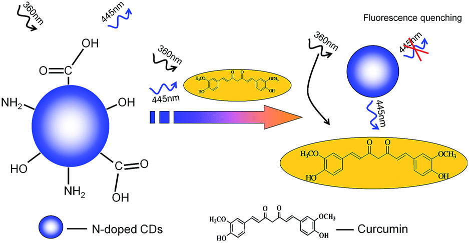 | ||
| Scheme 1 Schematic illustration for the sensing of curcumin based on IFE. | ||
2 Experimental
2.1 Chemicals
Curcumin was purchased from Shanghai Chemical Reagent (Shanghai, China). Citric acid and urea were from Sigma-Aldrich Chemical Co. (St. Louis, USA). The solutions of metal cations were prepared from their chlorine salts. A series of Briton–Robinson (BR) buffer solutions (50 mM) (H3PO4 + H3BO3 + HAc + NaOH) were used for the pH adjustment. All the chemicals used were of analytical reagents grade or above without further purification and double-distilled water (≥18.2 M cm−1) from the Millipore Milli-Q system was used throughout.2.2 Apparatus
All the fluorescence measurements were carried out with a F-4500 fluorescence spectrophotometer (Hitachi, Japan). UV-Vis absorption spectra were performed on a TU-1901 Double beam UV-Vis spectrophotometer (Hitachi, Japan). HPLC were recorded on an Agilent 1200 liquid chromatography system fitted with a analytical column C18 (Agilent SB, 2.1 mm × 50 mm × 1.8 μm). An Agilent 1260 series VWD detector was used for detection at a wavelength of 420 nm. Infrared spectroscopy was carried out using a FTIR-8400S Infrared spectrometer in the form of KBr pellets from 4000 to 500 cm−1. Fluorescence lifetime assays were conducted on a FLS-920 Edinburgh Fluorescence Spectrophotometer (Edinburgh Co., Ltd., England). Transmission electron microscopy (TEM) and high resolution TEM (HR-TEM) images were obtained on a FEI TECNAI G2 F20 U-TWIN transmission electron microscope at an accelerating voltage of 200 kV. The hydrodynamic diameter and zeta potential were measured with a Malvern Zetasizer Nano-ZS90 dynamic light scattering system. A FE20 pH meter (Mettler Toledo, Switzerland) was used to adjust the pH value.2.3 Preparation of N-doped CDs
Water-soluble N-doped CDs were synthesized via a simple-effective hydrothermal treatment according to a previously published method with slight modifications.40 In brief, citric acid (0.576 g) and urea (1.0809 g) were dissolved to distilled water (15 mL) to form a transparent solution. The solution was then heated in a stainless steel autoclave (50 mL) at 200 °C for 10 h. After the reaction, the vessel was cooled down naturally to obtain the brown solution, indicating the formation of CDs. Then the crude product was centrifuged at 13![[thin space (1/6-em)]](https://www.rsc.org/images/entities/char_2009.gif) 000 rpm for 15 min to remove the large or agglomerated solid particles. The aqueous solution of CDs was subject to further purify through a dialysis membrane (MWCO of 1000) in 1 L of Milli-Q water for 2 days. Finally, the resulting aqueous solution was collected from the dialysis membrane and freeze-dried to obtain the N-doped CDs product. The synthetic method can be used to prepare different types of N-doped CDs by tuning the precursors molar ratios as depicted in Fig. S1 and S2.†
000 rpm for 15 min to remove the large or agglomerated solid particles. The aqueous solution of CDs was subject to further purify through a dialysis membrane (MWCO of 1000) in 1 L of Milli-Q water for 2 days. Finally, the resulting aqueous solution was collected from the dialysis membrane and freeze-dried to obtain the N-doped CDs product. The synthetic method can be used to prepare different types of N-doped CDs by tuning the precursors molar ratios as depicted in Fig. S1 and S2.†
2.4 Fluorescence measurements
An aliquot of N-doped CDs (90 μL, 1 mg mL−1) was added to Britton–Robinson (BR) buffer (50 mM, pH 7.5, 910 μL). Different volumes of 1 × 10⁻³ M curcumin stock solution were successively pipetted and mixed thoroughly before determination. In BR buffer, the optimal excitation wavelength of as-prepared CDs was at 360 nm with the maximal emission peaks at the 445 nm for quantitative analysis. The excitation slit width was set as 2.5 nm and the emission slit was 5 nm. All the detections were performed in triplicate.3 Results and discussion
3.1 Characterization of N-doped CDs
The size distribution and morphology of N-doped CDs were characterized by TEM. As shown in Fig. 1A, CDs were well dispersed quasi-spherical nano-dots and meanwhile, the HR-TEM image of N-doped CDs (Fig. 1B) showed lattice spacing of 0.23 nm, which agreed with that of in-plane lattice spacing of graphene (100 facet).41 Fig. 1C clearly confirmed that CDs obtained here existed as a narrow distribution with diameters in the range of 4–6 nm and the average is about 5.23 nm (based on statistical analysis of more than 100 dots), in line with the DLS results (Fig. S4†).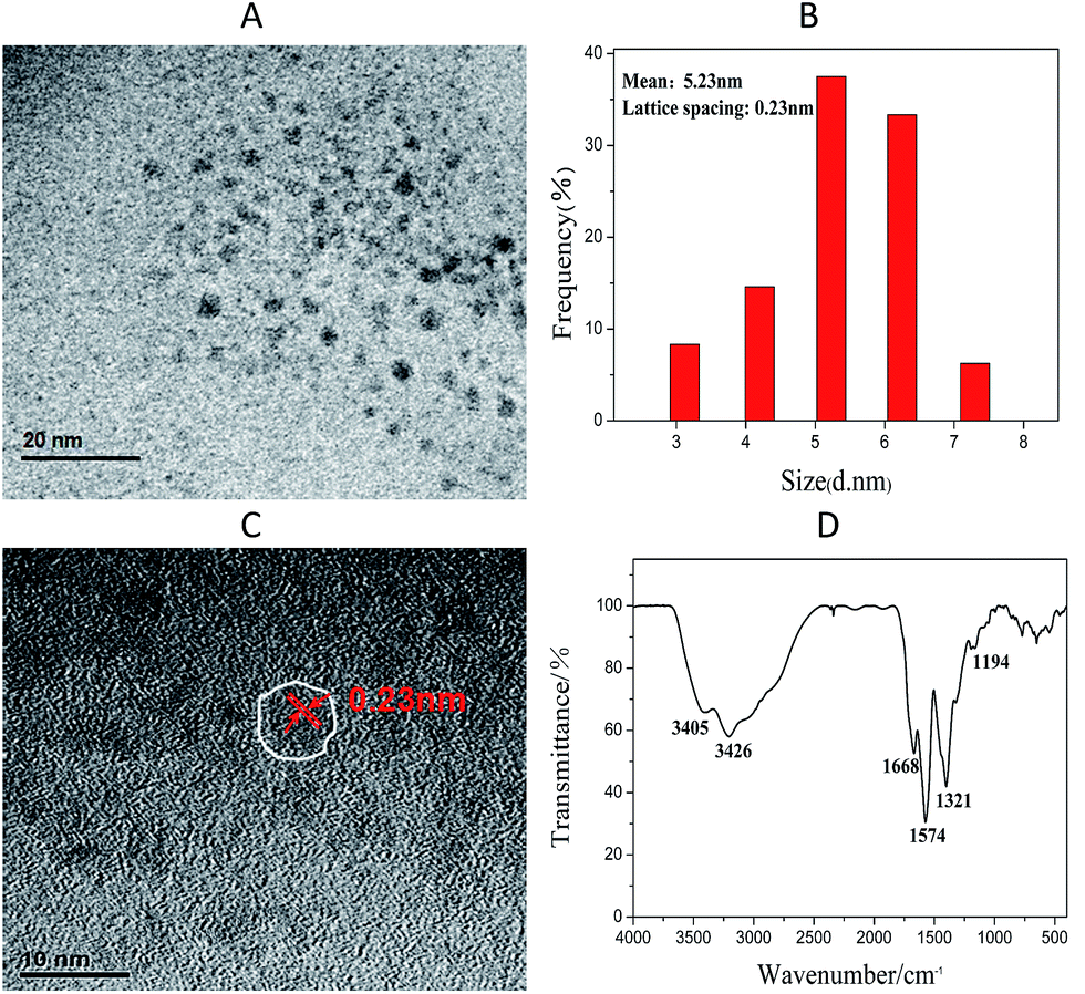 | ||
| Fig. 1 (A) TEM and (B) HRTEM images of the N-doped CDs; (C) particle size distribution of the N-doped CDs; (D) FT-IR spectra of the N-doped CDs. | ||
The surface functional groups of the N-doped CDs were characterized by FT-IR spectroscopy (Fig. 1D). The peak at 1321 and 1194 cm−1 were assigned to the asymmetric and symmetric stretching modes of C–O–C (ether or epoxy).42 Simultaneously, the peak at 1574 cm−1 was associated with the stretching vibration of C–N, indicating the formation of –CONR.43 C![[double bond, length as m-dash]](https://www.rsc.org/images/entities/char_e001.gif) O and CC stretching vibrations were at 1668 cm−1. In addition, the broad band at 3204–3405 cm−1 appeared because of the main absorption band of O–H and N–H stretching vibration.44 Similar to earlier reports,40 the FT-IR results gave the evidence that the surface of CDs was full of hydrophilic groups: hydroxyl and carboxylic groups, which can improve the hydrophilicity and stability of the CDs in water. In good agreement with FTIR, the zeta potential of the CDs in the aqueous solution was −21.2 mV. The surface of the prepared CDs was covered with carboxyl groups and was negatively charged. Because the same charges repel each other, CDs do not aggregate.
O and CC stretching vibrations were at 1668 cm−1. In addition, the broad band at 3204–3405 cm−1 appeared because of the main absorption band of O–H and N–H stretching vibration.44 Similar to earlier reports,40 the FT-IR results gave the evidence that the surface of CDs was full of hydrophilic groups: hydroxyl and carboxylic groups, which can improve the hydrophilicity and stability of the CDs in water. In good agreement with FTIR, the zeta potential of the CDs in the aqueous solution was −21.2 mV. The surface of the prepared CDs was covered with carboxyl groups and was negatively charged. Because the same charges repel each other, CDs do not aggregate.
The UV-Vis absorption and photoluminescence (PL) spectra of the as-prepared CDs were also investigated as shown in Fig. 2A. The absorption spectrum of N-doped CDs showed a weak shoulder band at 245 nm and a narrow peak at around 340 nm with a tail extending into the visible range. The two peaks were attributed to π–π* transition of the CC bond and n–π* transition of the CO bond, respectively. Simultaneously, the maximum excitation and emission wavelengths were located at 360 nm and 445 nm. The image of the N-doped CDs dispersion under UV light (365 nm) exhibited an obvious blue color (right) while appearing as light brown transparent under daylight (left).
 | ||
| Fig. 2 (A) PL spectra and UV-Vis absorption spectra of the N-doped CDs. Inset: photograph of the obtained CDs under illumination of white light (left) and UV (365 nm) light (right); (B) the PL spectra of N-doped CDs at different excitation wavelengths; (C) PL intensity of N-doped CDs against excitation time; (D) the effect of ionic strength on the N-doped CDs PL intensity. | ||
To further explore the optical properties of N-doped CDs, the excitation-wavelength dependent photoluminescence (PL) behavior was shown in Fig. 2B. With the increasing excitation wavelength from 310 to 410 nm, the PL first increases and then decreases with a red shift, indicating that PL of N-doped CDs are excitation-dependent. The quantum yield of N-doped CDs was measured to be about 25.4% by using quinine sulfate in 0.1 M sulfuric acid solution (quantum yield 54%) as the standard. By using various molar ratios of citric acid to urea, we found that the molar ratios play a significant role in determining the final quantum yield (QY) (Table S1†). In addition, the CDs exhibited excellent stability. Their PL intensity did not change even under continuous excitation for 6 h (Fig. 2C). And although the solution was preserved at 4 °C in the dark for more than three months, there was also no fade significantly in color and PL intensities. In Fig. 2D, it was observed that the PL intensity of the N-doped CDs remained constant over a wide concentrations of NaCl solutions (0–150 mM). The results revealed that N-doped CDs possess outstanding stability, tolerance for high ionic strength and photobleaching, which makes them a promising candidate for PL detection.
3.2 IFE between N-doped CDs and curcumin
IFE phenomenon of fluorescence usually occurs between the absorber and fluorophore resulting from the absorption of the excitation and/or emission light by absorbers in the detection system. The effective IFE requires the complementary overlap of the absorber's absorption band with the excitation and/or emission bands of the fluorophore. Therefore, it is important to choose a suitable absorber and fluorophore in the IFE-based fluorescent assays.45 As shown in Fig. 3A, curcumin possess a wide absorption peak at around from 300 to 550 nm. Our as-prepared N-doped CDs were chosen as the fluorophore because both the excitation and emission spectra of CDs showed quite precise overlapping with the absorption band of curcumin. Consequently, as expected from the original design (Scheme 1), curcumin can not only shield the excitation light from CDs but also absorb the emission light of CDs. Naturally, the absorbance enhancement of curcumin could be successfully converted to fluorescence quenching of CDs, which is favorable to enhance the detection sensitivity.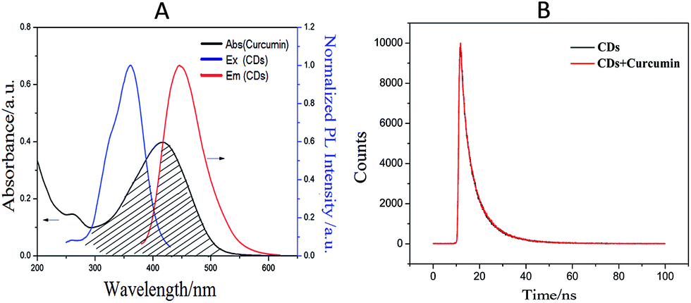 | ||
| Fig. 3 (A) The spectral overlap between the absorption band of the curcumin and the excitation and emission band of the N-doped CDs; (B) the fluorescence decay curve of N-doped CDs in the absence and presence of curcumin upon excitation at 360 nm. | ||
To demonstrate the fluorescence IFE between the N-doped CDs fluorophore and curcumin absorber, the time-resolved fluorescence spectroscopy was executed and the decay spectrum of the N-doped CDs fitted with bi-exponential curve were shown in Fig. 3B. In the absence of curcumin (the black curve), the average lifetime value of N-doped CDs was 7.61 ns (χ2 = 1.082) with lifetimes of τ1 = 3.96 ns (35.34%) and τ2 = 9.61 ns (64.66%). In the presence of 30 μM curcumin (the red curve), the average fluorescence lifetime of the N-doped CDs was 7.62 ns (χ2 = 1.076) with lifetimes of τ1 = 4.01 ns (35.41%) and τ2 = 9.60 ns (64.59%). The almost no lifetime change of the N-doped CDs indicated that there is no significant excited-state interaction between N-doped CDs and curcumin, demonstrating that the fluorescence change of N-doped CDs by curcumin resulted from the simple absorption of the excitation and emission light by the absorber.46 Also, curcumin is negatively charged, and no electrostatic interaction took place between N-doped CDs and curcumin. This can facilitate our demonstration of the present concept that the fluorescence sensing would be clearly based on the IFE rather than other possible approaches.
3.3 Optimization of experimental conditions
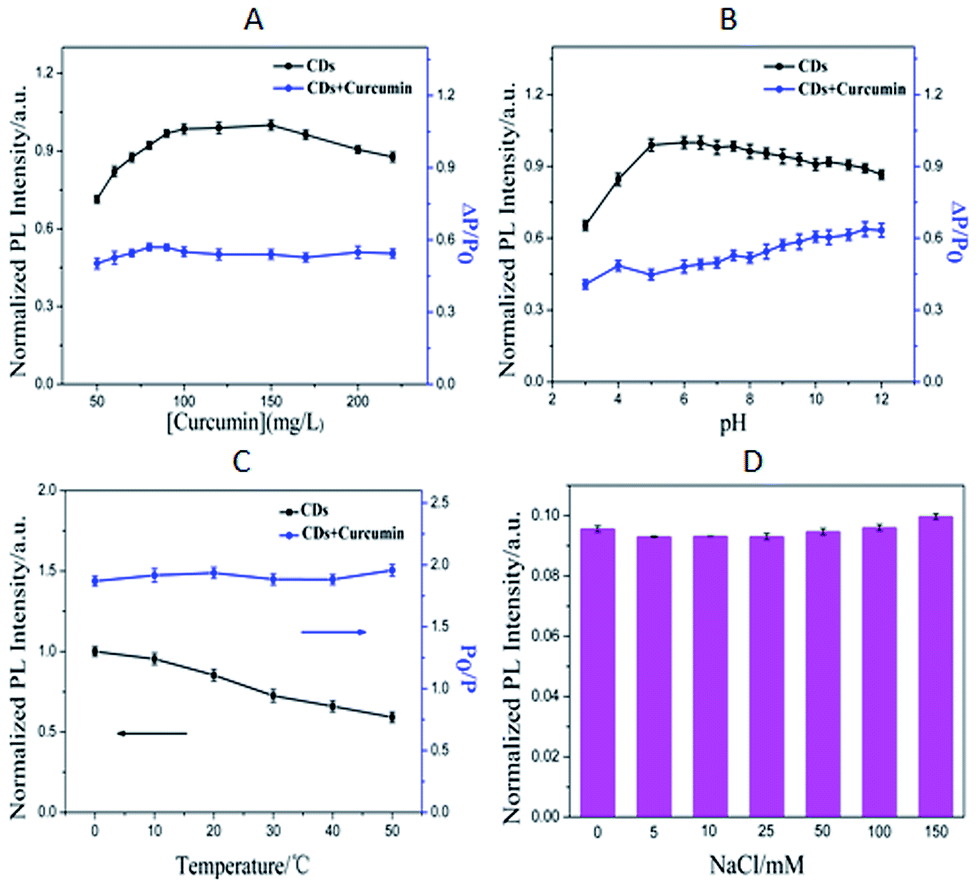 | ||
| Fig. 4 Effect of N-doped CDs concentrations (A) and pH (B) on the PL intensity of CDs (■) and ΔP/P0 in the presence of curcumin (●). ΔP = P0 − P, P0 and P are the PL intensity of N-doped CDs in the absence and presence of curcumin, respectively. (C) Effect of temperature on the PL intensity of N-doped CDs (■) and P0/P of the mixture of CDs with curcumin (●). (D) Effect of ionic strength on the PL intensity of N-doped CDs-curcumin. | ||
3.4 Detection selectivity
Selectivity is an important parameter to evaluate the anti-interference performance of the sensing system. The PL response when the sensing system was challenged with other potential interfering substances, including some common metal ions (Fe3+, Fe2+, Zn2+, Cu2+, Mg2+, Ca2+, Mn2+, K+, Na+, Hg2+, Pb2+, Cr6+, Cr3+, Co2+, Ni2+), anions (Cl−, I−, F−, Br−), small organic molecules (glucose, sucrose, fructose, lactose) and amino acid (L-valine, L-arginine, L-glutamate, L-asparagine, L-serine, L-methionine, L-phenylalanine, L-histidine, L-lysine, L-glycine, L-threonine) which possibly existed in the real samples was monitored. As shown in Fig. 5 (red histograms), 30 μM curcumin produced a more significant fluorescence quenching compared with that caused by other analytes mentioned above at ultrahigh concentrations (concentration is 100-fold of curcumin). Fig. 5 (black histograms) also revealed that the coexistence of curcumin and other ions and compounds did not affect detection by the N-doped CDs probe. These results demonstrated that the N-doped CDs shows excellent selectivity toward curcumin over other common ions and compounds. | ||
| Fig. 5 Selectivity of the sensing method against other ions and compounds. Concentrations of curcumin and other ions and compounds were 30 μM and 3 mM, respectively. Red bars represent the responses of the sensing system challenged with a specific ion or compound. Black bars show the responses of the sensing system in the presence of 30 μM curcumin and 3 mM corresponding ions or compounds. | ||
3.5 Analytical merits of curcumin
The PL intensity quenching of the N-doped CDs at various concentration of curcumin were displayed in Fig. 6A. Fig. 6B showed the Stern–Volmer plot of N-doped CDs with increasing concentration of curcumin (P0/P against concentration of curcumin). The quenching efficiency was fitted by the Stern–Volmer equation, P0/P = 1 + Ksv[Q], where Ksv is the Stern–Volmer quenching constant and [Q] is the curcumin concentration. The Ksv was calculated to be 0.10215 L mol−1 with a correlation coefficient r2 of 0.998. The P0/P curve was linearly related to the concentration of curcumin in the range of 2.0 × 10⁻⁷ to 1.0 × 10⁻⁵ mol L−1, indicating their excellent sensing properties in the detection of trace curcumin. And the relative standard deviation (RSD) of the proposed method was 3.17% (n = 11). The limit of detection (LOD) was calculated by taking the PL intensity equal to 3 times the standard deviation of the intensity at the blank (n = 10) divided by the slope of the calibration graph. The LOD of the proposed method was 8.48 × 10⁻⁸ mol L−1. The LOD was much lower than some other reported values (Table S2†). It was clearly demonstrated the present method with high sensitivity. | ||
| Fig. 6 (A) PL spectra of N-doped CDs in the presence of curcumin at various concentrations at pH 7.5. Amounts of curcumin were 0, 0.2, 0.5, 1.0, 1.4, 2.0, 3.0, 4.0, 5.0, 6.0, 7.0, 8.0, 9.0, 10.0, 11.0, 12.0, 14.0, 15.0, 16.0, 20.0, 24.0, 26.0, 28.0 and 30.0 μM from top to bottom, respectively. Inset: the relationship between the concentration of curcumin and P0/P; (B) linear calibration plot for curcumin detection. | ||
3.6 Analytical application of curcumin
For testing the practicality of this developed approach, recovery experiments were performed in human urine samples obtained from the volunteer students (Table 1). As listed, the recoveries of curcumin were in range of 95.71–103.80%, revealing satisfactory consistencies between the actual and the measured concentrations.| Urine sample | Added (μM) | Found (μM) | Recovery (%) | RSD (n = 6) of proposed method (%) | ||
|---|---|---|---|---|---|---|
| Proposed method | HPLC | Proposed method | HPLC | |||
| 1 | 0.70 | 0.67 | 0.71 | 95.71 | 101.43 | 2.17 |
| 5.00 | 5.19 | 5.13 | 103.80 | 102.60 | 0.79 | |
| 2 | 0.70 | 0.71 | 0.72 | 101.43 | 102.86 | 2.94 |
| 5.00 | 4.89 | 5.19 | 97.72 | 103.80 | 3.25 | |
| 3 | 0.70 | 0.72 | 0.73 | 102.86 | 104.28 | 1.78 |
| 5.00 | 5.11 | 5.24 | 102.20 | 104.80 | 1.44 | |
Also, to check the accuracy of the method, a high performance liquid chromatography (HPLC) was used to measure the curcumin in the human urine samples (Fig. S6†).51 As shown in Table 1, there was a good agreement between the results obtained by our developed method and by HPLC, indicating that the proposed method may broaden ways for practical detection of curcumin in real samples.
4 Conclusions
In summary, we have developed and validated a simple, convenient, high sensitive and selective method for curcumin analysis using N-doped CDs as a fluorescent probe based on the inner filter effect. Curcumin can not only hide the excitation light of CDs but also absorb the emission light from CDs, and naturally, the PL intensity of the N-doped CDs was quenched obviously with increasing curcumin concentration. This method has been applied towards the detection of curcumin contents in human urine samples with satisfactory results. To the best of our knowledge, this is the first demonstration of a CDs-based nanosensor for curcumin via fluorescent inner filter effect. We believed that this method might pave a new way for the further exploration and practical application in analytical detection and chemical sensing area.Acknowledgements
Financial supports from the National Natural Science Foundation of China (No. 21475080 and 21575084), Shanxi Province Hundred Talents Project, Shanxi Scholarship Council of China (No. 2013 - key 1).Notes and references
- T. H. Cooper, G. Clark and J. Guzinski, J. Am. Chem. Soc., 1994, 547, 231–236 CAS.
- P. Basnet and N. Skalko-Basnet, Molecules, 2011, 16, 4567–4598 CrossRef CAS PubMed.
- S. Bengmark, JPEN, J. Parenter. Enteral Nutr., 2006, 30, 45–51 CrossRef CAS.
- R. A. Sharma, S. A. Euden, S. L. Platton, D. N. Cooke, A. Shafayat and H. R. Hewitt, Clin. Cancer Res., 2004, 10, 6847–6854 CrossRef CAS PubMed.
- R. A. Sharma, A. J. Gescher and W. P. Steward, Eur. J. Cancer, 2005, 41, 1955–1968 CrossRef CAS PubMed; H. Zhou, C. S. Beevers and S. Huang, Curr. Drug Targets, 2011, 12, 332–347 CrossRef PubMed.
- H. Zhou, C. S. Beevers and S. Huang, Curr. Drug Targets, 2011, 12, 332–347 CrossRef CAS PubMed.
- G. K. Ziyatdinova, A. M. Nizamova and H. C. Budnikov, J. Anal. Chem., 2012, 67, 591–594 CrossRef CAS.
- W. H. Chan, H. Y. Wu and W. H. Chang, Food Chem. Toxicol., 2006, 44, 1362–1371 CrossRef CAS PubMed.
- F. Wang, W. Huang and Y. W. Wang, J. Lumin., 2008, 128, 110–116 CrossRef CAS.
- F. Wang and W. Huang, J. Pharm. Biomed. Anal., 2007, 43, 393–398 CrossRef CAS PubMed.
- A. Navas Diad and M. C. Ramos Peinado, J. Agric. Food Chem., 1992, 40, 56–59 CrossRef.
- B. Tang, L. Ma, H. Y. Wang and G. Y. Zhang, J. Agric. Food Chem., 2002, 50, 1355–1361 CrossRef CAS PubMed.
- G. K. Ziyatdinova, A. M. Nizamova and H. C. Budnikov, J. Anal. Chem., 2012, 67, 591–594 CrossRef CAS.
- M. Lechtenberg, B. Quandt and A. Nahrstedt, Phytochem. Anal., 2004, 15, 152–158 CrossRef CAS PubMed.
- M. Rahimi, P. Hashemi and F. Nazari, Anal. Chim. Acta, 2014, 826, 35–42 CrossRef CAS PubMed.
- J. Li, Y. Y. Jiang, J. Wen, G. R. Fan, Y. T. Wu and C. Zhang, Biomed. Chromatogr., 2009, 23, 1201–1207 CrossRef CAS PubMed.
- J. H. Lee and M. G. Choung, Food Chem., 2011, 124, 1217–1222 CrossRef CAS.
- D. D. Heatha, M. A. Pruittb, D. E. Brennerc and C. L. Rockd, J. Chromatogr. B: Anal. Technol. Biomed. Life Sci., 2003, 783, 287–295 CrossRef.
- Z. G. Chen, L. Zhu, T. H. Song, J. H. Chen and Z. M. Guo, Spectrochim. Acta, Part A, 2009, 72, 518–522 CrossRef PubMed.
- X. J. Zhao, F. X. Lia, Q. Y. Zhang, Z. B. Li, Y. H. Zhou, J. Yang, C. Dong, J. P. Wang and S. M. Shuang, RSC Adv., 2015, 5, 21504–21510 RSC.
- S. K. Bhunia, A. Saha, A. R. Maity, S. C. Ray and N. R. Jana, Sci. Rep., 2013, 3, 1473–1478 CrossRef PubMed.
- H. Li, Z. Kang, Y. Liu and S. T. Lee, J. Mater. Chem., 2012, 22, 24230–24253 RSC.
- J. Shen, Y. Zhu, X. Yang and C. Li, Chem. Commun., 2012, 48, 3686–3699 RSC.
- Y. F. Wang and A. G. Hu, J. Mater. Chem. C, 2014, 2, 6921–6939 RSC.
- W. P. Wang, Y. C. Lu, H. Huang, J. J. Feng, J. R. Chen and A. J. Wang, Analyst, 2014, 139, 1692–1696 RSC.
- W. P. Wang, Y. C. Lu, H. Huang, A. J. Wang, J. R. Chen and J. J. Feng, Biosens. Bioelectron., 2015, 64, 517–522 CrossRef CAS PubMed.
- X. H. Gao, Y. Z. Lu, R. Z. Zhang, S. J. He, J. Ju, M. M. Liu, L. Li and W. Chen, J. Mater. Chem. C, 2015, 3, 2302–2309 RSC.
- J. Ju and W. Chen, Biosens. Bioelectron., 2014, 58, 219–225 CrossRef CAS PubMed.
- Y. Dong, H. Pang, H. B. Yang, C. Guo, J. Shao, Y. Chi, C. M. Li and T. Yu, Angew. Chem., Int. Ed., 2013, 52, 7800–7804 CrossRef CAS PubMed.
- P. Ayala, R. Arenal, A. Loiseau, A. Rubio and T. Pichler, Rev. Mod. Phys., 2010, 82, 1843–1885 CrossRef CAS.
- M. J. Krysmann, A. Kelarakis, P. Dallas and E. P. Giannelis, J. Am. Chem. Soc., 2012, 134, 747–750 CrossRef CAS PubMed.
- J. Ju, R. Z. Zhang, S. J. He and W. Chen, RSC Adv., 2014, 4, 52583–52589 RSC.
- J. Ju and W. Chen, Anal. Chem., 2015, 87, 1903–1910 CrossRef CAS PubMed.
- R. Z. Zhang and W. Chen, Biosens. Bioelectron., 2014, 55, 83–90 CrossRef CAS PubMed.
- A. Zhao, C. Zhao, M. Li, J. Ren and X. Qu, Anal. Chim. Acta, 2014, 809, 128–133 CrossRef CAS PubMed.
- Q. Wang, X. Liu, L. C. Zhang and Y. Lv, Analyst, 2012, 137, 5392–5397 RSC.
- N. Shao, Y. Zhang, S. M. Cheung, R. H. Yang, W. H. Chan, T. Mo, K. A. Li and F. Liu, Anal. Chem., 2005, 77, 7294–7303 CrossRef CAS PubMed.
- L. Shang and S. J. Dong, Anal. Chem., 2009, 81, 1465–1470 CrossRef CAS PubMed.
- M. Zheng, Z. G. Xie, D. Qu, D. Li, P. Du, X. B. Jing and Z. C. Sun, ACS Appl. Mater. Interfaces, 2013, 5, 13242–13247 CAS.
- B. B. Wang, Y. F. Wang, H. Wu, X. J. Song, X. Guo, D. M. Zhang, X. J. Ma and M. Q. Tan, RSC Adv., 2014, 4, 49960–49963 RSC.
- Y. Q. Dong, H. C. Pang, H. B. Yang, C. X. Guo, J. W. Shao, Y. W. Chi, C. M. Li and T. Yu, Angew. Chem., Int. Ed., 2013, 52, 7800–7804 CrossRef CAS PubMed.
- Y. Liu, N. Xiao, N. Gong, H. Wang, X. Shi, W. Gu and L. Ye, Carbon, 2014, 68, 258–264 CrossRef CAS.
- X. Zhai, P. Zhang, C. Liu, T. Bai, W. Li, L. Dai and W. Liu, Chem. Commun., 2012, 48, 7955–7957 RSC.
- J. Jiang, Y. He, S. Y. Li and H. Cui, Chem. Commun., 2012, 48, 9634–9636 RSC.
- L. Shang and S. J. Dong, Anal. Chem., 2009, 81, 1465–1470 CrossRef CAS PubMed.
- D. W. Zhang, Z. Y. Dong, X. Z. Jiang, M. Y. Feng, W. Li and G. H. Gao, Anal. Methods, 2013, 5, 1669–1675 RSC.
- W. Kong, H. Wu, Z. Ye, R. Li, T. Xu and B. Zhang, J. Lumin., 2014, 148, 238–242 CrossRef CAS.
- Q. Zhao, D. X. Kong and H. Y. Zhang, Nat. Prod. Commun., 2008, 3, 229–232 CAS.
- Y. J. Wang, M. H. Pan, A. L. Cheng, L. I. Lin, Y. S. Ho, C. Y. Hsieh and J. K. Lin, J. Pharm. Biomed. Anal., 1997, 15, 1867–1876 CrossRef CAS PubMed.
- P. T. Burks, A. D. Ostrowski, A. A. Mikhailovsky, E. M. Chan, P. S. Wagenknecht and P. C. Ford, J. Am. Chem. Soc., 2012, 134, 13266–13275 CrossRef CAS PubMed.
- G. K. Jayaprakasha, L. J. Rao and K. K. Sakariah, J. Agric. Food Chem., 2002, 50, 3668–3672 CrossRef CAS PubMed.
Footnote |
| † Electronic supplementary information (ESI) available: Chemical structure of curcumin, hydrodynamic diameter distribution of CDs, the spectral overlap different pH values. Comparison of different methods for curcumin detection. HPLC method for determination of curcumin. See DOI: 10.1039/c5ra18176c |
| This journal is © The Royal Society of Chemistry 2015 |