Influence of magnetic nanoparticle arrangement in ferrogels for tunable biomolecule diffusion

Ting-Yu Liu*, Tzu-Yi Chan, Kuan-Syun Wang and Hui-Ming Tsou
Department of Materials Engineering, Ming Chi University of Technology, New Taipei City 24301, Taiwan. E-mail: tyliu0322@gmail.com

Received 26th August 2015 , Accepted 7th October 2015

First published on 7th October 2015


Abstract

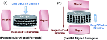
Magnetic sensitive hydrogels (ferrogels) with tunable nanochannels were prepared with poly (vinyl alcohol) (PVA) and iron oxide magnetic nanoparticles under a uniform magnetic field in a freezing–thawing process, evaluated by differential scanning calorimetry (DSC), and the influence of magnetic nanoparticle arrangement on the diffusion behavior of biomolecules was investigated. Nanochannels self-assembled by the arranged magnetic nanoparticles could be tuned by manipulating the direction of the magnetic field, which results in the formation of “needle-like” structures from the magnetic nanoparticles aligned parallel or perpendicular to the permeation direction (anisotropic ferrogels). The effect of biomolecule diffusion between the anisotropic and isotropic ferrogels was observed in a diffusion diaphragm cell (using random nanoparticle dispersions without a magnetic field, as a control). It was established that the parallel-aligned ferrogels exhibit a higher drug diffusion rate compared to the isotropic ferrogels, whereas the perpendicular-aligned ferrogels display the lowest biomolecule diffusion rate. The novel ferrogels were expected to be suitable for application in bio-membranes, and the nanochannels for biomolecule (MW: ca. 0.1–10 kDa) diffusion could be constructed precisely by the magnetic nanoparticle arrangement.


1. Introduction

Intelligent polymer based nanocomposites have the unique ability to change their swelling behavior, permeability and elasticity in a reversible manner in response to external stimuli, such as pH, temperature, electric fields and magnetic fields.1–19 Magnetically sensitive polymers are superior to traditional stimuli responsive polymers because magnetic stimulation is an action-at-distance force (non-contact force) which is easier to adapt to biomedical devices.19–24 Polymeric nanocomposites called ferrogels, consisting of polymeric hydrogels filled with magnetic nanoparticles have been the subject of many studies in the past two decades. These ferrogels have been successfully developed with regard to their applications in several biomedical and industrial fields such as muscle-like sift linear actuators, controlled drug release, and dialysis membranes.

Membranes are practical tools for molecular separation because they provide an energy efficient green technology. Nanoscale manipulation of the membrane nanochannels is an innovative idea to control the membrane permeability. Csetneki et al.4 reported a thermal/magnetic sensitive nanocomposite polymer membrane (magnetic polystyrene latex-poly(N-isopropylacrylamide)) with on/off switching control through thermal manipulation, where nanochannels could be opened when the temperature is above the collapse transition temperature, but closed when the temperature is below the collapse transition temperature. In our previous studies,19–24 we also fabricated smart magnetic hydrogels for the development of a new magnetically induced drug delivery system. By applying direct current (DC) magnetic fields, we were able to switch the drug release profile of the hydrogels between “on” and “off” modes with “random-dispersion” and “aggregation” of the magnetic nanoparticles, respectively.

Recently, Varga et al.25,26 reported a fascinating result, tuning the elastic modulus with controlled anisotropy of the ferrogel. It was established that the uniaxial field structured composites exhibited a larger excess modulus compared to the random magnetic particle dispersions. The most significant effect found was that the mechanical stress of the ferrogel would be modulated under controlled anisotropy (the applied magnetic field is parallel or perpendicular to the particle alignments). Against this background, we will try to design anisotropic ferrogels, in which the magnetic nanoparticles are parallel or perpendicular aligned to diffusion direction. Also, the drug permeability of anisotropic and isotropic (using random magnetic nanoparticles without magnetic field control as a control) ferrogels is studied in this work.

The main purpose of the magnetic nanoparticle arrangement is to control the direction of the nanochannels (related to the diffusion direction) in the ferrogel using an external magnetic field (parallel or perpendicular to the particle alignments) and thus control the permeability of the ferrogels to biomolecules. The characterizations of PVA physically cross-linked by freezing and thawing cycles could be examined carefully by DSC analysis. The pearl-chain structures of the ferrogels developed by the magnetic field were observed by optical microscopy. The diffusion properties of the ferrogels were evaluated using some biomolecules of varied molecular weights, such as creatinine, vitamin B12, cytochrome c and bovine serum albumin.

2. Methods

2.1 Fabrication of ferrogels with various sizes of magnetic nanoparticles

Intermolecular interactions like hydrogen bond-bridges or polymer microcrystals are responsible for the formation of a three-dimensional network structure. A so-called freezing–thawing process was used to prepare the ferrogel.27 First, 0.05 g mL−1 poly(vinyl alcohol) (PVA, Fluka, MW: 72[thin space (1/6-em)]000, degree of hydrolysation: 97.5–99.5 mol%) was dissolved in 10 mL dimethylsulfoxide (DMSO) at 80 °C and stirred for 6 h, and then mixed with 0.17 g mL−1 of magnetic particles (ca. 150–250 nm, purchased from Aldrich) at 60 °C under ultrasonication for 6 h to ensure that the magnetic particles were well dispersed. The resulting solution was then poured into a plastic dish and kept frozen at −20 °C for 16 h. Subsequently, the gels were thawed at 25 °C for 5 h. This cyclic process including freezing and thawing was repeated 5 times. Finally, prior to the release test, the ferrogels were washed five times and then immersed in water for 24 h to completely remove DMSO.

2.2 Fabrication of anisotropic ferrogels

The main purpose of the present work was to establish the effect of the direction of magnetic fields on drug diffusion behavior. As shown in Fig. 1, PVA hydrogels loaded with randomly, perpendicularly, and parallelly distributed iron oxide nanoparticles regarding to the drug diffusion direction, were modulated by a uniform magnetic field. Fig. 1a and c show that the iron oxide nanoparticles of the ferrogels were arranged by left-to-right electronic magnets, referred to as “perpendicular” to the drug diffusion direction. Fig. 1b and d illustrate that the iron oxide nanoparticles of the ferrogels were arranged by top-to-down electronic magnets, referred to as “parallel” to the drug diffusion direction.
image file: c5ra17306j-f1.tif
Fig. 1 Schematic drawing of the experimental set-up for ferrogel preparation under a uniform magnetic field: (a) and (c) the iron oxide nanoparticles of the ferrogels were arranged by left-to-right electronic magnets (“perpendicular” to the drug diffusion direction); (b) and (d) the iron oxide nanoparticles of the ferrogels were arranged by top-to-down electronic magnets (“parallel” to the drug diffusion direction).

2.3 Drug diffusion test

The diffusion coefficients of the solutes were measured by a diffusion diaphragm cell (side-by-side cell).21 The solution in the donor side is 80 mL of isotonic phosphate buffer (PBS) (pH 7.4) containing 200 ppm of a model drug (creatinine, vitamin B12, cytochrome c, and bovine serum albumin). The receptor compartment, separated by the ferrogel, was filled with 80 mL of PBS solution. The concentration of each compound in the receptor compartment was determined using a UV spectrophotometer. The permeation coefficient (P, cm2 min−1) was calculated according to the following equation for the diaphragm cell:
 
image file: c5ra17306j-t1.tif(1)
where Cd0 is the initial concentration of the permeant in the donor compartment; Cd and Cr are indicative of the concentrations in the donor side and receptor side, respectively; D is the diffusion coefficient (cm2 min−1);28–31 H is the partition coefficient; A is the effective area of the ferrogel; δ is the thickness of the ferrogel; V is the volume of solution in the donor or receptor compartment (both are 80 mL). By plotting ln[Cd0/(CdCr)] versus time (t), the permeability coefficient (P) can be calculated from the slope of the line by using eqn (1). Each data point was obtained by averaging at least three measurements.

Moreover, the drug-free ferrogel of dry weight (Wdry) was immersed in the release medium until an equilibrium state was reached and then the wet weight (Wwet) was recorded. Subsequently, the ferrogel was immersed in 10 mL of vitamin B12-containing medium. The partition coefficient (H) was determined from the initial (C0) and equilibrium (Ce) concentrations of vitamin B12-containing media using eqn (2).30,31

 
image file: c5ra17306j-t2.tif(2)

2.4 Pearl-chain structure of anisotropic ferrogels test

0.01 g mL−1 of magnetic particles were dissolved in a 0.1 g mL−1 PVA solution (dissolved in DMSO at 80 °C beforehand). Further, the behavior of the magnetic particles was observed directly by an optical microscope (Olympus, Japan) in the absence and presence of a magnetic field.

3. Results and discussion

3.1 Physical cross-linking (freezing and thawing process) of the ferrogel

The PVA ferrogel was fabricated by a physical cross-linking method (freezing and thawing process) and displayed elastic properties due to the hydroxyl groups of PVA molecules participating in hydrogen bonding. A continuous thermal analysis observed by differential scanning calorimetry (DSC) was used to evaluate the PVA crystal growth during freezing–thawing cycles. As shown in Fig. 2, the number of cross-linking points and the crystal region of the PVA hydrogels expanded with increasing freezing–thawing cycles. DSC measurement is based on the fabrication process of the ferrogels (freezing and thawing process). The control parameter in the DSC is (1) cooling from 25 °C to −20 °C (cooling rate: 1 °C min−1); (2) kept frozen at −20 °C for 16 h; (3) heating from −20 °C to 25 °C (heating rate: 1 °C min−1); (4) kept thawed at 25 °C for 5 h. This cyclic process including freezing and thawing was repeated 6 times.
image file: c5ra17306j-f2.tif
Fig. 2 DSC analysis of PVA physical cross-linking by freezing and thawing cycles: (a) cooling curve; (b) change of integrated area of heat flow and crystal temperature (Tc) with different cycles.

The result (Fig. 2a) shows that the crystal temperature (Tc) of the PVA ferrogel increases from −5.3 to 1.1 °C when increasing the number of freezing and thawing cycles (cycle 1–6, c1–c6), indicating that plenty of cross-linked points are introduced to the solid network of the PVA ferrogel. Furthermore, the area of crystallization (integrated peak area) also enlarges with increasing cycle number. It also demonstrated that the higher crystallization of the PVA ferrogel and the stronger network would be found in the higher cycles of the freezing and thawing process. However, it seems to reach saturation during the 5th and 6th cycles, with only a slight increase in Tc and crystallized area (Fig. 2b), which means the process of PVA crystallization and the hydrogen bonding have been stabilized. Therefore, five cycles of the freezing and thawing process were used in this study.

3.2 Pearl-chain structure of anisotropic ferrogels

While the imposed field induces magnetic dipoles, mutual particle interactions occur if the particles are so closely packed that the local field can influence their neighbors. The particles attract with each other when aligned in an end-to-end configuration and thus a “pearl-chain structure” was developed via the attractive forces. From the optical microscopy images (Fig. 3), it was demonstrated that 0.01 g mL−1 magnetic particles were randomly distributed in the 0.1 g mL−1 PVA solution (dissolved in DMSO at 80 °C beforehand) in the absence of a magnetic field. However, the magnetic particles attract with each other to line up in an ordered form in the presence of a magnetic field. By controlling the magnetic nanoparticle arrangement to be perpendicular or parallel to the drug diffusion direction, pearl-chain structure ferrogels were prepared for a further drug diffusion test. The isotropic ferrogel (random direction of magnetic nanoparticles) was used as a reference.
image file: c5ra17306j-f3.tif
Fig. 3 The pearl-chain structure developed in the presence of the magnetic field as seen by optical microscopy (scale bar: 5 μm).

3.3 Biomolecule permeation behavior in the anisotropic ferrogel

The biomolecule diffusion behavior is described in Fig. 4. The lower initial diffusion state (the slope in Fig. 4) of the biomolecules (vitamin B12) is because the biomolecules would pass through the bumpy nanochannels in the ferrogel, which makes it difficult for the biomolecules to get across the membrane. The diffusion rate of the biomolecules increased at ca. 40 min, when the biomolecules passed though the membrane. On the other hand, the result shows that the drug diffusion rate would change with the arrangement of iron oxide nanoparticles, implying the channel parallel to the drug diffusion direction exhibits the highest rate of biomolecule diffusion, whereas the perpendicular direction exhibits the lowest. It suggests that the biomolecules’ mobility would be restricted in the perpendicular-aligned ferrogel and thus it would be difficult for them to pass through, but it is unrestricted in the parallel-aligned ferrogel. Therefore, it was anticipated that control of the arrangement of magnetic nanoparticles in the ferrogel could modulate the biomolecule diffusion rate. It would be useful for an application in dialysis membranes containing nanochannels which can be controlled by a magnetic field.
image file: c5ra17306j-f4.tif
Fig. 4 Biomolecule (vitamin B12) permeation behavior in the various ferrogels; the arrangement of iron oxide nanoparticles in the ferrogels are random, perpendicular and parallel to the drug diffusion direction, respectively. There are two slopes (permeation coefficient, P) in the permeation experiment. The first one is the slower initial state of biomolecule permeation and the second one is the higher general state of biomolecule permeation. P was determined from the second linear slope.

The influence of the respective constituent components of the ferrogels on the permeability coefficient (P), the diffusion coefficient (D), and the partition coefficient (H) of the model drug (vitamin B12) was systematically investigated in this work. When the nanochannels of the ferrogel are parallel to the direction of drug permeation, it caused a considerable increase (2.2 times) in the P value (480 × 10−6 cm2 min−1) compared to the value when they are perpendicular to the direction of drug permeation (223 × 10−6 cm2 min−1), and 1.6 times an increase compared to the random magnetic particles of the ferrogels (306 × 10−6 cm2 min−1), as shown in Table 1 and Fig. 4. It was believed that a relationship exists between the P and H values of the drug inside the membrane (see eqn (1), where P = DH). The H value in the parallel ferrogel exhibits the highest drug permeation, whereas the perpendicular ferrogel exhibits the lowest, which is in the order: parallel (0.151) > random (0.142) > perpendicular (0.135) ferrogel, as related to the formation of nanochannel morphology and the direction of drug permeation, as illustrated in Table 1.

Table 1 Permeability coefficient (P), diffusion coefficient (D) and partition coefficient (H) of vitamin B12 in the various ferrogels; the arrangement of iron oxide nanoparticles in the ferrogels are random, perpendicular and parallel to the drug diffusion direction, respectively
  Random Perpendicular Parallel
P (cm2 min−1) × 106 306 223 480
H 0.142 0.135 0.151
D (cm2 min−1) × 106 2155 1726 3179


The value of D would be used to determine the interaction between Fe3O4 nanochannels and drug diffusion. The diffusion coefficient (D) will play a rather important role to evaluate the behavior of the drug inside the ferrogel. It could be found that the D curves show a similar trend to those for P. The D value decreased rapidly when the ferrogel is perpendicular to the direction of drug permeation (1726 × 10−6 cm2 min−1), compared to those for the parallel (3179 × 10−6 cm2 min−1) and random (2155 × 10−6 cm2 min−1) ferrogels. It is because the biomolecules inside the ferrogel were obstructed more strongly in the perpendicular ferrogels compared to in the parallel and random ones.

Creatinine (MW 113), vitamin B12 (MW 1355), cytochrome c (MW 12[thin space (1/6-em)]327) and bovine serum albumin (BSA, MW 65[thin space (1/6-em)]000) were used to verify the pore size, porosity and pore geometry of the ferrogels (Fig. 5). The results show that creatinine and vitamin B12 could rapidly pass through the ferrogels and displayed a 1.9–2.2 times difference in permeability coefficient (P) between the perpendicular and parallel ferrogels. However, the biomolecules with a larger molecular weight such as cytochrome c and BSA display lower diffusion coefficients, which means the larger biomolecules would be blocked inside the pores of the ferrogels. Especially in BSA, the permeability coefficient is lower than 10 × 10−6 cm2 min−1, indicating that the huge molecular weight of the protein could not pass through the pores of the ferrogels. Biomolecules with molecular weights in the range of 100–100[thin space (1/6-em)]000 Da are suitable for use in these novel nanohybrid ferrogels.


image file: c5ra17306j-f5.tif
Fig. 5 Permeability coefficient (P) of the varied molecular weight (MW) of biomolecules through the PVA ferrogel.

4. Conclusions

We successfully developed a novel method to control nanochannel formation in magnetic nanohybrid membranes by applying a magnetic field. The direction of the nanochannels could be manipulated by magnetic nanoparticle self-assembly (pearl-chain structure) under different directions of external magnetic fields. The results of the drug release behavior study show the parallel-aligned ferrogels exhibit the highest rate of drug diffusion, whereas the perpendicular-aligned ferrogel shows the lowest, which is in the order: parallel (anisotropic) > random (isotropic) > perpendicular (anisotropic)-aligned ferrogel. It correlates with the morphology of magnetic nanochannels and the direction of drug diffusion. It suggests biomolecules of molecular weight lower than 100k Da could be used in this ferrogel platform. This effective and rapid method to arrange magnetic nanoparticles to form the nanochannels in the membrane would be expected to be applicable in bio-membranes and drug carriers, such as in controlled drug release.

Acknowledgements

This work was financially supported by Ministry of Science and Technology of Taiwan (MOST 104-2221-E-131-010).

References

  1. Y. Deng, W. Yang, C. Wang and S. Fu, Adv. Mater., 2003, 15, 1729–1732 CrossRef CAS PubMed.
  2. P. M. Xulu, G. Filipcsei and M. Zrínyi, Macromolecules, 2000, 33, 1716–1719 CrossRef CAS.
  3. J. Y. Kim, S. B. Lee, S. J. Kim and Y. M. Lee, Polymer, 2002, 43, 7549–7558 CrossRef CAS.
  4. I. Csetneki, G. Filipcsei and M. Zrínyi, Smart nanocomposite polymer membranes with on/off switching control, Macromolecules, 2006, 39, 1939–1942 CrossRef CAS.
  5. R. Fernandes, L. Q. Wu, T. Chen, H. Yi, G. W. Rubloff, R. Ghodssi, W. E. Bentley and G. F. Payne, Langmuir, 2003, 19, 4058–4062 CrossRef CAS.
  6. S. Y. Kim and Y. M. Lee, J. Appl. Polym. Sci., 1999, 74, 1752–1761 CrossRef CAS.
  7. K. Haraguchi, T. Takehisa and S. Fan, Macromolecules, 2002, 35, 10162–10171 CrossRef CAS.
  8. R. Mohr, K. Kratz, T. Weigel, M. Lucka-Gabor, M. Moneke and A. Lendlein, Proc. Natl. Acad. Sci. U. S. A., 2006, 103, 3540–3545 CrossRef CAS PubMed.
  9. E. C. Muniz and G. Geuskens, Macromolecules, 2001, 34, 4480–4484 CrossRef CAS.
  10. T. G. Park and H. K. Choi, Macromol. Rapid Commun., 1998, 19, 167–172 CrossRef CAS.
  11. H. Guo, Q. Lai, W. Wang, Y. Wu, C. Zhang, Y. Liu and Z. Yuan, Int. J. Pharm., 2013, 451, 1–11 CrossRef CAS PubMed.
  12. T. Y. Liu and Y. L Lin, Acta Biomater., 2010, 6, 1423–1429 CrossRef CAS PubMed.
  13. S. M. Lee, K. H. Liu, Y. Y. Liu, Y. P. Chang and C. C. Lin, Materials, 2013, 6, 1391–1402 CrossRef CAS PubMed.
  14. K. H. Liu, T. Y. Liu, S. Y. Chen and D. M. Liu, Acta Biomater., 2007, 3, 919–926 CrossRef CAS PubMed.
  15. K. H. Liu, T. Y. Liu, S. Y. Chen and D. M. Liu, Acta Biomater., 2008, 4, 1038–1045 CrossRef CAS PubMed.
  16. J. C. Ribot, C. Guerrero-Sanchez, T. L. Greaves, D. F. Kennedy, R. Hoogenboom and U. S. Schubert, Soft Matter, 2012, 8, 1025–1032 RSC.
  17. T. Yamada, R. Kumai, Y. Takahashi and T. Hasegawa, J. Mater. Chem., 2010, 20, 5810–5812 RSC.
  18. J. C. Ribot, C. Guerrero-Sanchez, R. Hoogenboom and U. S. Schubert, Chem. Commun., 2010, 46, 6971–6973 RSC.
  19. C. Guerrero-Sanchez, C. Fabrie and U. S. Schubert, Proc. SPIE, 2009, 7289, 72891U CrossRef PubMed.
  20. T. Y. Liu, S. H. Hu, K. H. Liu, D. M. Liu and S. Y. Chen, J. Controlled Release, 2008, 126, 228–236 CrossRef CAS PubMed.
  21. T. Y. Liu, S. H. Hu, T. Y. Liu, D. M. Liu and S. Y. Chen, Langmuir, 2006, 22, 5974–5987 CrossRef CAS PubMed.
  22. T. Y. Liu, S. H. Hu, K. H. Liu, D. M. Liu and S. Y. Chen, J. Magn. Magn. Mater., 2006, 304, e397–e399 CrossRef CAS PubMed.
  23. S. H. Hu, T. Y. Liu, D. M. Liu and S. Y. Chen, J. Controlled Release, 2007, 121, 181–189 CrossRef CAS PubMed.
  24. T. Y. Liu, S. H. Hu, D. M. Liu, S. Y. Chen and I. W. Chen, Nano Today, 2009, 4, 52–65 CrossRef CAS PubMed.
  25. Z. Varga, G. Filipcsei and M. Zrínyi, Polymer, 2006, 47, 227–233 CrossRef CAS PubMed.
  26. Z. Varga, G. Filipcsei and M. Zrínyi, Polymer, 2005, 46, 7779–7787 CrossRef CAS PubMed.
  27. T. Hatakeyema, J. Uno, C. Yamada, A. Kishi and H. Hatakeyama, Thermochim. Acta, 2005, 431, 144–148 CrossRef CAS PubMed.
  28. D. K. Singha and A. R. Raya, J. Membr. Sci., 1999, 155, 107–112 CrossRef.
  29. M. C. Yang and T. Y. Liu, J. Membr. Sci., 2003, 226, 119–130 CrossRef CAS PubMed.
  30. M. Miyajima, A. Koshika, J. Okada and M. Ikeda, J. Controlled Release, 1999, 60, 199–209 CrossRef CAS.
  31. T. Y. Liu, S. Y. Chen, J. H. Li and D. M. Liu, J. Controlled Release, 2006, 112, 88–95 CrossRef CAS PubMed.

This journal is © The Royal Society of Chemistry 2015
Click here to see how this site uses Cookies. View our privacy policy here.