DOI:
10.1039/C5RA17121K
(Paper)
RSC Adv., 2015,
5, 86990-86996
Nano-pesticide formulation based on fluorescent organic photoresponsive nanoparticles: for controlled release of 2,4-D and real time monitoring of morphological changes induced by 2,4-D in plant systems†
Received
24th August 2015
, Accepted 7th October 2015
First published on 7th October 2015
Abstract
In recent times, nano-pesticide formulations have gained great popularity since they enable effective usage of smaller quantities of the pesticides without creating much damage to the environment. The benefits of a nano-pesticide formulation can be further expanded by adding to its arsenal both tracking ability and precise control over the pesticide release. Recently, fluorescent photoresponsive nanocarriers have gained considerable momentum in the field of drug delivery since they can act as both a “fluorophore” for cell luminescence imaging and a “phototrigger” for regulated drug release by external light stimuli. Hence, we report for the first time a nano-pesticide formulation based on fluorescent photoresponsive organic nanoparticles of perylene-3-ylmethanol for regulated release of pesticide 2,4-dichlorophenoxyacetic acid (2,4-D). Further, the fluorescent nature of the photoresponsive organic nanoparticles was used to study the morphological changes induced by 2,4-D inside the plant system using confocal imaging studies. Additionally, the fluorescent colour change by photoresponsive organic nanoparticles before and after photorelease was exploited for real time monitoring of 2,4-D release inside the plants. Bioassay experiments revealed that nano-pesticide, Pe-2,4-D efficiently delivered 2,4-D inside the plant tissues improving its herbicidal activity. Such photoresponsive multifunctional nanocarriers with good fluorescence, cellular uptake property and efficient photoregulated release ability will be of great benefit in the construction of nano-pesticide formulations.
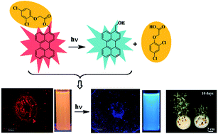
Nano-pesticide formulations have recently gained considerable importance in agriculture since they provide unique advantages like (i) increasing the apparent solubility of poorly soluble pesticides (ii) releasing the pesticides in a slow/targeted manner (iii) protecting the pesticides against premature degradation.1 The major goal in developing nano-pesticide formulations is to design smart nanocarriers that will combine various functionalities, like fluorescence, targeting ability and precise control over the pesticide release. To date, several nanocarriers have been fabricated for the delivery of pesticides which include polymeric nanospheres2–4 and nanocapsules,5 solid lipid nanoparticles,6,7 porous hollow silica nanoparticles,8–11 layered double hydroxides and clays,12–14 and nano-sized metals and metal oxides.15–19 Although nano-pesticide formulations have exhibited low toxicity, good biocompatibility and higher bioavailability, their applications are still restricted. In order to expand the possible benefits of applying nano-pesticide formulations to agriculture and plant science research, we need to develop nano-pesticide formulations with unique properties like (i) being able to visualize and track the transport and deposition of nano-pesticides inside the plant system, (ii) helping to image the morphological changes induced by the nano-pesticides to the plant, and (iii) having precise control over the pesticide release including location, timing and dosage. Hence, it is essential to look for suitable fluorescent nanoparticles that can endow to the nano-pesticide formulations with above said properties.
Recently, fluorescent photoresponsive nanocarriers are being used in the area of drug delivery systems (DDSs) because of their key abilities to perform both as “fluorophore” for cell luminescence imaging and “phototrigger” for regulated drug release by external light stimuli.20,21 Generally, photoresponsive nanocarriers for DDS are constructed using two main ingredients: biocompatible nanocarrier and a small organic molecule “phototrigger”. Specific types of recently developed photoresponsive nanocarriers include gold nanoparticles attached with phototrigger o-nitrobenzyl moiety,22 mesoporous silica grafted with two-photon coumarin based phototrigger,23 photoresponsive block copolymer micelles constructed using phototrigger [7-(diethylamino)coumarin-4-yl]methyl.24
Our group recently demonstrated perylen-3-ylmethanol, a phototrigger can be directly made as single component photoresponsive nanocarriers for controlled drug delivery excluding the need of external nanocarriers.25 In the above system, perylene-3-ylmethanol nanoparticles performed four important roles: (i) “nanocarriers” for drug delivery; (ii) “phototriggers” for the drug release; (iii) fluorescent chromophores for cell imaging; and (iv) detectors for real time-monitoring of drug release. Inspired by the multifunctional performance of perylene-3-ylmethanol nanoparticles, we aimed to develop nano-pesticide formulation of 2,4-D using perylene-3-ylmethanol as nanocarrier so that tracking of morphological changes induced by the pesticide 2,4-D and control over the 2,4-D release can be simultaneously accomplished (Scheme 1).
 |
| Scheme 1 Schematic representation of the entry and photoinduced release of the pesticide (2,4-D) in plant system by perylene-3-ylmethanol nanoparticles. | |
Materials and methods
Materials
Perylene, 2,4-dichlorophenoxy acetic acid, potassium tri bromide and others reagents were purchased from Sigma Aldrich and used without further purification. 1H NMR (400 MHz) spectra were recorded on a BRUKER-AC 400 MHz spectrometer. Chemical shifts are reported in ppm from tetramethylsilane with the solvent resonance as the internal standard (deuterodimethyl sulfoxide: 2.54 ppm). Data are reported as follows: chemical shifts, multiplicity (s = singlet, d = doublet, t = triplet, m = multiplet), coupling constant (Hz). 13C NMR (100 MHz) spectra were recorded on a BRUKER-AC 400 MHz spectrometer with complete proton decoupling. Chemical shifts are reported in ppm from tetramethylsilane with the solvent resonance as the internal standard (deuterochloroform: 77.0 ppm). Chromatographic purification was done with 60–120 mesh silica gel (Merck). For reaction monitoring, precoated silica gel 60 F254 TLC sheets (Merck) were used. UV/vis absorption spectra were recorded on a Shimadzu UV-2450 UV/vis spectrophotometer and fluorescence emission spectra were recorded on a Hitachi F-7000 fluorescence spectrophotometer. FT-IR spectra were recorded on a Perkin Elmer RXI spectrometer. High-resolution mass spectra were recorded using QTOF Micro YA 263 mass spectrometer. RP-HPLC was performed using Waters 2489 liquid chromatography on a C18 column (4.6 mm × 250 mm) with a UV-vis detector. Transmission Electron Microscopy (TEM) was measured on a EFI Tecnai G220S-Twin at 200 kV. The sample was prepared by dispersing compounds in water and dropping on the surface of a copper grid.
Synthesis of perylene-2,4-D ester conjugate
In 5 ml dry N,N-dimethylformamide (DMF) 3-(bromomethyl)perylene (0.100 g, 0.29 mmol) was dissolved, potassium carbonate (0.048 g, 0.34 mmol), potassium iodide (0.022 g, 0.12 mmol) and 2,4-dichlorophenoxy acetic acid (0.088 g, 0.29 mmol) were added to it. The reaction mixture was stirred at room temperature for a period of 8 h and the reaction was monitored by TLC. After completion of the reaction solvent was removed and crude reaction mixture was purified by column chromatography using 15% EtOAc in pet ether. Perylene-3-ylmethyl 2-(2,4-dichlorophenoxy)acetate: yellow solid, mp: 126–128 °C; UV/vis (EtOH 50
:
water 50): λmax (ε M−1 cm−1) 288 (0.6 × 10⁴), 315 (0.8 × 10⁴); FTIR (KBr) νmax (cm−1): 2959, 2923, 2851, 1739, 1479, 1386, 1261, 1189, 1083, 1019, 810, 766; 1H NMR (CDCl3, 400 MHz) δ: 4.73 (s, 2H); 5.63 (s, 2H); 6.65 (d, 1H, J = 2.4, 8.8); 7.33 (d, 1H, J = 2.4); 7.516 (dd, 4H, J = 8, 16); 7.71 (m, 3H); 8.22 (m, 4H). 13C NMR (CDCl3, 100 MHz) δ: 168.1, 152.15, 134.5, 132.8, 132.6, 131.8, 130.9, 130.6, 130.2, 129.7, 128.9, 128.8, 128.3, 128.3, 128.0, 127.8, 127.3, 127.3, 127.1, 127.0, 126.6, 126.5, 122.9, 120.6, 120.3, 119.4, 114.4, 66.3, 65.5. HRMS (ES+) m/z calcd for C29H18Cl2O3 [M + H]+: 350.0984: found: 350.0995.
Preparation of Pe-2,4-D nano-pesticides
Next, we prepared Pe-2,4-D nano-pesticides following the reprecipitation technique26 by the means of slow addition of 10 μl of 3 mM acetone solution of Pe-2,4-D ester conjugate into 25 ml water with constant sonication for 30 min at room temperature. The shape and size of the resulting Pe-2,4-D nano-pesticides were determined by transmission electron microscopy (TEM).
Preparation of perylene-3-yl methanol nanoparticles
Preparation of nanoparticles of perylene-3-yl methanol was carried out using the same procedure as described earlier for Pe-2,4-D nano pesticide.
Photolysis of Pe-2,4-D nano-pesticides
A suspension of the Pe-2,4-D nano-pesticides in water (1.5 × 10⁻⁴ M) was irradiated under visible light (≥410 nm) by 125 W medium pressure Hg lamp using a suitable filter (1 M NaNO2 solution). At a regular interval of time (4 min) 100 μL of the aliquots was taken and 100 μL of methanol was added to it and the whole solution was centrifuged for 5 min. This centrifuged solutions were used to follow the photorelease of 2,4-D by reverse phase HPLC using methanol as the mobile phase keeping the flow rate of 1 ml min−1. The course of the photorelease of the pesticide was then quantified by plotting the HPLC peak area obtained for 2,4-D. The reaction was followed until the consumption of the Pe-2,4-D nano-pesticides is less than 5% of the initial area.
Stability of Pe-2,4-D nano pesticide under dark
To check the stability of the Pe-2,4-D nano-pesticides the suspension of the Pe-2,4-D nano-pesticides in water (1.5 × 10⁻⁴ M) were incubated at 35 °C in the dark for 7 days. After 7 days we carried out UV-vis and fluorescence spectroscopy of the incubated solution. 2 ml of acetonitrile was added to the 2 ml of incubated solution and centrifuged for 10 min. The supernatant solution was analysed by reverse phase HPLC.
Plant root cross section imaging using confocal laser scanning microscope
Brown peas (C. arietinum) obtained from the local market in Midnapore, West Bengal, India. The seeds were surface sterilized with 95% ethanol and 2.5% sodium hypochlorite, and were washed several times with distilled water, dried and stored in sterile condition in closed container before being used in the treatment. Similar-sized 10 no. of germinated seeds of brown pea were selected and placed on Petri dish containing Whatman No. 1 filter paper moisten with distilled water. Ten ml of 6 × 10⁻⁶ M Pe-2,4-D nano-pesticides solution was added into Petri dish. In parallel, control plants were grown in similarly prepared Petri dish where instead of Pe-2,4-D nano-pesticides solution, 10 ml distilled water was used during experiment. The Petri dishes containing germinated seeds were incubated for 1 day at 35 °C in dark. From these growing seedlings, the cross sections were cut following standard technique described below. The images of cross sections of growing seedlings were taken in Olympus confocal microscope (FV1000, Olympus) using the respective filter. The imaging system was used to overlay pictures taken of the same sample in up to three different illumination conditions.
Sectioning and staining of plant root cross section
For microscopic observation, plant root cross sections were prepared by standard freehand sectioning.27 All root samples were washed with distilled water. The root cross sections were cut with smooth stokes and transferred from the blade into a drop of water on a microscope slide. The water from the drop was then partially absorbed by tissue paper, and staining solution was subsequently added to the sections. For staining the cross sections, the Safranin O (Sigma Aldrich), 0.5% (w/v) dissolved in 50% EtOH solution28 was used.
Bioassay of 2,4-D, perylene-3-yl methanol nanoparticles and Pe-2,4-D nano-pesticides
Three sets of growth experiments were carried out by using surface sterilized brown pea's seeds. In experimental set 1, we tested the effect of different concentrations (10−4 to 10−7 M) of 2,4-D on brown pea seedling. In experimental set 2, 10−4 to 10−6 M concentrations of perylene-3-yl methanol nanoparticles were used to study their effect on the root and shoot growth of the plant species at a stipulated time interval. After completion of experimental sets 1 and 2, experimental set 3 was conducted for perylene-2,4-D nano-pesticides to understand the effect of released 2,4-D on growth. For the experimental set 3, 10−4 to 10−6 M concentrations of perylene-2,4-D nano-pesticides were considered based on the results obtained from experimental sets 1 and 2.
For all three sets of experiments similar-sized germinated seeds (10 nos) of brown pea were selected, and placed on each Petri dish containing Whatman No. 1 filter paper moisten with distilled water. Ten ml of required stock solution of test compound was added into Petri dish. Control plants were grown in similarly prepared Petri dishes using distilled water instead of any test compound during each experiment. The Petri dishes were incubated in the constant temperature-light room. The plants were grown for 10 days at 35 °C in a 8 h photoperiod with a light intensity of 70 μmol photon m−2 s−1 provided by fluorescent lamps. Each sample was moistened with an equal volume of distilled water for daily watering. A regular time interval such as every 2 days the root and shoot length of tested seedlings were measured by using centimeter-scale. Other morphological and anatomical changes of the seedlings were also observed.
Each treatment was replicated thrice. The data were analyzed by analysis of variance of shoot and root length using ANOVA (analysis of variance: a collection of statistical models, and their associated procedures, useful in comparing two, three, or more means) followed by Duncan's multiple range tests to delineate the treatment means using SPSS (Statistical Package for the Social Sciences: a software system for data management and analysis) computer software. The percentage stimulation or inhibition activity was assessed in comparison with the control experiment.
Results and discussion
Synthesis of perylene 2,4-D ester conjugate
The pesticide 2,4-D was attached with phototrigger, perylen-3-ylmethanol as shown in Scheme 2. First, bromination of perylen-3-ylmethanol (1) by reaction with phosphorus tribromide (PBr3) in carbon tetrachloride (CCl4) afforded compound 2, which on treatment with pesticide 2,4-D in the presence of K2CO3/KI in dry N,N-dimethylformamide (DMF) at room temperature for a period of 8 h resulted the perylene 2,4-D ester conjugate 3 (Pe-2,4-D) in excellent yield (86%). The Pe-2,4-D ester conjugate was characterized by 1H, 13C NMR, FTIR and mass spectral analysis.
 |
| Scheme 2 Synthesis of Pe-2,4-D ester conjugate from perylen-3-ylmethanol. | |
Preparation of Pe-2,4-D nano-pesticides and perylen-3-ylmethanol nanoparticles
We prepared Pe-2,4-D nano-pesticides following the reprecipitation technique by means of the slow addition of 5 μl of 3 mM acetone solution of Pe-2,4-D ester conjugate into 25 ml water with constant sonication for 30 min. The shape and size of the resulting Pe-2,4-D nano-pesticides were determined by transmission electron microscopy (TEM), which showed the particles to be globular in shape with an average particle size of 25 nm (Fig. 1a). By using similar reprecipitation technique, phototrigger perylen-3-ylmethanol nanoparticles were also prepared, and the particle size was found to be ∼10 nm (Fig. 1c).
 |
| Fig. 1 TEM image of (a) Pe-2,4-D nano-pesticides (∼25 nm), (b) Pe-2,4-D nano-pesticides after photolysis, (c) perylene-3-ylmethanol nanoparticles (∼10 nm). | |
UV-vis absorption and fluorescence spectra of Pe-2,4-D nano-pesticides and perylen-3-ylmethanol nanoparticles
After successful preparation of Pe-2,4-D nano-pesticides, we recorded UV-vis and fluorescence spectroscopy of Pe-2,4-D ester conjugate (2 × 10⁻⁶ M in absolute ethanol), Pe-2,4-D nano-pesticides (20 μM in water), and perylene-3-ylmethanol nanoparticles (20 μM in water). UV-vis and fluorescence spectra of the above said compounds are displayed in Fig. 2a–c. The absorption and emission spectra of Pe-2,4-D nano-pesticides were found to be quite different from both perylene-3-ylmethanol nanoparticles and Pe-2,4-D ester conjugate. Interestingly, perylene-3-ylmethyl nanoparticles showed blue fluorescence (Fig. 2d(ii)), however after attachment to pesticide 2,4-D it turned into strong red fluorescence (Fig. 2d(i)). The broad absorption of the Pe-2,4-D nano-pesticides from 350 nm to 550 nm and strong emission at 625 nm indicate that our nano-pesticides can be used for both cell imaging and the precise release of the 2,4-D under visible light irradiation.
 |
| Fig. 2 Normalized absorption and emission spectra of (a) Pe-2,4-D ester conjugate (2 × 10⁻⁶ M in absolute ethanol), (b) Pe-2,4-D nano-pesticides (20 μM in water), (c) perylene-3-ylmethanol nanoparticles (20 μM in water), (d) fluorescence colour of (i) Pe-2,4-D nano-pesticides and (ii) perylene-3-ylmethanol nanoparticles. | |
Photolysis of Pe-2,4-D nano-pesticides under visible light
A suspension of the Pe-2,4-D nano-pesticides in water (1.5 × 10⁻⁴ M) was irradiated using 125 W medium pressure Hg lamp as a visible light source (≥410 nm) and 1 M NaNO2 solution as UV cut-off filter. The course of reaction was monitored by reverse phase HPLC using methanol as the mobile phase. HPLC profile of the photolysis of Pe-2,4-D nano-pesticide (Fig. 3) at regular interval of 4 min showed a clean photocleavage of the Pe-2,4-D nano-pesticides into corresponding photoproducts 2,4-D and perylene-3-ylmethanol. The peak at retention time 5.78 min represented Pe-2,4-D nano-pesticides, and peaks at retention time 3.1 min, 4.18 min corresponds to photoproducts 2,4-D and perylene-3-yl methanol, respectively.
 |
| Fig. 3 HPLC profile of Pe-2,4-D nano-pesticides (1.5 × 10⁻⁴ M) in water at regular time intervals of photolysis under visible light (≥410 nm). (X axis is offset by 6 s and the Y axis is offset by 10 mAU) for better visualization. Peaks at Rt 3.1 min, 4.18 min and 5.78 min corresponds to released 2,4-D, perylene-3-ylmethanol and Pe-2,4-D respectively. | |
Further, during the course of photolysis we noted fluorescent colour change from red to blue. Pe-2,4-D nano-pesticides are of size 25 nm and shows strong emission at 625 nm, which is responsible for the red fluorescence of Pe-2,4-D nano-pesticides. Upon photolysis, Pe-2,4-D nano-pesticides releases 2,4-D and produces perylen-3-ylmethanol as photoproduct of size 10 nm and has emission band at 450 nm which is responsible for its blue fluorescence. Hence the variation in the particle size during the course of reaction from 25 nm to 10 nm is the responsible for the change in fluorescence from red to blue (Fig. S2†). The above distinct fluorescent colour change on photolysis will be exploited for real time monitoring of 2,4-D release.
Precise control over the photolytic release of the pesticide 2,4-D
After successful photorelease of pesticide 2,4-D by Pe-2,4-D nano-pesticides under visible light, we were interested to evaluate the precise control over the photolytic release of the pesticide 2,4-D. Hence, we monitored the release of 2,4-D after periods of exposure of Pe-2,4-D nano-pesticides to light and dark conditions. Fig. 4 clearly indicates that the 2,4-D release proceeds only under illumination.
 |
| Fig. 4 Release of 2,4-D (%) from Pe-2,4-D nano-pesticides (1.5 × 10⁻⁴ M in water) under visible light (≥410 nm) irradiation (inset: progress of release of 2,4-D under bright and dark conditions. “On” indicates the beginning of light irradiation and the “OFF” indicates the ending of light irradiation). | |
Stability of Pe-2,4-D nano pesticides under dark
The stability of the Pe-2,4-D nano-pesticides was also tested by keeping them in water at 35 °C in dark for a period of 7 days. We observed insignificant (3–5%) release of the pesticide 2,4-D, which proves that the nano-pesticides are quite stable under the dark condition.
Confocal imaging of the uptake of Pe-2,4-D nano-pesticides by the plant
To investigate the uptake of Pe-2,4-D nano-pesticides inside the plant system, plant tissue imaging study was carried out using one day old seedlings of brown pea (Cicer arietinum). The seedlings were treated with 2 × 10⁻⁶ M Pe-2,4-D nano-pesticides in dark for 6 h. The images of the cross sections of seedlings were taken in Olympus confocal microscope (FV1000, Olympus) using the respective filter. The images of cellular uptake study reveals that Pe-2,4-D nano-pesticides are easily internalized by the plant epidermal cells and the concentration of sample is found to be higher in xylem–phloem region (Fig. 5).
 |
| Fig. 5 Confocal fluorescence and brightfield images of plant root cross section under (i) control condition and (ii) treatment with 2 × 10⁻⁶ M Pe-2,4-D nano-pesticide in dark for 6 h [(a) brightfield, (b) fluorescence and (c) overlay]. | |
Photorelease of 2,4-D by Pe-2,4-D nano-pesticides inside the plant
After successful documentation of the pronounced accumulation of Pe-2,4-D nano-pesticides in xylem–phloem region, in planta photorelease of 2,4-D was monitored. For the light exposure experiment, the fresh one day seedlings were treated with 2 × 10⁻⁶ M Pe-2,4-D nano-pesticide in dark for 6 h, and then irradiated for 30 min under visible light (≥410 nm). We noticed change in fluorescent colour from red to blue (Fig. 6), suggesting photorelease of 2,4-D from nano-pesticides (since, Pe-2,4-D nano-pesticides exhibits red fluorescence whereas perylene-3-ylmethyl nanoparticles has strong blue fluorescence).
 |
| Fig. 6 Confocal fluorescence and brightfield images of plant root cross section under (i) control condition and (ii) 30 min irradiation (≥410 nm) after treatment with 2 × 10⁻⁶ M Pe-2,4-D nano-pesticide in dark for 6 h [(a) brightfield, (b) fluorescence and (c) overlay]. | |
Real time monitoring of photorelease of 2,4-D by Pe-2,4-D nano-pesticides inside the plant
Further, we were also interested to exploit the unique fluorescent colour change exhibited by Pe-2,4-D nano-pesticides before and after photo release for the real time monitoring of 2,4-D release inside the plants. Hence, we treated fresh one day seedlings with 2 × 10⁻⁶ M Pe-2,4-D nano-pesticides in dark for 6 h, and the cross sections of the seedlings were imaged at regular time intervals of photolysis. Fig. 7 displays image of cross section of seedlings obtained after 15 minutes of irradiation. We observed the cross section of seedlings exhibited both red and blue fluorescence, indicating partial release of 2,4-D by Pe-2,4-D nano-pesticides.
 |
| Fig. 7 Confocal microscopic images showing real time nano-pesticide release in plant (i) 15 min irradiation (≥410 nm) after treatment with 2 × 10⁻⁶ M Pe-2,4-D nano-pesticide in dark for 6 h (ii) control condition [(a) brightfield, (b) fluorescence (625 nm emission channels) (c) fluorescence (445 nm emission channels) and (d) overlay]. | |
Bioassay of 2,4-D, perylene-3-yl methanol nanoparticles and Pe-2,4-D nano-pesticides
After successful demonstration of penetration and photo release of Pe-2,4-D nano-pesticides inside the plants seedling, we were interested to study the effect of nano-pesticides on the growth of roots and shoots. Hence, C. arietinum seedlings were grown in the presence of different concentrations, ranging from 10−4 to 10−6 M, of 2,4-D, perylene-3-ylmethanol nano particles and perylene-2,4-D nano-pesticides. The results on the shoot and root length inhibition of C. arietinum obtained from the experiments are shown in Fig. 8 (Tables S1 and S2†). Although the nano-pesticide inhibited main root growth effectively (Fig. 8a), at concentration of 10−6 M, Pe-2,4-D nano-pesticides delayed inhibition of main root elongation, but promotes lateral root formation as compared to control. On the other hand, there was no significant inhibition of root growth was observed with the plants treated with perylene-3-ylmethanol nanoparticles (Fig. 8a), indicating the inhibition effect was likely caused by the released pesticide, 2,4-D. On comparison with the same concentration of 2,4-D to that of Pe-2,4-D nano-pesticides (Fig. 8b), Pe-2,4-D nano-pesticides showed much lower root length inhibition compared to 2,4-D at initial days. However at 10 days, Pe-2,4-D nano-pesticides showed an improved root length inhibition in comparison to 2,4-D alone, because of the efficient photo release of 2,4-D. The similar effect was also observed in case of shoot length growth experiment (Fig. 8c and d). Thus, the perylene-3-ylmethanol serves as an effective nanocarrier for the controlled release of the 2,4-D inside the plants.
 |
| Fig. 8 Effect of 2,4-D, Pe-2,4-D nano-pesticides and perylene-3-ylmethanol nano particles (10−4 to 10−6 M) on the root and shoot length of C. arietinum at regular time intervals. | |
Further, Pe-2,4-D nano-pesticide was found to induce certain change in the morphological features of C. arietinum (Fig. 9) which were found to be similar with the previous studies involving 2,4-D in plant system.29,30 At initial 2 days, plants treated with nano-pesticides produced seedling with thickened and fascinated roots. After 4 days, small increase of primary roots were observed but with inhibited lateral root growth. In addition, swelling of both lateral root and base of culms were also noted. A major portion of root swelling was the result of enlargement of cortical cell. Development of roots was almost completely inhibited after 6 days treatment of nano-pesticides. On the other hand, the roots of the control plants appeared healthy and nicely grown.
 |
| Fig. 9 Effect of 6 × 10⁻⁶ M Pe-2,4-D nano-pesticides after regular time intervals of irradiation in visible light (in each set left side represents control and right side treatment). | |
Tracking of anatomical changes in root tissues induced by 2,4-D after released from Pe-2,4-D nano-pesticides
The slow release of 2,4-D resulted in characteristic anatomical changes in brown pea roots. Anatomical changes were observed 3 and 6 days after incubation of brown pea seeds in water (as control) or in different concentrations of Pe-2,4-D nano-pesticides (10−4 to 10−6 M). The overall tissue architecture was found to be changed due to the effects of released 2,4-D (Fig. 10). Development and arrangement of xylem tissue was markedly affected along with the alteration in the structure of pericycle. Change in the phloem structure was also observed. Moreover, a cell death like phenomenon was recorded in the root tip regions during the incubation of brown pea seeds in presence of different concentrations of caged 2,4-D. Presently, we are investigating the physiological and biochemical consequences of the controlled release of 2,4-D in vivo, where we have already obtained some preliminary results indicating the effect of controlled release of 2,4-D on reactive oxygen species (ROS) homoeostasis in plant system.
 |
| Fig. 10 Anatomical changes induced in the root tissues of C. arietinum by photoreleased 2,4-D (10−6 to 10−4 M). Here Pc, Ph and Xy represents pericycle, phloem and xylem respectively. | |
Conclusions
We have developed excellent nano-pesticide formulation based on fluorescent photoresponsive organic nanoparticles, perylene-3-ylmethanol for controlled release of pesticide 2,4-D. The strong fluorescence of Pe-2,4-D nano-pesticides have been explored for the in vivo plant cell imaging application. Photoregulated 2,4-D release ability of Pe-2,4-D nano-pesticides has been established by the means of periodic exposure to light and dark condition. Ten days growth experiment using different concentration (10−4 to 10−6 M) of Pe-2,4-D nano-pesticides showed the growth inhibition effect of released 2,4-D on the root and shoot systems of brown pea (C. arietinum) plant. Thus our newly developed nano-pesticides formulation provides following advantages compared to the simple 2,4-D like (i) Pe-2,4-D nano-pesticides can easily be absorbed by plant cell because of its nano size (ii) Pe-2,4-D nano-pesticides can be formulated in water so that they can overcome the limitation of low water solubility of 2,4-D and (iii) distinct fluorescent colour change of Pe-2,4-D nano-pesticides upon irradiation can be utilized for real time monitoring of 2,4-D release.
Acknowledgements
We thank DST-SERB for financial support, DST-FIST for 400 MHz NMR, Sanghamitra is grateful to UGC (India) and Ikbal to CSIR (India) for their research fellowship.
References
- M. Kah, S. Beulke, K. Tiede and T. Hofmann, Crit. Rev. Environ. Sci. Technol., 2013, 43, 1823–1867 CrossRef CAS PubMed.
- A.-L. Le Roy Boehm, R. Zerrouk and H. Fessi, J. Microencapsulation, 2000, 17, 195–205 CrossRef CAS PubMed.
- Y. Liu, L. Yan, P. Heiden and P. Laks, J. Appl. Polym. Sci., 2001, 79, 458–465 CrossRef CAS.
- U. Salma, N. Chen, D. L. Richter, P. B. Filson, B. Dawson-Andoh, L. Matuana and P. Heiden, Macromol. Mater. Eng., 2010, 295, 442–450 CAS.
- N. Anton, J. P. Benoit and P. Saulnier, J. Controlled Release, 2008, 128, 185–199 CrossRef CAS PubMed.
- H. K. Frederiksen, H. G. Kristenson and M. Pedersen, J. Controlled Release, 2003, 86, 243–252 CrossRef CAS.
- F. Lai, S. A. Wissing, R. H. Muller and A. M. Fadda, AAPS PharmSciTech, 2006, 7, E10–E18 CrossRef PubMed.
- Z. Z. Li, J. F. Chen, F. Liu, A. Q. Liu, Q. Wang, H. Y. Sun and L. X. Wen, Pest Manage. Sci., 2007, 63, 241–246 CrossRef CAS PubMed.
- F. Liu, L. X. Wen, Z. Z. Li, W. Yu, H. Y. Sun and J. F. Chen, Mater. Res. Bull., 2006, 41, 2268–2275 CrossRef CAS PubMed.
- A. G. S. Prado, A. O. Moura and A. R. Nunes, J. Agric. Food Chem., 2011, 59, 8847–8852 CrossRef CAS PubMed.
- F. Torney, B. G. Trewyn, V. S. Y. Lin and K. Wang, Nat. Nanotechnol., 2007, 2, 295–300 CrossRef CAS PubMed.
- M. Z. Bin Hussein, A. H. Yahaya, Z. Zainal and L. H. Kian, Sci. Technol. Adv. Mater., 2005, 6, 956–962 CrossRef CAS PubMed.
- M. Park, C. I. Lee, Y. J. Seo, S. R. Woo, D. Shin and J. Choi, Environ. Sci. Pollut. Res., 2010, 17, 203–209 CrossRef CAS PubMed.
- D. P. Qiu, Y. H. Li, X. Y. Fu, Z. Jiang, X. Y. Zhao, T. Wang and W. G. Hou, Chin. J. Chem., 2009, 27, 445–451 CrossRef CAS PubMed.
- H.-N. Guan, D.-F. Chi, J. Yu and S.-Y. Zhang, Colloids Surf., B, 2011, 83, 148–154 CrossRef CAS PubMed.
- A. Sooresh, F. Kwon, R. Tayor, P. Pietrantonio, M. Pine and C. M. Sayes, ACS Appl. Mater. Interfaces, 2011, 3, 3779–3787 CAS.
- Y. K. Jo, B. H. Kim and G. Jung, Plant Dis., 2009, 93, 1037–1043 CrossRef CAS.
- J.-H. Jung, S.-W. Kim, J.-S. Min, Y.-J. Kim, K. Lamsal, K. S. Kim and Y. S. Lee, Mycobiology, 2010, 38, 39–45 CrossRef CAS PubMed.
- H. J. Park, S. H. Kim, H. J. Kim and S. H. Choi, Plant Pathol. J., 2006, 22, 295–302 CrossRef.
- M. S. Yavuz, Y. Cheng, J. Chen, C. M. Cobley, Q. Zhang, M. Rycenga, J. Xie, C. Kim, K. H. Song, A. G. Schwartz, L. V. Wang and Y. Xia, Nat. Mater., 2009, 8, 935–939 CrossRef CAS PubMed.
- S. Angelos, Y.-W. Yang, N. M. Khashab, J. F. Stoddart and J. I. Zink, J. Am. Chem. Soc., 2009, 131, 11344–11346 CrossRef CAS PubMed.
- S. S. Agasti, A. Chompoosor, C.-C. You, P. Ghosh, C. K. Kim and V. M. Rotello, J. Am. Chem. Soc., 2009, 131, 5728 CrossRef CAS PubMed.
- Q. Lin, Q. Huang, C. Li, C. Bao, Z. Liu, F. Li and L. Zhu, J. Am. Chem. Soc., 2010, 132, 10645–10647 CrossRef CAS PubMed.
- J. Babin, M. Pelletier, M. Lepage, J. F. Allard, D. Morris and Y. Zhao, Angew. Chem., Int. Ed., 2009, 48, 3329–3332 CrossRef CAS PubMed.
- A. Jana, S. P. Devi, T. K. Maiti and N. D. P. Singh, J. Am. Chem. Soc., 2012, 134, 7656–7659 CrossRef CAS PubMed.
- K. Baba, H. Kasai, A. Masuhara, H. Oikawa and H. Nakanishi, Jpn. J. Appl. Phys., 2009, 48, 117002–117005 CrossRef.
- Plant microtechnique and microscopy, ed. S. E. Ruzin, Oxford University Press, New York, 1999, pp. 87–116 Search PubMed.
- A. Lux, S. Morita, J. Abe and K. Ito, Ann. Bot., 2005, 96, 989–996 CrossRef PubMed.
- M. H. Wilde, Am. J. Bot., 1951, 38, 79–91 CrossRef CAS.
- S. Kumar and A. K. Singh, J. Chem. Pharm. Res., 2010, 2, 118–124 CAS.
Footnote |
† Electronic supplementary information (ESI) available. See DOI: 10.1039/c5ra17121k |
|
This journal is © The Royal Society of Chemistry 2015 |
Click here to see how this site uses Cookies. View our privacy policy here.