DOI:
10.1039/C5RA17046J
(Paper)
RSC Adv., 2015,
5, 88583-88589
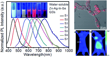
Water-soluble Zn–Ag–In–Se quantum dots with bright and widely tunable emission for biomedical optical imaging†
Received
23rd August 2015
, Accepted 12th October 2015
First published on 13th October 2015
Abstract
Quantum dots (QDs), as a new fluorescent reagent, should have potential applications in biomedical optical imaging; however their biological applications are limited by the toxicity of the component elements. Hence, in this work, water-soluble Zn–Ag–In–Se (ZAISe) QDs were synthesized without using highly toxic heavy metal elements. The as-prepared quaternary QDs exhibit bright and widely composition-tunable photoluminescence (PL) emission (namely, maximum PL quantum yield (QY) reaching 30%; PL peak from 450 to 760 nm). MTT assay proved that these QDs have much less cytotoxicity than Cd-based QDs. After being further modified by DHLA–PEG–Suc–RGD ligands, water-soluble ZAISe QDs were explored as a fluorescent probe for tumor cell-targeted optical imaging. In vitro and in vivo results demonstrate that ZAISe QDs prepared here should be a promising substitute for Cd-based QDs in biomedical optical imaging.
Introduction
In the last ten years, quantum dots (QDs) have been widely anticipated to find applications in many fields, making them an important class of materials in the nanotechnology toolbox. Nowadays, researchers have realized the applications of QDs in solar cells, LEDs, diode lasers, quantum computing etc.1–5 In the case of biomedical imaging, QDs possess many advantages compared to organic dyes or genetically engineered fluorescent proteins. For example, the narrow emission width makes it easier to perform multicolor imaging with minimal spectral overlap. Besides, better resistance to photo-bleaching allows a longer time period for viewing the same region.6–9 As a result, QDs might have the potential to partly replace the traditional fluorescent reagents for biomedical optical imaging.
The original investigations were focused on binary Cd- or Pb-based QDs, resulting in their well-established synthetic chemistry. Although these binary QDs are highly luminescent with PL QY up to 60% to 80%,10,11 they are not suitable for biological research. This is because it is extremely difficult to prevent the leakage of toxic Pb2+ or Cd2+.12 Therefore, synthesizing QDs with low toxic elements has been an urgent research task in recent years. Here, worth mentioning is that, ternary QDs – a complex system relative to binary ones, e.g., CuInS2 QDs without highly toxic heavy metal elements could be synthesized by employing 1-dodecanethiol to balance the activities of various metal ions.13,14 However, the as-prepared naked group I–III–VI2 QDs suffered from relatively weak PL emission, needing further ZnS or CdS overcoating.15–17 Different from the naked ternary QDs, the synthesized quaternary I–II–III–VI3 QDs showed favorable PL properties,18,19 for instance, oil-soluble Zn–Ag–In–Se (ZAISe) QDs with 50% of PL QY.18
Most of high-quality QDs were prepared in organic media,20,21 whereas it is not facile to transfer them into water for biomedical applications, and the transfer process might decrease their PL intensity.18,22–25 To address this problem, recently, quaternary Ag doped ZnInSe QDs have been fabricated in water with mercaptopropionic acid (MPA) as the stabilizing agent, exhibiting adjustable emission from 504 to 585 nm.26 As MPA is a nocuous and carcinogenic reagent with horrid stink, it is necessary to improve the synthetic method using the biocompatible ligands. Furthermore, water-soluble quaternary ZAISe QDs with NIR PL emission have not been prepared, although it is favorable for fabricating NIR-emitting QDs (AgInSe2, Eg = 1.24 eV (∼1000 nm)).18,20,21 NIR light (specifically, Herschel-spectra region, 700–1000 nm) is desirable for in vivo imaging because of the deeper tissue penetration.27
In this work, quaternary ZAISe QDs were synthesized directly in aqueous phase, in which glutathione, a much more biocompatible agent than MPA, was used as the stabilizing agent. These QDs all show strong PL emission while their PL peaks could be conveniently tuned from 450 (blue) to 760 nm (NIR) by varying the precursor feed ratio. Meanwhile, after being further modified by the DHLA–PEG–Suc–RGD ligand, these QDs showed promising applications in biomedical optical imaging serving as tumor targeting fluorescent probes.
Experimental
Chemicals
Zinc acetate (Zn(Ac)2, 99%), silver nitrate (AgNO3, 99%), indium acetate (In(Ac)3, 99.99%), Se powder (100 mesh, 99.5%), reduced glutathione (GSH, ≥98%), diamino polyethylene glycol 1000, succinic anhydride (≥99%), thioctic acid (TA, ≥98%), monomeric cyclic c(RGDyk) peptide (RGD, 98%), 1-ethyl-3(3-dimethylaminopropyl) carbodiimide hydrochloride (EDCI, 98.5%), N-hydroxysuccinimide (NHS, ≥99%), DMEM medium, calf serum, 3-(4,5-dimethylthiazol-2-yl)-2,5-diphenyltetrazolium bromide (MTT). All these reagents were purchased from commercial sources and used without further purification. The water used in this study was double distilled. Water-soluble CdTe QDs with red PL emission were prepared according to our previously reported method.28
Human malignant glioma cell line (U87MG), human breast cancer cell line (MCF-7) and normal human liver cell line (L02) were purchased from American Type Culture Collection (ATCC, Manassas, VA, USA). BALB/c nude mice bearing U87MG tumors were purchased from KeyGEN Biotech. Co. Ltd. (Nanjing, China).
Synthesis of aqueous ZAISe QDs
Aqueous quaternary ZAISe QDs were synthesized as below. Briefly, the mixture of 1 mL of 0.05 M Zn(Ac)2 solution, 1 mL of 0.05 M AgNO3 solution, 2 mL of 0.05 M In(Ac)3 solution, and 20 mL of 0.05 M GSH was adjusted to pH 7.5 using 1 M NaOH solution. Then, a certain amount of freshly prepared selenium sodium sulfite (Na2SeSO3) solution was injected under stirring. Here, the reaction solution volume was set to 50 mL. Subsequently, the resulting reaction mixture was heated to 100 °C under N2 atmosphere and refluxed to allow the growth of ZAISe QDs. To monitor the growth of ZAISe QDs, aliquots were taken out from the reaction mixture and quenched in cold water for further characterization. The detailed parameters for the synthesis of aqueous ZAISe QDs were tabulated in Table 1.
Table 1 Summary of feed ratios, PL peaks and PL QYs of samples S1 to S7
Sample (PL peak) |
Zn (mM) |
Ag (mM) |
In (mM) |
Se (mM) |
QY (%) |
S1 (450 nm) |
20 |
0.8 |
0 |
4 |
25 |
S2 (490 nm) |
18 |
0.2 |
0.2 |
4 |
15 |
S3 (583 nm) |
10 |
0.2 |
0.2 |
4 |
21 |
S4 (630 nm) |
1.8 |
0.2 |
2 |
4 |
26 |
S5 (660 nm) |
1.5 |
0.5 |
2 |
4 |
30 |
S6 (694 nm) |
1 |
1 |
2 |
4 |
25 |
S7 (760 nm) |
0.2 |
1.8 |
2 |
4 |
23 |
Synthesis of RGD modified QDs
Modification of RGD was realized using dihydrolipoic acid–diamino polyethylene glycol 1000–succinyl (DHLA–PEG1000–Suc) as a bridging ligand, which could be anchored onto the surface of QD due to the strong complex between the dithiol of DHLA and the cations on the QD surface.29,30 Specifically, 0.1 mmol RGD was mixed with stoichiometric succinic anhydride in DMSO and stirred at room temperature for 12 h to yield RGD–succinic acid which was then activated by 1.5 stoichiometric NHS/EDCI. Meanwhile, 0.1 mmol thioctic acid NHS ester in DMSO was added dropwise into stoichiometric diamino-PEG1000–DMSO solution.29,30 Silica column chromatography using CH2Cl2/CH3OH (10–15%) as the eluent was used to purify the TA–PEG1000–NH2. Then, RGD–succinic acid NHS ester was further reacted with the terminal amine of TA–PEG1000–NH2 in DMSO for 12 h, after which the TA–PEG1000–Suc–RGD was obtained. The end product was extracted from DMSO via ether precipitation and centrifugation. The resulting precipitation was further dried via evaporation and then redissolved in water. Subsequently, the reductive ring of TA–PEG1000–Suc–RGD was opened via reacting with NaBH4 in water at 0 °C for 4 h, followed by the ligand exchange with GSH-capped ZAISe QDs to obtain QD–DHLA–PEG1000–Suc–RGD fluorescence probe (denoted ‘QD–RGD probe’ hereafter). Unreacted precursors were removed with ultrafiltration using a filter of 5000 g mol−1 MWCO.
Characterization
A Lambda 25 UV-Vis spectrophotometer (Perkin Elmer, America) was used for the detection of UV-Vis absorption spectra. LS55 fluorescence spectrophotometer (Perkin Elmer, America) and S2000 eight-channel optical fiber spectrophotometer (Ocean Optics Corporation, America) were employed to obtain the PL emission spectra. In the calculation of PL QY of QDs, Rhodamine 6G in ethanol (95%) and fluorescence in ethanol (97%) were used as references and the optical densities of all solutions at the excitation wavelength were less than 0.1 to minimize re-absorption effects. All above optical measurements were performed at room temperature. To take TEM images, a JEM-2100 (JEOL, Japan) transmission electron microscope with an acceleration voltage of 200 kV was utilized, and carbon-coated copper grids were dipped in QDs solution to deposit QDs on the film. X-ray diffraction (XRD) measurement was carried out with a D8 Advance X-ray diffractometer (Bruker, Germany). Elemental composition of QDs was assayed using a PHI 5000 VersaProbe (UIVAC-PHI, Japan) X-ray photoelectron spectrometer (XPS). Here, for XRD and XPS measurements, the ZAISe QDs were separated via ethanol precipitation and centrifugation, and then dried at 50 °C in air to obtain the QD powder.
In vitro cell cytotoxicity
MTT assays were carried out to evaluate the potential cytotoxicity of ZAISe QDs in vitro. The L02 cells were seeded onto 96-well plates at a density of 1 × 10⁴ cells per well and then cultured for 24 h. Subsequently, the cells were further maintained at 37 °C after treatment of aqueous ZAISe QDs and CdTe QDs at a wide final concentration range of 0–100 μg mL−1 (0 μg mL−1 for control). Add 20 μL of MTT solution into each well. After incubating for another 4 h, the medium containing MTT was carefully removed, followed by the addition of 150 μL of DMSO into each well. The optical density (OD) of each well at 570 nm was measured after gently shaking the plates for 10 min.
In vitro cell microscopy imaging
In the evaluation of the tumor targeting capability of the QD–RGD probe, αvβ3-positive U87MG and αvβ3-negative MCF-7 cells were used. Each cell line was cultured in RGD-free DMEM medium with 10% (v/v) calf serum at 37 °C in a humidified atmosphere containing 5% CO2, and then seeded in confocal Petri dishes. The cells would attach themselves to the bottom of the dishes with 24 h cultivation, after which 200 μL QD–RGD solution (∼0.5 mg mL−1) was added. After incubating for 2 h, the medium was aspirated and the cells were washed with phosphate buffered saline (PBS) for three times. Fluorescence images of cells were taken using a laser confocal scanning microscope (LCSM, Olympus Fluoview 1000, Japan).
In vivo animal imaging
To establish the tumor-bearing mice models, U87MG cells were injected subcutaneously into the right armpit of each male nude mouse (n = 4, ∼20 g of body weight for each). The mice would be ready for in vivo imaging experiments when the tumors grew to 100–150 mm3 in volume. In a typical NIR imaging experiment, 200 μL of QD–RGD solution (∼1 mg mL−1) was administrated through tail vein into each tumor-bearing nude mouse. Then, a homemade in vivo animal NIR imaging system was applied to monitor the distribution of the probes in mice bodies at different time points (0–6 h) post administration.
Results and discussion
Water-soluble quaternary Zn–Ag–In–Se QDs
In our recent work, oil-soluble Zn–Ag–In–Se (ZAISe) QDs with excellent PL property have been fabricated.18 However, with regard to their biological applications, the complicated phase transfer procedures are needed, besides the complex synthetic steps. Hence, in this work, we tried to prepare quaternary Zn–Ag–In–Se QDs directly in water using glutathione (GSH) as the stabilizing agent. Reduced glutathione, an important tripeptide in human body, is implicated in a variety of molecular reactions.31 Besides the best known role as an antioxidant, GSH can detoxify organism by binding heavy metal ion clusters.32 Thus, here, biocompatible GSH was used to replace toxic sulfhydryl reagents, e.g., mercaptoacetic acid (TGA) and MPA as the stabilizing agent for the synthesis of aqueous ZAISe QDs.
The synthesis of water-soluble GSH-stabilized ZAISe QDs was investigated systematically (see Fig. S1–S4†). It was found that the absence of Zn in the reaction mixture resulted in decreased PL emission, while the absence of any of Ag, In and Se did not show PL emission (Fig. S1†). This observation suggests that the host QDs should possess quaternary elemental composition. Single-factor experiments were performed upon optimizing the synthetic conditions. The corresponding experimental results in Fig. S2–S4† indicate that pH 7.5 and 12 mmol L−1 of GSH are favorable for ZAISe QDs to achieve the strong PL emission, while the optimal refluxing time depends on the Zn/Ag/In/Se/GSH feed ratio.
Fig. 1 presents the UV-Vis absorption and PL spectra of the prepared ZAISe QDs (i.e., the samples S1–S7). Detailed descriptions of precursor feed ratios and PL properties of samples S1–S7 have been tabulated in Table 1. As shown, by increasing the ratio of Ag component, the absorption edge and PL peak of ZAISe QDs gradually shift toward a longer wavelength. Meanwhile, a large Stokes shift between the absorption onset and the PL peak was observed regardless the precursor feed ratio, indicating that the radiative transition is not excitonic recombination but the recombination of two different trap states instead.33–35 In agreement with previous reports, no sharp absorption peak in Fig. 1a was observed because the discrete electronic states have been masked by large inhomogeneous broadening in the linear absorption spectra of chemically-grown QDs.36,37 Fig. 1b illustrates that (i) by varying the precursor feed ratio, the PL emission has been conveniently tuned in the range of 450 to 760 nm; (ii) the PL spectra are broad with a full width at half maximum (FWHM) of 80–120 nm. The broadened PL spectra could be ascribed mainly to the inhomogeneities of the elemental distribution and the stoichiometry,9,33–35 especially for ZAISe QDs with quaternary chemical composition (the coexistent inhomogeneities in size and geometry in an ensemble could be secondary reason).36
 |
| Fig. 1 Absorption (a) and PL (b) spectra of aqueous quaternary ZAISe QDs obtained from different precursor feed ratios (samples S1–S7). The inset of panel (b) is the corresponding digital photograph of the samples S1–S7 taken under UV lamp with 365 nm irradiation. | |
Next, the as-prepared ZAISe QDs were further characterized by TEM, XRD and XPS. TEM and HRTEM images (Fig. 2) reveal that the ZAISe QDs are generally spherical, and have a relatively narrow size distribution (3.5–4 nm), which could be helpful for demonstrating that the broadened PL spectra should not be caused mainly by the size inhomogeneity. The XRD patterns in Fig. 3 indicate that the obtained quaternary ZAISe QDs have a cubic crystal structure. The diffraction peaks of all samples are broad and relatively weak because the size of the samples is in nano-scale. As a typical indicative characteristic of the quaternary elemental composition of the fabricated QDs, all three major diffraction peaks shift toward higher angles with the Ag/Zn feed ratio decreasing.27,35 XPS patterns of sample S5 (Fig. 4a and S5†) may indicate the composition and chemical electronic state of the ZAISe QDs. As shown, the elements Zn, Ag, In, Se and S have been detected, which confirms further the quaternary elemental composition of the ZAISe QDs and suggests the efficient capping of GSH. Fig. 4b and c present the variation of the elemental content in QDs with feed ratio (samples S5–S7). These results could be used to account for the shifts of UV-Vis absorption and PL spectra observed in Fig. 1. That is, with the feed ratio of Zn/Ag increasing, the elemental composition of QDs approaches to ZnSe (Eg = 2.7 eV), leading to the blue shift of the spectra. On the contrary, elemental composition approximates AgInSe2 (Eg = 1.24 eV) when decreasing the feed ratio of Zn/Ag, resulting in the red shift of the spectra.
 |
| Fig. 2 TEM (a) and HRTEM (b) images of the sample S5. | |
 |
| Fig. 3 The XRD patterns of samples prepared from various precursor feed ratios (the patterns of chalcopyrite AgInSe2 and cubic ZnSe have been given for comparison). | |
 |
| Fig. 4 (a) XPS survey spectrum of quaternary ZAISe QDs (sample S5), (b) XPS spectra of Zn 2p of ZAISe QDs synthesized with different Zn/Ag ratios and (c) the corresponding XPS spectra of Ag 3d. | |
Here, we investigated further the cytotoxicity of water-soluble ZAISe QDs, in which aqueous CdTe QDs were selected as a comparison since CdTe QDs prepared in water showed strongest PL emission and often were used for biomedical optical imaging.4,38,39 The corresponding MTT assay results (Fig. S6†) suggest that the Cd-free ZAISe QDs exhibit much lower cytotoxicity compared with aqueous CdTe QDs, especially at high concentrations (40–100 μg mL−1). Consequently, next, our focus was concentrated to explore the in vitro and in vivo imaging of ZAISe QDs as a potential substitute for Cd-based QDs.
Water-soluble quaternary ZAISe QDs for biomedical imaging via RGD modification
On the surface of cancer cells, some specific receptors will be overexpressed.40–42 Among them, the αvβ3 integrin receptor, a family of cell surface glycoprotein consisting of two noncovalently bound transmembrane subunits (α and β), is receiving extensive attention.41–43 Previous studies have shown that RGD peptide can specifically bind to the αvβ3 integrin receptor.43 With such specific binding, anti-cancer drug or contrast agent will accumulate at the tumor sites when covalently coupled with RGD peptide. In this study, QD–DHLA–PEG–Suc–RGD fluorescent probe was prepared (see Fig. 5 and S7†),29,30 in which the bridging between RGD and QD, especially the PEG spacer, is crucial to maintain the targeting capability.44,45 The DHLA–PEG–Suc–RGD ligand could sufficiently bond with QD due to the strong chelation between the dithiol of DHLA and cations on the QD surface.33–35 After RGD modification, the PL intensity of initial ZAISe QDs slightly increased (Fig. 6).
 |
| Fig. 5 Schematic diagrams of the applications of ZAISe QDs in biomedical optical imaging (a) and the synthesis of TA–PEG1000–Suc–RGD ligand ((b); n = 20–25). Reagents and conditions: (1) 8 h, DMSO; (2) NHS, EDCI, 12 h, DMSO; (3) NHS, EDCI, 12 h, DMSO. | |
 |
| Fig. 6 Absorption (a) and PL (b) spectra of ZAISe QDs before and after RGD modification. The slight increase of PL intensity after modified with RGD could be ascribed to the better surface passivation provided by the dithiol in DHLA. | |
The representative LCSM images of U87MG and MCF-7 cells co-incubated with QD–RGD probe are presented in Fig. 7. As shown, no obvious fluorescence signal was observed in αvβ3-negative MCF-7 cells (Fig. 7a). Conversely, clear fluorescence signal could be detected in αvβ3-positive U87MG cells (Fig. 7b), indicating that after co-incubated for 2 h, a significant number of QD–RGD probes were internalized by U87MG cells and mainly accumulated in the cytoplasm. These results indicate that the αvβ3 integrin receptor plays a vital role in the phagocytosis of QD–RGD probes. This also demonstrates the potential of the ZAISe QDs for in vitro cell imaging. Subsequently, we explored further the tumor-targeting capability of the QD–RGD probe in vivo. QD–RGD probes were administrated into nude mice bearing αvβ3-positive U87MG tumor via intravenous injection, followed by the capture of NIR fluorescence images at different time points post injection (P.I.) (Fig. S8†). As shown in Fig. 8, under excitation of 660 nm laser light, the background fluorescence (before injection) was quite weak; 4 h P.I., significant accumulation of fluorescence signal was detected at the liver and the tumor site. These results confirm further the high tumor-targeting capability of the QD–RGD probe.
 |
| Fig. 7 The representative laser confocal scanning microscopy (LCSM) images of MCF-7 (a) and U87MG cells (b) co-incubated with QD–RGD probe (PL peak at 660 nm) for 2 h. | |
 |
| Fig. 8 The NIR images of U87MG tumor-bearing nude mouse under 660 nm laser light excitation (a) before the injection (background) and (b) after intravenous injection with the QD–RGD probe (PL peak at 760 nm) for 4 h. | |
Conclusions
In summary, water-soluble quaternary GSH-capped ZAISe QDs exhibiting bright and widely composition-tunable PL emission (namely, PL QY reaching 30%; PL peak, from 450 to 760 nm) have been fabricated. These quaternary ZAISe QDs possess much low cytotoxicity compared with Cd-based QDs due to the absence of highly toxic heavy metal elements. Importantly, by fabricating QD–DHLA–PEG–Suc–RGD tumor specific fluorescent probes, we demonstrate that these biocompatible ZAISe QDs have great potential in biomedical imaging, as a promising substitute for Cd-based QDs.
Acknowledgements
This work was financially supported by the National Natural Science Foundation of China (81371627 and 81220108012), the Program for New Century Excellent Talents (NCET-12-0974) in University of the Ministry of Education of China, and A Project Funded by the Priority Academic Program Development of Jiangsu Higher Education Institutions (PAPD).
Notes and references
- Z. Pan, I. Mora-Seró, Q. Shen, H. Zhang, Y. Li, K. Zhao, J. Wang, X. Zhong and J. Bisquert, J. Am. Chem. Soc., 2014, 136, 9203–9210 CrossRef CAS PubMed.
- C. Gimbert-Suriñach, J. Albero, T. Stoll, J. Fortage, M. N. Collomb, A. Deronzier, E. Palomares and A. Llobet, J. Am. Chem. Soc., 2014, 136, 7655–7661 CrossRef PubMed.
- K. Srinivasan and O. Painter, Nature, 2007, 450, 862–865 CrossRef CAS PubMed.
- G. Fan, L. Han, J. Zhang and J. Zhu, Anal. Chem., 2014, 86, 10877–10884 CrossRef CAS PubMed.
- V. Sukhovatkin, S. Hinds, L. Brzozowski and E. H. Sargent, Science, 2009, 324, 1542–1544 CrossRef CAS PubMed.
- S. Bhandari, R. Khandelia, U. N. Pan and A. Chattopadhyay, ACS Appl. Mater. Interfaces, 2015, 7, 17552–17557 CAS.
- F. Zhao, Z. Li, L. Wang, C. Hu, Z. Zhang, C. Li and L. Qu, Chem. Commun., 2015, 51, 13201–13204 RSC.
- X. Tang, W. B. A. Ho and J. M. Xue, J. Phys. Chem. C, 2012, 116, 9769–9773 CAS.
- D. Deng, L. Qu, S. Achilefu and Y. Gu, Chem. Commun., 2013, 49, 9494–9496 RSC.
- R. E. Bailey and S. Nie, J. Am. Chem. Soc., 2003, 125, 7100–7106 CrossRef CAS PubMed.
- J. Y. Kim, O. Voznyy, D. Zhitomirsky and E. H. Sargent, Adv. Mater., 2013, 25, 4986–5010 CrossRef CAS PubMed.
- S. P. Samuel, M. J. Santos-Martinez, C. Medina, N. Jain, M. W. Radomski, A. Prina-Mello and Y. Volkov, Int. J. Nanomed., 2015, 10, 2723–2734 CAS.
- R. Xie, M. Rutherford and X. Peng, J. Am. Chem. Soc., 2009, 131, 5691–5697 CrossRef CAS PubMed.
- S. Liu and X. Su, RSC Adv., 2014, 4, 43415–43428 RSC.
- J. Park and S.-W. Kim, J. Mater. Chem., 2011, 21, 3745 RSC.
- T. Torimoto, S. Ogawa, T. Adachi, T. Kameyama, K. Okazaki, T. Shibayama, A. Kudo and S. Kuwabata, Chem. Commun., 2010, 46, 2082–2084 RSC.
- D. Deng, Y. Chen, J. Cao, J. Tian, Z. Qian, S. Achilefu and Y. Gu, Chem. Mater., 2012, 24, 3029–3037 CrossRef CAS.
- D. Deng, L. Qu, J. Zhang, Y. Ma and Y. Gu, ACS Appl. Mater. Interfaces, 2013, 5, 10858–10865 CAS.
- J. Zhang, R. G. Xie and W. S. Yang, Chem. Mater., 2011, 23, 3357–3361 CrossRef CAS.
- P. M. Allen and M. G. Bawendi, J. Am. Chem. Soc., 2008, 130, 9240–9241 CrossRef CAS PubMed.
- M. Langevin, A. M. Ritcey and C. N. Allen, ACS Nano, 2014, 8, 3476–3482 CrossRef CAS PubMed.
- H. G. Bagaria, G. C. Kini and M. S. Wong, J. Phys. Chem. C, 2010, 114, 19901–19907 CAS.
- D. Dorokhin, N. Tomczak, M. Han, D. N. Reinhoudt, A. H. Velders and G. J. Vancso, ACS Nano, 2009, 3, 661–667 CrossRef CAS PubMed.
- D. A. G. Navarro, D. F. Watson, D. S. Aga and S. Banerjee, Environ. Sci. Technol., 2009, 43, 677–682 CrossRef CAS.
- G. Palui, T. Avellini, N. Zhan, F. Pan, D. Gray, I. Alabugin and H. Mattoussi, J. Am. Chem. Soc., 2012, 134, 16370–16378 CrossRef CAS PubMed.
- C. Wang, S. Xu, Y. Shao, Z. Wang, Q. Xu and Y. Cui, J. Mater. Chem. C, 2014, 2, 5111–5115 RSC.
- D. Deng, L. Qu and Y. Gu, J. Mater. Chem. C, 2014, 2, 7077–7085 RSC.
- D. Deng, L. Qu and Y. Gu, RSC Adv., 2012, 2, 11993–11999 RSC.
- R. Kikkeri, B. Lepenies, A. Adibekian, P. Laurino and P. H. Seeberger, J. Am. Chem. Soc., 2009, 131, 2110–2112 CrossRef CAS PubMed.
- W. Liu, M. Howarth, A. B. Greytak, Y. Zheng, D. G. Nocera, A. Y. Ting and M. G. Bawendi, J. Am. Chem. Soc., 2008, 130, 1274–1284 CrossRef CAS PubMed.
- C. Tortiglione, A. Quarta, A. Tino, L. Manna, R. Cingolani and T. Pellegrino, Bioconjugate Chem., 2007, 18, 829–835 CrossRef CAS PubMed.
- E. M. Ali, Y. Zheng, H. H. Yu and J. Y. Ying, Anal. Chem., 2007, 79, 9452–9458 CrossRef CAS PubMed.
- L. Li, A. Pandey, D. J. Werder, B. P. Khanal, J. M. Pietryga and V. I. Klimov, J. Am. Chem. Soc., 2011, 133, 1176–1179 CrossRef CAS PubMed.
- F. Meinardi, H. McDaniel, F. Carulli, A. Colombo, K. A. Velizhanin, N. S. Makarov, R. Simonutti, V. I. Klimov and S. Brovelli, Nat. Nanotechnol., 2015, 10, 878–885 CrossRef CAS PubMed.
- D. Deng, J. Cao, L. Qu, S. Achilefu and Y. Gu, Phys. Chem. Chem. Phys., 2013, 15, 5078–5083 RSC.
- J. Sun, M. Ikezawa, X. Wang, P. Jing, H. Li, J. Zhao and Y. Masumoto, Phys. Chem. Chem. Phys., 2015, 17, 11981–11989 RSC.
- H. Zhong, S. S. Lo, T. Mirkovic, Y. Li, Y. Ding, Y. Li and G. D. Scholes, ACS Nano, 2010, 4, 5253–5262 CrossRef CAS PubMed.
- J. D. Major, R. E. Treharne, L. J. Phillips and K. Durose, Nature, 2014, 511, 334–337 CrossRef CAS PubMed.
- P. Singh, K. Joshi, D. Guin and A. A. Prabhune, RSC Adv., 2013, 3, 22319 RSC.
- M. Watanabe, K. D. Moon, M. S. Vacchio, K. S. Hathcock and R. J. Hodes, Immunity, 2014, 40, 681–691 CrossRef CAS PubMed.
- Y. Ye, S. Bloch, B. Xu and S. Achilefu, J. Med. Chem., 2006, 49, 2268–2275 CrossRef CAS PubMed.
- Y. P. Yu, Q. Wang, Y. C. Liu and Y. Xie, Biomaterials, 2014, 35, 1667–1675 CrossRef CAS PubMed.
- E. S. Mittra, M. L. Goris, A. H. Iagaru, A. Kardan, L. Burton, R. Berganos, E. Chang, S. Liu, B. Shen, F. T. Chin, X. Chen and S. S. Gambhir, Radiology, 2011, 260, 182–191 CrossRef PubMed.
- X. He, C. S. Alves, N. Oliveira, J. Rodrigues, J. Zhu, I. Bányai, H. Tomás and X. Shi, Colloids Surf., B, 2015, 125, 82–89 CrossRef CAS PubMed.
- Q. Chen, K. Li, S. Wen, H. Liu, C. Peng, H. Cai, M. Shen, G. Zhang and X. Shi, Biomaterials, 2013, 34, 5200–5209 CrossRef CAS PubMed.
Footnote |
† Electronic supplementary information (ESI) available: Additional characterization of ZAISe QDs and QD–RGD probe. See DOI: 10.1039/c5ra17046j |
|
This journal is © The Royal Society of Chemistry 2015 |
Click here to see how this site uses Cookies. View our privacy policy here.