DOI:
10.1039/C5RA16952F
(Paper)
RSC Adv., 2015,
5, 98004-98009
Controlled protein separation based on pressure–voltage (P–V) coupling effects in a nanopore based device
Received
21st August 2015
, Accepted 3rd November 2015
First published on 9th November 2015
1. Introduction
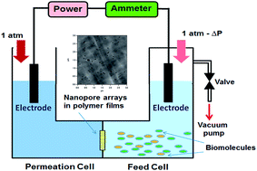
The separation and/or purification of biomolecules are of great importance in biotechnological analysis, characterization and applications, especially in in vitro analysis, antibody generation, binding assays and structural studies, which require pure protein samples with different sizes, shapes, charges, and hydrophobicities.1–10 Generally, ultrafiltration,11 electrophoretic separation,12 liquid chromatography, magnetic nanoparticles based separation13 and membrane chromatography14,15 are common methods for biomolecule separation or purification, among which membrane separation and ultrafiltration have been extensively adapted.16–19 In membrane based separation, critical structures and properties of membrane are required, especially for definite pore sizes. However, the general use of membranes with a broad distribution of pore size and heterogeneous matrices leads to low protein adsorption capacities. From another point of view, if separation or purification can be determined by other factors out of pore size, more precisely controlled separations may be expected.
In recent years, nanopore or nanopore array based analytical technology have provided potentials to fabricate novel nanofluidic or microfluidic devices for biomolecule sensing and separation.20–27 The general picture of nanopore or nanopore array based nanofluidic devices can be illustrated as follows: a pair of separated liquid cells with certain electrolyte solutions are linked by a chip containing a nanopore or nanopore array; along the length direction of the nanopore a certain voltage is applied, and charged biomolecules and ions can migrate from one cell to another through the nanopores by electrophoresis. If other factors are introduced to the nanopore based device, the movements of the biomolecules can be controlled more precisely. For example, Golovchenko and his coworkers28 showed that the threading and detection functions could be decoupled by the addition of a pressure bias across a voltage-biased solid-state pore. In a pressure–voltage (P–V)-biased pore, the net force (and hence the speed) of a biomolecule could be reduced by an order of magnitude without a similar reduction in the ionic current through the nanopores. The movement of the biomolecules depends on an experimentally adjustable balance of the applied pressure and voltage gradients.
These also provide novel ideals for biomolecule separation. In this work, a set of “U”-type devices containing a feed cell and a permeation cell connected by nanopore arrays was designed and employed for controlled protein separation. The voltage and pressure difference (ΔP) between two liquid cells can be changed in a certain range, thus protein separation can be achieved based on this device via a pressure–voltage (P–V) coupling control.
2. Experimental
2.1 Materials
Polycarbonate membranes containing nanopore arrays (pore diameter: 50 nm, pore density: 6 pores per μm2) were purchased from Whatman, Inc. Bovine serum albumin (BSA, pI = 4.9) and bovine hemoglobin (Hb, pI = 7.0)29 were obtained from Shi Feng Inc. Ultra-pure water (resistivity 18.25 MΩ cm) was used for all the solutions and rinsing.
2.2 Device
A “U”-type device containing a feed cell (on the right side) and a permeation cell (on the left side) was designed and employed in our experiments, as shown in Fig. 1. Two separated cells with certain solutions were linked by a piece of PC membrane containing nanopore arrays and they were sealed by PDMS. In our experiment, an electric field was applied to transport proteins in the feed cell to the permeation cell through the nanopore arrays, and the values of ΔP generated by a vacuum pump were adjusted in order to produce a resistant force. The direction of the electric field can be adjusted to guarantee that the electric field force is serving as the driving force. When the protein was positively charged, the anode and the cathode were set in the feed cell and the permeation cell respectively; when the protein was negatively charged, the electrodes were reversed.
 |
| Fig. 1 “U”-Type reaction device containing a feed cell and a permeation cell. The pressure difference (ΔP) and the voltage between the two cells can be modulated in order to achieve pressure–voltage (P–V) coupling effect based separation of proteins. | |
2.3 Parameters for pressure–voltage (P–V) coupling experiments
BSA and Hb were employed in our work for protein separation experiments. The applied voltage was 1.0 V to 3.0 V, the pH value for the main experiments was controlled at 7.4 or 4.0, the ion strength was controlled at 0.105 mol L−1, and ΔP was controlled at 0 MPa, 0.02 MPa, 0.04 MPa, 0.06 MPa and 0.08 MPa. All of the experiments were carried out at room temperature, and the time for individual protein translocation or protein separation was 3 hours. The concentration of protein in the feed solution was 1 μM (66
400 μg mL−1 for BSA, 65
000 μg mL−1 for Hb), the concentration of protein in the permeation cell, which reflected the separation efficiency, was determined by the strength of UV-vis absorbance. The characteristic UV-vis wavelengths were at 291 nm and 405 nm for Hb, and 291 nm for BSA. In our experiments, the concentration for Hb was determined as CHb = 241.23483 × A405 nm + 4.4, and the concentration for BSA was determined as CBSA = 6922.81006 × ABSA 291 nm − 44.91. In the last equation, ABSA 291 nm = A291 nm − AHb 291 nm = A291 nm − 0.24861 × A405 nm + 0.00179. Here CHb (μg mL−1) and CBSA (μg mL−1) stand for the calculated concentrations of Hb and BSA respectively; A405 nm, A291 nm, AHb 291 nm, ABSA 291 nm stand for the total absorbance at 405 nm, the total absorbance at 291 nm, the absorbance at 291 nm contributed by Hb and the absorbance at 291 nm contributed by BSA.
3. Results and discussion
3.1 Pressure–voltage (P–V) coupling effects
The basic idea for controlled protein separation based on pressure–voltage (P–V) coupling effects is illustrated in Fig. 2. Protein A will transfer from the feed cell into the permeation cell driven by the voltage; at the same time, ΔP can generate a resistance for its translocation. If the force generated by ΔP is bigger than that generated by the voltage, protein A could theoretically transfer back into the feed cell. That is to say, there must be a P–V equilibrium point (EPA) for protein A, at which the driving force is equal to the resistance. Similarly, there must be a P–V equilibrium point (EPB) for protein B. Generally, the conformation and charge state of the proteins are different from each other, so EPA is not the same as EPB. At EPA, the main movement of protein A is free diffusion because of the equilibrium of the driving force and resistance, while protein B can move from the feed cell into the permeation cell under the residual driving force (the resultant of forces generated by voltage and ΔP). In this way, the separation of protein A and protein B can be achieved by pressure–voltage (P–V) coupling effects. Of course, finding out the P–V equilibrium point of the single protein is the basis for the separation of the proteins by pressure–voltage (P–V) coupling effects.
 |
| Fig. 2 An illustration of the mechanism of protein separation based on a pressure–voltage (P–V) coupling effect. | |
Fig. 3 shows the pressure modulation in the translocation process of BSA and Hb through the nanopore arrays over time. The black and green points stand for protein translocation driven only by the voltage, while the red and blue points stand for protein translocation under the synergically driven by the voltage and ΔP. The concentration in the permeation cell can be determined by the changes in the UV-vis spectra of the solution, as shown in the inserted image ① in Fig. 3. Here the translocation ratio (the concentration ratio of the two proteins obtained from individual translocation experiments, BSA
:
Hb or Hb
:
BSA) is defined to evaluate the relative translocation abilities of BSA and Hb. Based on the data in Fig. 3, the translocation ratio (BSA
:
Hb) at different times can be calculated, as shown in the inserted image ② in Fig. 3. Driven by the voltage or synergically driven by the voltage and ΔP, the protein concentrations in the permeation cell increase with time approximately linearly, which indicates that their migration rates are nearly constant under given conditions. Obviously, the migration rates for both BSA and Hb are decreased under the resistance from a ΔP of 0.04 MPa, which indicates that the protein’s translocation can be modulated by the comprehensive control of voltage and ΔP. In addition, the translocation ratio (BSA
:
Hb) seems to increase with time, especially under the comprehensive control of 3.0 V voltage and a ΔP of 0.04 MPa.
 |
| Fig. 3 The translocation process of BSA and Hb through nanopore arrays with time: the black and blue points stand for protein translocation driven by voltage, while the red and green points stand for protein translocation driven by the pressure–voltage (P–V) coupling effect. The concentration in the permeation cell can be determined by the UV-vis spectra of the solution from the cell. Inserted image ①: as an example, the UV-vis spectra for Hb translocation at different times are shown in the inserted picture. Inserted image ②: the translocation ratio (BSA : Hb). | |
3.2 Protein translocations
Fig. 4 shows the individual voltage-driven translocation behaviors of BSA and Hb modulated by ΔP when the pH value of the solution is 7.4 or 4.0, and the total time for each experiment is 3 hours. Here the applied voltage (3.0 V or 1.5 V) works as a driving force and ΔP works as the resistance. With increasing ΔP, the average translocation velocities of BSA and Hb are both decreased, no matter if the voltage is 3.0 V and 1.5 V (as shown in Fig. 4a and b). At the same time, it can be found that the translocation abilities of BSA and Hb are different from each other. According to the experimental results shown in Fig. 4a and b, the translocation ratios of BSA to Hb (BSA
:
Hb) obtained under the driving voltage of 3.0 V are bigger than those obtained under the driving voltage of 1.5 V. With increasing ΔP, the translocation ratios of BSA to Hb (BSA
:
Hb) obtained under a voltage of 3.0 V continue to increase, while those obtained under a voltage of 1.5 V decrease after reaching the maximum value at about ΔP = 0.04 MPa. According to these data, the biggest translocation ratio of BSA to Hb (BSA
:
Hb) is 23.75 when the pH value, applied voltage and ΔP are controlled at 7.4, 3.0 V and 0.08 MPa respectively.
 |
| Fig. 4 The translocation ratio for BSA and Hb (the blue line and point stand for BSA : Hb; the red line and point stand for Hb : BSA) obtained from independent translocation. (a) 3.0 V, pH = 7.4; (b) 1.5 V, pH = 7.4; (c) 3.0 V, pH = 4.0; (d) 3.0 V, pH = 7.4; the inserted picture shows the concentrations of BSA and Hb in the permeation cell after a 3 hour experimental time. The driven voltage was controlled at 3.0 V or 1.5 V, and the pH value was controlled at 7.4 or 4.0. | |
As reported in previous literature, changing the pH value can change the charged amounts of proteins, which will alter the electrostatic force applied to proteins. Definitely, the alternation in pH value of the solution will change the voltage-driven translation behaviors of proteins. Fig. 4c and d show the individual translocation behaviors of BSA and Hb modulated by ΔP when the pH value of the solution are 7.4 and 4.0 respectively, and the total time for each experiment is 3 hours. The applied voltage of 3.0 V works as a driving force and ΔP works as the resistance. Obviously, net charges belonging to these two proteins are changed because of the pH value changing. Similarly, with increasing ΔP, the average translocation velocities of BSA and Hb are decreased, regardless of whether the pH value is 4.0 or 7.4. With ΔP increasing, the translocation ratios of Hb to BSA (Hb
:
BSA) obtained in the solution with the pH value of 4.0 continue to increase, while those obtained in the solution with the pH value of 7.4 are almost unchanged. In our experiment, the biggest translocation ratio of Hb to BSA (Hb
:
BSA) is 8.0 when the pH value, applied voltage and ΔP are controlled at 4.0, 3.0 V and 0.08 MPa respectively.
3.3 Protein separation
In separation experiments, two kinds of proteins (BSA and Hb, their initial concentrations in the feed cell were both 1 μM) were added into the feed cell of the device. Here the separation ratio (the concentration ratio of the two proteins obtained from mixture translocation experiments, BSA
:
Hb or Hb
:
BSA) is defined to evaluate the relative separation abilities. Fig. 5 shows the separation ratio with increasing ΔP, which is similar to the translocation ratio changing tendency shown in Fig. 4. The separation ratios of BSA to Hb (BSA
:
Hb) obtained under the driving voltage of 3.0 V are bigger than those obtained under the driving voltage of 1.5 V. Similarly, with increasing ΔP, the separation ratios of BSA to Hb (BSA
:
Hb) obtained under the driving voltage of 3.0 V continues to increase, while those obtained under the driving voltage of 1.5 V decrease after reaching the maximum value at ΔP = 0.04 MPa. The biggest separation ratio of BSA to Hb (BSA
:
Hb) is 12.5 when the pH value, voltage and ΔP are controlled at 7.4, 3.0 V and 0.08 MPa respectively. When the pH value of the solution is 7.4, both proteins are negatively charged. However, because BSA has a lower isoelectric point, the charge belonging to BSA is bigger than the charge belonging to Hb. This point (pH = 7.4, voltage = 3.0 V, ΔP = 0.08 MPa) can be regarded as an approximate P–V equilibrium point for Hb (EPHb), at which Hb stays in the feed cell due to the balance of the driving force and resistance, while BSA can migrate from the feed cell into the permeation cell. In this case, the P–V equilibrium point for Hb (EPHb) is the basis for the separation. The pH value, voltage and ΔP are three important factors for P–V equilibrium. If one of them is changed, the equilibrium will be destroyed; but a new equilibrium can be reached by changing the other one or two factors. Fig. 5 indicates that for the best separation value, ΔP increases from 0.02 MPa to 0.08 MPa with the driving voltage increasing from 1.0 V to 3.0 V. For Hb (EPHb), the increasing voltage means a larger driving force, and it needs a bigger ΔP for the next P–V equilibrium, which is the reason that the ΔP corresponding to the best separation shifts to a bigger value with the increasing driving voltage. BSA is always in a nonequilibrium state (the driving force is always bigger than the resistance). The increment in the driving force generated by the increasing voltage is bigger than the resistance increment generated by ΔP, which results in the increasing tendency of the best separation (from 6.3 to 12.5, as shown in the insert in Fig. 5) value with the driving voltage varying from 1.0 V to 3.0 V.
 |
| Fig. 5 The separation ratio for BSA and Hb (BSA : Hb) obtained from mixed translocation under different driving voltages after an experimental time of 3 hours. The insert shows ΔP corresponding to the best separation and the value of the best separation under different driving voltage. The pH value was controlled at 7.4. The driving voltage was changed from 1.0 V to 3.0 V. | |
On the other hand, the pH value can determine the net charges on the protein and thus influence the driving force generated by the voltage. Definitely, the alternation in the pH value of the solution will change the P–V equilibrium points of the proteins and thus change the parameters for protein separation based on pressure-coupling effects. In the following experiment, the pH value of the solution is decreased to 4.0, at which BSA and Hb are both positively charged and the net charges belonging to Hb is bigger than the net charges belonging to BSA. As shown in the insert in Fig. 6, when ΔP is less than 0.06 MPa, the BSA concentration in the permeation cell is greater than the Hb concentration. This may be attributed to the shape of BSA, which is more advantageous to diffuse than Hb, and the electrophoretic contribution is not big enough to change the diffusion influence of BSA, which has been mentioned by Chun and Stroeve.30 When ΔP was 0.06 MPa and 0.08 MPa, the concentrations of Hb in the permeation cell were greater than BSA. With increasing ΔP, the resultant forces applied to BSA became smaller and smaller, the amount of BSA transported across the nanopores decreased greatly, while Hb could still be transported into the permeation cell because of a relatively larger electrical force. The biggest separation ratio of Hb to BSA (Hb
:
BSA) is 14.3 when the pH value, applied voltage and ΔP are controlled at 4.0, 3.0 V and 0.08 MPa respectively. Similarly, this point (pH = 4.0, voltage = 3. V, ΔP = 0.08 MPa) can be regarded as an approximate P–V equilibrium point for BSA (EPBSA), at which BSA stays in the feed cell due to the balance of the driving force and the resistance, while Hb can migrate from the feed cell into the permeation cell. Similarly, if the pH is changed now, the equilibrium will be destroyed, and the related separation ratio will also be changed, as shown in the insert of Fig. 6. Taking into account their isoelectric points, if the pH value of the solution is between 4.0 and 7.4, the two proteins will be differently charged (positive and negative), and the separation by pressure–voltage (P–V) coupling effects will be difficult to achieve.
 |
| Fig. 6 The separation ratio for Hb and BSA (Hb : BSA) obtained from mixed translocation after an experimental time of 3 hours. The driving voltage was controlled at 3.0 V, and the pH value was controlled at 7.4 or 4.0. The insert shows the separation ratio (SR) of Hb : BSA when the pH value was controlled at 7.4, 9.0, 10.0 or 4.0, 3.0, 2.0. | |
4. Conclusion
In summary, we provide a new way to achieve protein separation based on pressure–voltage (P–V) coupling effects in a nanopore based fluidic device, which provides a new possibility and choice for protein selection or separation. The resultant driving forces applied to the proteins can be modulated by changing the parameters such as voltage, pH value and ΔP between two cells, thus their translocation velocity can be controlled. By finding the P–V equilibrium points for BSA and Hb, the separation ratios (mass ratio) for BSA and Hb can be achieved as BSA
:
Hb = 12.5 and Hb
:
BSA = 14.3 receptively.
It should be pointed out that the separation speed will be reduced under these equilibrium points. However, the separation efficiency can be improved by optimizing the experimental apparatus and method (for example, to find a new P–V equilibrium point under a smaller value of ΔP; to enlarge the effective working areas of the polycarbonate membranes to provide more nanopores for protein translocation). Better parameters for more efficient separation are now in progress.
Acknowledgements
This work is financially supported by the Natural Science Foundation of China (U1332134), the National Basic Research Program of China (2011CB707601 and 2011CB707605), the Natural Science Foundation of Suzhou (SYG201329), the Fundamental Research Funds for the Central Universities, the Qing Lan Project and the International Foundation for Science, Stockholm, Sweden, the Organization for the Prohibition of Chemical Weapons, The Hague, Netherlands, through a grant to Lei Liu (F/4736-2).
References
- W. Zhai, H. Jeong, L. Cui, D. Krainc and R. Tjian, In vitro analysis of Huntingtin-Mediated Transcriptional Repression Reveals Multiple Transcription Factor Targets, Cell, 2005, 123, 1241–1253 CrossRef CAS PubMed.
- C. Smith, Striving for Purity: Advances in Protein Purification, Nat. Methods, 2005, 2, 71–77 CrossRef CAS.
- J. E. Schnitzer, D. P. McIntosh, A. M. Dvorak, J. Liu and P. Oh, Separation of Caveolae from Associated Microdomains of GPI-Anchored Proteins, Science, 1995, 269, 1435–1439 CrossRef CAS PubMed.
- C. R. Martin, Nanomaterials: A Membrane-based Synthetic Approach, Science, 1994, 266, 1961–1966 CAS.
- K. B. Jirage, J. C. Hulteen and C. R. Martin, Nanotubule-based Molecular-Filtration Membranes, Science, 1997, 278, 655–658 CrossRef CAS.
- S. Yu, S. B. Lee, M. Kang and C. R. Martin, Size-based Protein Separations in Poly(ethylene glycol)-Derivatized Gold Nanotubule Membranes, Nano Lett., 2001, 1, 495–498 CrossRef CAS.
- R. Reis and A. L. Zydney, Membrane Separations in Biotechnology, Curr. Opin. Biotechnol., 2001, 12, 208–211 CrossRef PubMed.
- H. Yang, M. Bitzer and M. R. Etzel, Analysis of Protein Purification Using Ion Exchange Membranes, Ind. Eng. Chem. Res., 1999, 38, 4044–4050 CrossRef CAS.
- K. H. Kim and M. H. Moon, Development of a Multilane Channel System for Nongel-Based Two-Dimensional Protein Separations Using Isoelectric Focusing and Asymmetrical Flow Field-flow Fractionation, Anal. Chem., 2009, 81, 1715–1721 CrossRef CAS PubMed.
- J. Kim, Y. Piao, N. Lee, Y. I. Park, I. H. Lee, J. H. Lee, S. R. Paik and T. Hyeon, Magnetic Nanocomposite Spheres Decorated with NiO Nanoparticles for a Magnetically Recyclable Protein Separation System, Adv. Mater., 2010, 22, 57–60 CrossRef CAS PubMed.
- A. G. Fane, C. J. D. Fell and A. Suki, The Effect of pH and Ionic Environment on the Ultrafiltration of Protein Solutions with Retentive Membranes, J. Membr. Sci., 1983, 16, 195–199 CrossRef CAS.
- H. Stutz, G. Bordin and A. R. Rodriguez, Separation of Selected Metal-binding Proteins with Capillary Zone Electrophoresis, Anal. Chim. Acta, 2003, 477, 1–19 CrossRef CAS.
- S. H. Kim, M. J. Kim and Y. H. Choa, Fabrication and Estimation of Au-Coated Fe3O4 Nanocomposite Powders for the Separation and Purification of Biomolecules, Mater. Sci. Eng., A, 2007, 449, 386–388 CrossRef.
- M. I. Shukoor, F. Natalio, M. N. Tahir, V. Ksenofontov, H. A. Therese, P. Theato, H. C. Schroder, W. E. G. Müller and W. Tremel, Superparamagnetic Gamma-Fe2O3 Nanoparticles with Tailored Functionality for Protein Separation, Chem. Commun., 2007, 4677–4679 RSC.
- C. C. Striemer, T. R. Gaborski, J. L. McGrath and P. M. Fauchet, Charge- and Size-Based Separation of Macromolecules Using Ultrathin Silicon Membranes, Nature, 2007, 445, 749–753 CrossRef CAS PubMed.
- T. M. Przybycien and N. S. Pujar, Alternative Bioseparation Operations: Life Beyond Packed-Bed Chromatography, Curr. Opin. Biotechnol., 2004, 15, 469–478 CrossRef CAS PubMed.
- L. Wang, G. Hale and R. Ghosh, Non-size-based Membrane Chromatographic Separation and Analysis of Monoclonal Antibody Aggregates, Anal. Chem., 2006, 78, 6863–6867 CrossRef CAS PubMed.
- A. Saxena, M. Kumar, B. P. Tripathi and V. K. Shahi, Organic–Inorganic Hybrid Charged Membranes for Proteins Separation: Isoelectric Separation of Proteins under Coupled Driving Forces, Sep. Purif. Technol., 2010, 70, 280–290 CrossRef CAS.
- R. Ghosh, Protein Separation Using Membrane Chromatography: Opportunities and Challenges, J. Chromatogr. A, 2003, 952, 13–27 CrossRef.
- K. Kant, M. Kurkuri, J. Yu, J. G. Shapter, C. Priest and D. Losic, Impedance Spectroscopy Study of Nanopore Arrays for Biosensing Applications, Sci. Adv. Mater., 2014, 6, 1375–1381 CrossRef CAS.
- K. Kant, J. X. Yu, C. Priest, J. G. Shapter and D. Losic, Impedance Nanopore Biosensor: Influence of Pore Dimensions on Biosensing Performance, Analyst, 2014, 139, 1134–1140 RSC.
- H. Im, N. J. Wittenberg, A. Lesuffleur, N. C. Lindquist and S. H. Oh, Membrane Protein Biosensing with Plasmonic Nanopore Arrays and Pore-spanning Lipid Membranes, Chem. Sci., 2010, 1, 688–696 RSC.
- L. A. Baker, Y. S. Choi and C. R. Martin, Nanopore Membranes for Biomaterials Synthesis, Biosensing and Bioseparations, Curr. Nanosci., 2006, 2, 243–255 CrossRef CAS.
- L. Q. Gu, B. Ritzo and Y. Wang, Biosensing When Less is More in a Nanopore, Nat. Nanotechnol., 2012, 7, 212–213 CrossRef CAS PubMed.
- M. Taniguchi, Selective Multidetection Using Nanopores, Anal. Chem., 2015, 87, 188–199 CrossRef CAS PubMed.
- W. J. Lan, D. A. Holden and H. S. White, Pressure-Dependent Ion Current Rectification in Conical-Shaped Glass Nanopores, J. Am. Chem. Soc., 2011, 133, 13300–13303 CrossRef CAS PubMed.
- B. N. Miles, A. P. Ivanov, K. A. Wilson, F. Dogan, D. Japrung and J. B. Edel, Single molecule sensing with solid-state nanopores: novel materials, methods, and applications, Chem. Soc. Rev., 2013, 42, 15–28 RSC.
- B. Lu, D. P. Hoogerheide, Q. Zhao, H. Zhang, Z. Tang, D. Yu and J. A. Golovchenko, Pressure-Controlled Motion of Single Polymers through Solid-State Nanopores, Nano Lett., 2013, 13, 3048–3052 CrossRef CAS PubMed.
- S. F. Yu, S. B. Lee and C. R. Martin, Electrophoretic Protein Transport in Gold Nanotube Membranes, Anal. Chem., 2003, 75, 1239–1244 CrossRef CAS PubMed.
- K. Y. Chun and P. Stroeve, Protein Transport in Nanoporous Membranes Modified with Self-Assembled Monolayers of Functionalized Thiols, Langmuir, 2002, 18, 4653–4658 CrossRef CAS.
|
This journal is © The Royal Society of Chemistry 2015 |
Click here to see how this site uses Cookies. View our privacy policy here.