pH and stability of the α-gel phase in glycerol monostearate–water systems using sodium stearoyl lactylate and sodium stearate as the co-emulsifier

Fan C. Wang and Alejandro G. Marangoni*
Department of Food Science, University of Guelph, Guelph, ON N1G2W1, Canada. E-mail: amarango@uoguelph.ca

Received 15th August 2015 , Accepted 3rd November 2015

First published on 5th November 2015


Abstract

Changing the environmental pH alters the melting profiles of monostearate–water systems and affects the stability of the α-gel phase using sodium stearoyl lactylate (SSL) and sodium stearate (NaS) as co-emulsifiers. NaS in MG-gels may be present both in a micellar phase and in a lamellar phase. Once above the critical micellar concentration of NaS, the NaS solution and diluted MG-gels remained at a stable pH.
1. Introduction

Monoglycerides (MGs) are commonly used emulsifiers in food and personal care products due to their ability to structure both water and liquid oil. Saturated MGs display polymorphic and mesomorphic properties, and the phase behavior of MG–water systems has been studied extensively.1–6 However, distilled MGs alone are not able to structure water, and the hydrated lamellar α-gel phase can only be formed with the assistance of a co-emulsifier.7 The stability of the α-gel phase of MG–water systems (MG-gels) is affected by various factors. Higher α-gel phase stability can be achieved by storing the gels at refrigeration temperatures, using α-tending co-emulsifiers, and incorporating anionic co-emulsifiers.8–10 The polymorphic form of MG-gel systems can be characterized using powder X-ray diffraction (XRD). The α-gel phase shows small angle X-ray diffraction (SAXS) reflections representing the (001) plane at ∼52 Å, and wide angle X-ray diffraction (WAXS) spacing at 4.1 Å; while the coagel phase is characterized by SAXS spacing at 49 Å and WAXS spacings between 3.6 and 4.6 Å.8,11

Previous work done by our group examined factors that affect the stability of MG–water systems structured with glycerol monostearate (GMS) without adjusting the environmental pH.6,8,9 However, in application of MG-gels in food and personal care products, the pH changes upon addition of various ingredients. Such changes in the environmental pH could affect the structure and stability of MG-structured systems. The literature suggests that the balance between the pH and ionic strength of the environment needs to be considered in order to attain optimal stability.1,10,12 This work therefore further investigates how changes in environmental pH affects the stability of the α-gel phase of GMS–water systems using sodium stearoyl lactylate (SSL) and sodium stearate (NaS) as co-emulsifiers.

2. Experimental

The distilled GMS used was Alphadim 90 SBK from Caravan Ingredients (Lenexa, KS, USA). The co-emulsifiers were Emplex sodium stearoyl lactylate (Caravan Ingredients, Lenexa, KS, USA) and sodium stearate, minimum 99% purity (Sigma-Aldrich Inc., St. Louis, MO, USA). MG-gels containing 20% (w/w) solids were prepared following the sample preparation method previously published by our group.8 GMS and co-emulsifier powders were mixed at 19[thin space (1/6-em)]:[thin space (1/6-em)]1 GMS[thin space (1/6-em)]:[thin space (1/6-em)]co-emulsifier (w/w) in water, heated above the Krafft transition temperature of GMS (57 °C), kept in a hot water bath until the powders were fully dissolved or melted, and then cooled on a bench top without shear. NaS solution were prepared by mixing the powder with distilled water, heated and stirred on a hot plate set at 125 °C until the powders are fully dissolved. The pH of the MG-gels was adjusted with 0.1 M HCl or 0.1 M NaOH and measured with an Oakton pH 310 Waterproof Handheld Meter Kit (Cole-Parmer Canada, Montreal, QC, Canada). Samples were prepared and analyzed in duplicates. All the samples were stored at 45 °C in capped glass vials for stimulated shelf life tests.

The melting and crystallization profiles of MG-gels were measured with a Mettler Thermal Analysis DSC 1 (Mettler Toledo Canada, Mississauga, Canada). Two heating cycles from 1 °C to 75 °C at 10 °C per minute were applied to the gels. Peak integrations were performed with Stare Software equipped with the DSC unit to determine the enthalpy of melting obtained from the two heating cycles. The Coagel Index (CI) was then calculated by taking the ratio of the melting enthalpy at the first heating cycle and the second heating cycle, based on Cassin et al.4

The lamellar spacings and polymorphic forms of MG-gels were determined using a Rigaku Multiflex X-ray diffractometer (RigakuMSC Inc., The Woodlands, TX, USA). Experimental set up and method were adapted from Wang and Marangoni (2015).9

3. Results and discussion

The pH of MG-gels using SSL and NaS as the co-emulsifiers are summarized in Table 1. The pKa of lactic acid and stearic acid are 3.86 and 10.15 respectively.13 Undiluted MG-gels with SSL had a pH of 3.77, close to the pKa of lactic acid, while the 1[thin space (1/6-em)]:[thin space (1/6-em)]10 and 1[thin space (1/6-em)]:[thin space (1/6-em)]100 dilutions resulted in a pH of 4.62 and 5.62. The pH of gels using SSL increased by ∼1 when diluted 10 fold, implying that changes in pH were solely caused by dilution. This dilution mediated pH change was also observed for the 1[thin space (1/6-em)]:[thin space (1/6-em)]100 and 1[thin space (1/6-em)]:[thin space (1/6-em)]1000 dilutions of MG-gels made using NaS, characterized by a difference of 1.05. On the other hand, lower dilutions of MG-gels made using NaS displayed similar pH between 8.85 and 9.37, which is possibly caused by a mechanism other than dilution.
Table 1 The pH of undiluted and diluted MG-gels
Co-emulsifier Dilution pH
SSL Original 3.77
1[thin space (1/6-em)]:[thin space (1/6-em)]10 4.62
1[thin space (1/6-em)]:[thin space (1/6-em)]100 5.62
NaS Original 8.85
1[thin space (1/6-em)]:[thin space (1/6-em)]2 9.19
1[thin space (1/6-em)]:[thin space (1/6-em)]5 9.55
1[thin space (1/6-em)]:[thin space (1/6-em)]10 9.37
1[thin space (1/6-em)]:[thin space (1/6-em)]100 9.01
1[thin space (1/6-em)]:[thin space (1/6-em)]1000 7.96


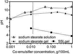
The pH of diluted gels using NaS as the co-emulsifier was compared with the pH of NaS solutions with similar NaS concentration in solution (Fig. 1). NaS alone forms micelles in water above the critical micellar concentration (CMC) of 0.0004 M (0.0123 g/100 mL), and each micelle containing 78 monomers.14,15 When the concentration of NaS dropped below the CMC, both the pH of NaS-MG-gels and NaS solution decreased by ∼1 when diluted ten fold. Above the CMC, the pH of NaS solutions remained stable around 10.5, closer to the pKa of stearic acid, while the pH of MG-gels with NaS remained around 9. In MG-gel systems with NaS concentration above its CMC, some NaS molecules acted as co-emulsifiers and incorporated into the lamellar structure formed by GMS, while others were predicted to form micelles. The stable pH above the CMC in both NaS solutions and MG-gels was possibly a result of micellisation of the co-emulsifier, as no free monomers were in solution to cause changes in pH.


image file: c5ra16457e-f1.tif
Fig. 1 The pH of diluted MG-gels and NaS solutions. The critical micellar concentration of NaS is marked with dotted line.

The d-spacings of MG-gels obtained from XRD experiments are summarized in Table 2. Freshly prepared MG-gels using SSL as the co-emulsifier displayed SAXS spacing representing the (001) plane at 53 Å and a single WAXS spacing at 4.2 Å under all the pH, indicating they are initially in the α-gel phase. MG-gels using NaS as the co-emulsifier under all the pH were also in the α-gel phase when freshly prepare, suggested by single WAXS spacing at 4.1 Å; however they showed slightly longer SAXS spacings compared with gels structured with SSL. After four weeks of incubation at 45 °C, all the MG-gels displayed SAXS spacings at 49 Å, suggesting that the MG-bilayers are all become more densely packed. However samples using NaS and SSL displayed different polymorphic forms, as suggested by WAXS spacings, at various environmental pH. MG-gel using SSL as the co-emulsifier at pH 5 displayed WAXS spacing at 4.1 Å and multiple spacings between 3.6 and 4.6 Å, meaning that the α-gel phase and the cogel phase coexist in the systems. At pH 7 and pH 10, only WAXS spacings between 3.6 and 4.6 Å were observed, meaning that the gel systems were all in the coagel phase. In MG-gels structured with NaS, multiple spacings between 3.6 and 4.6 Å, spacing at 4.1 Å and multiple spacings between 3.6 and 4.6 Å, and a single spacing at 4.1 Å were observed at pH 5, pH 7, and pH 10 respectively. The MG-gels structured with NaS transformed to the coagel phase at pH 5, formed a mixture of the α-gel phase and the coagel phase at pH 7, and was in the α-gel phase at pH 10.

Table 2 d-spacings of MG-gels obtained form small angle X-ray diffraction (SAXS) and wide angle X-ray diffraction (WAXS) experiments when samples were freshly prepared and after storage at 45 °C for four weeks
Co-emulsifier pH Day 0 Week 4
SAXS (Å) WAXS (Å) SAXS (Å) WAXS (Å)
SSL 5 53 4.2 49 4.1, 3.6–4.6
7 53 4.2 49 3.6–4.6
10 53 4.2 49 3.6–4.6
NaS 5 57 4.1 49 3.6–4.6
7 55 4.1 49 4.1, 3.6–4.6
10 55 4.1 49 4.1


The melting profiles of pH-adjusted MG-gels were examined with DSC during storage, and the melting profiles of MG-gels using SSL are summarized in Fig. 2. Gels adjusted to pH 5, 7 and 10 displayed similar melting profiles at day 0, indicating that differences in pH did not change the initial structure of the gels. When the pH was increased slightly to 5 (Fig. 2a), gels preserved similar melting curves after five weeks of incubation, during which the solid-state phase transition from the sub-α-gel phase to the α-gel phase (∼13 °C) was observed and the area under the melting peak of the α-gel phase (∼60 °C) increased slightly. When increasing the pH to 7 (Fig. 2b), the area under the peak of the sub-α-gel phase decreased faster and the area under the α-gel phase increased faster than pH 5 samples, but both peaks were observed for five weeks. Further increasing the pH to 10 (Fig. 2c) led to the disappearance of the peak representing the sub-α-gel phase after two weeks, and the area under the melting peak at ∼60 °C increased dramatically throughout this period of time. The melting profiles showed a trend that adjusting the pH further from the original pH of the gels leads to a faster change of their melting profile upon aging, in agreement with XRD results. The relationship between pH and rate of coagel formation in MG-gels is also confirmed by the calculated Coagel Index (Fig. 2d). The CI of MG-gels adjusted to pH 5 displayed the slowest increment in CI, which was 1.4 after four weeks; while gels adjusted to pH 7 and 10 displayed higher CIs and reached 1.7 and 1.8 respectively after four weeks of storage at 45 °C.


image file: c5ra16457e-f2.tif
Fig. 2 Melting curves of MG-gels structured using SSL as the co-emulsifier. The pH of the gels was adjusted to (a) 5, (b) 7, and (c) 10, and (d) their Coagel Indices were calculated.

Samples prepared with NaS had a gel-like texture when adjusted to pH 7 and 10; when adjusted to pH 5, the system displayed a loosely packed grainy structure instead of a gel-like structure, indicating that the pH was too low to form a desirable α-gel phase. The calculated CI of samples structured with NaS were all lower than 1.5 when storing the MG-gels for four weeks, suggesting that NaS helped structure a stable α-gel phase formed by GMS, in agreement with previous works published by our group.9 NaS as a free fatty acid salt therefore helps increase the water swelling capacity and stability of the α-gel phase.10

Interestingly, MG-gels using NaS as the co-emulsifier adjusted to different pH showed different melting profiles when prepared fresh (Fig. 3a). MG-gels adjusted to pH 5 and 7 showed similar initial melting profiles with the gels using SSL; gels adjusted to pH 10 displayed a distinct peak representing the sub-α-gel phase, however the peak profile was not complete at 1 °C. No DSC cycles were conducted below 0 °C because ice crystal formation affects the structural properties of MG-gels. Adjusting the pH of the gel systems changed the degree of neutralization of NaS, possibly affecting the electrostatic repulsion between the co-emulsifier molecules, thus changing the microstructure of the sub-α-gel phase and showing different melting profiles. After four weeks of incubation at 45 °C, the melting curves of the gels showed similar shape and area under the melting peak at ∼60 °C compared with freshly prepared samples structured with NaS. However the shape of the endothermic peak at ∼13 °C representing the polymorphic transformation from the sub-α-gel phase to the α-gel phase displayed different shape from freshly prepared samples. No well-defined endothermic peak was observed at ∼13 °C; instead, the baseline at that temperature range became wavy. Changes in the endothermic peak representing the phase transformation from the sub-α-gel phase to the α-gel phase upon aging further suggested that environmental pH changed the structured of the sub-α-gel phase but did not affect the α-gel phase.


image file: c5ra16457e-f3.tif
Fig. 3 Melting profiles of MG-gels using NaS as the co-emulsifier adjusted to various pH when (a) freshly prepared and (b) after four weeks of incubation at 45 °C.

ΔHsubα and ΔHα of MG-gels using SSL and NaS co-emulsifier are both affected by pH, as summarized in Table 3. In both MG-gels using SSL and NaS, higher ΔHsubα and ΔHα were associated with slower increase in CI (Fig. 2d), i.e. the CI of gels after four weeks of storage at 45 °C increased in the order of NaS adjusted to all the pH values, SSL at pH 5, SSL at pH 7, and SSL at pH 10. Higher ΔHsubα and ΔHα in MG-gels represents the lager energy gradient of phase transition, leading to higher stability of the α-gel phase. In MG-gels using SSL, increasing the pH caused a decrease in ΔHsubα and ΔHα. Such drops in the enthalpy are indicative of a diminished energy gradient of the polymorphic transformation from the α-gel phase to the coagel phase, and thus, the gels adjusted to a higher pH displayed a faster increase in CI. MG-gels with NaS displayed a more complex change in their enthalpy while adjusting the pH. The possible mechanism is that the dissociation and micellisation of the co-emulsifiers at various pH values not only changed the energy gradient of the phase transition but also changed the packing structure of the MG-gels. Found similar in the two MG-gels was that higher α-gel stability was obtained with environmental pH closer to the pKa of the conjugated acid (lactic acid and stearic acid) of the co-emulsifier incorporated in the system. Adjusting the environmental pH of the MG-gels away from the pKa of these co-emulsifiers may have altered the dissociation of these co-emulsifiers, which led to excess amount of electrostatic repulsion between the MG-bilayers resulting in water release and gel destabilization.

Table 3 Enthalpy (J g−1) obtained from the solid-state phase transition from the sub-α-gel phase to the α-gel phase (ΔHsubα), and from melting the α-gel phase (ΔHα) after adjusting the MG-gels' pH. Measurements were taken when the gels were freshly prepared
  SSL NaS
ΔHsubα ΔHα ΔHsubα ΔHα
pH 5 6.35 ± 0.40 16.79 ± 0.85 7.04 ± 0.68 19.37 ± 1.79
pH 7 5.96 ± 0.92 16.55 ± 1.43 5.28 ± 0.64 16.66 ± 0.95
pH 10 3.17 ± 0.38 13.65 ± 1.15 2.04 ± 1.27 20.11 ± 1.11


4. Conclusion

This work examined the effect of pH on the stability of the α-gel phase in GMS-structured gel systems using SSL and NaS as co-emulsifiers. Adjusting the pH altered the melting profile of MG-gels, where SSL formed a stable α-gel phase in MG-gels at slightly acidic pH. On the other hand, NaS formed a stable α-gel phase in MG-gels at neutral to alkaline pH. MG-gels prepared using NaS formed a more stable α-gel phase, but the co-emulsifiers possibly arranged in micelles and incorporated into the lamellar structure of GMS at the same time. In practice, environmental pH should be taken into consideration together with the type and concentration of co-emulsifiers used in MG-structured systems.

References

  1. N. Krog and K. Larsson, Chem. Phys. Lipids, 1968, 2, 129–143 CrossRef CAS PubMed .
  2. N. Krog and A. P. Borup, J. Sci. Food Agric., 1973, 24, 691–701 CrossRef CAS .
  3. K. Larsson, K. Gabrielsson and B. Lundberga, J. Sci. Food Agric., 1978, 909–914 CrossRef CAS .
  4. G. Cassin, C. de Costa, J. P. M. van Duynhoven and W. G. M. Agterof, Langmuir, 1998, 14, 5757–5763 CrossRef CAS .
  5. A. Sein, J. a. Verheij and W. G. M. Agterof, J. Colloid Interface Sci., 2002, 249, 412–422 CrossRef CAS PubMed .
  6. A. Zetzl, M. Ollivon and A. Marangoni, Cryst. Growth Des., 2009, 9, 3928–3933 CAS .
  7. G. Y. Brokaw, W. C. Lyman and D. P. Industries, J. Am. Oil Chem. Soc., 1958, 35, 5–8 CrossRef .
  8. F. C. Wang and A. Marangoni, RSC Adv., 2014, 4, 50417–50425 RSC .
  9. F. C. Wang and A. Marangoni, RSC Adv., 2015, 5, 43121–43129 RSC .
  10. I. Heertje, E. Roijers and H. A. C. Hendrickx, LWT--Food Sci. Technol., 1998, 31, 387–396 CrossRef CAS .
  11. N. Krog, in Food Emulsions, ed. S. E. Friberg and K. Larsson, Marcel Dekker, New York, 3rd edn, 1997 Search PubMed .
  12. L. Mao, S. Calligaris, L. Barba and S. Miao, Frin, 2014, 58, 81–88 CAS .
  13. J. R. Kanicky and D. O. Shah, J. Colloid Interface Sci., 2002, 256, 201–207 CrossRef CAS PubMed .
  14. H. A. Capelle, L. G. Britcher and G. E. Morris, J. Colloid Interface Sci., 2003, 268, 293–300 CrossRef CAS PubMed .
  15. Introduction to Colloid & Surface Chemistry, ed. D. J. Shaw, Butterworth Heinemann, Oxford, 4th edn, 1992, pp. 84–93 Search PubMed .

This journal is © The Royal Society of Chemistry 2015
Click here to see how this site uses Cookies. View our privacy policy here.