DOI:
10.1039/C5RA15793E
(Paper)
RSC Adv., 2015,
5, 76607-76614
Effects of spermine on the morphology, digestive enzyme activities, and antioxidant status of jejunum in suckling rats
Received
6th August 2015
, Accepted 3rd September 2015
First published on 4th September 2015
Abstract
Spermine is a ubiquitous cellular component that plays vital roles in the maintenance of nucleic acids, regulation of kinase activities, protein synthesis, control of ion channel activities and renewal of the gut epithelium. However, knowledge of the effects of spermine supplementation and its duration extension on intestinal growth and antioxidant capacity is lacking. The present work aims to investigate the effects of spermine administration and its extended supplementation on the morphology, digestive enzyme activities, and antioxidant status of the jejunum in suckling rats. The rats received 0.2 μmol g−1 body weight of either spermine or saline solution via intragastric ingestion for 3 or 7 d, and jejunum samples obtained 24 h after the last spermine supplementation were analyzed. The results demonstrated that the specific activity of maltase and the total antioxidant capacity (T-AOC) in rat jejunum were significantly increased by 105.5% and 11.1%, respectively; in contrast, lactase activity was significantly decreased by 34.8% (spermine group versus the control group). Time extension of spermine administration (7 d) significantly increased the villus height, villus width, surface area, and crypt depth in rat jejunum by 11.2%, 18.2%, 5.9%, and 50%, respectively, but significantly decreased the specific activities of lactase, maltase, alkaline phosphatase, and malondialdehyde content by 26.8%, 36.4%, 41.3%, and 26.0% (P < 0.05), respectively. Protein content, sucrase, catalase, T-SOD, and anti-superoxide anion activities were also increased by 23.1%, 424.2%, 45.7%, 11.7%, and 26.4% (P < 0.05), respectively, relative to the levels observed during spermine administration for 3 d. Taken together, the results suggest that spermine administration and extension of its supplementation duration can accelerate gut development and enhance antioxidant properties.
Introduction
The suckling period is an important phase of intestinal growth, and the small intestine develops at a rate faster than many other organs (e.g., stomach) in the body during this stage.1 The intestine is a major interface for substance and energy exchange between an animal and its environment; this part of the digestive system is also the first barrier of the body's defense system against hazardous substances including carcinogens, dietary-derived mutagens, and oxidants. Besides its vital action in nutrient digestion and absorption, the intestine can maintain an organism's health.2–4 This organ plays a crucial role in nutrient supply and is related to the functions of the entire organism associated with its epithelium.5 The gut epithelium is constantly renewed, but the proliferation and migration of small intestinal epithelial cell in suckling animals is slow. Upon weaning, the dynamics and morphology of the intestinal epithelium exhibits significant changes. In fact, villous atrophy and crypt hyperplasia, which can induce transient decreases in the digestive and absorptive ability of the small intestine, have been observed in the gut structure after weaning. Many health problems during the animal growth process are associated with the status and structures of the small intestine, and poor small intestinal development in lactating animals can limit the performance of the animal during weaning. Thus, maintaining the normal functions of the small intestine or promoting intestinal maturity is essential for animal growth and development in livestock production. A previous study has indicated that problems associated with immature intestinal development in animals can be alleviated through nutritional regulation.6
Spermine, a novel natural antioxidant agent in vitro, can be found in the cells of all animate organisms as a low-molecular weight aliphatic cation. It contributes to synthesizing proteins, stabilizing cellular macromolecules (e.g., nucleic acids), controlling cell growth and differentiation, alleviating intestinal dysfunction, promoting immune function, protecting from oxygen toxicity, adjusting cell membrane transport, and regulating calcium-related signal transduction.7–9 Spermine administration to neonatal rats makes the gradual alteration of the structure, and thus results in maturation of the small intestine. Previous experiments demonstrate that administration of appropriate doses of spermine promotes small intestine epithelium migration and damage repair.10 Spermine can also affect enzyme activities in the small intestine of rat.11 Considering its many health benefits, spermine has received considerable attention as an important bioactive antioxidant and anti-inflammatory agent to protect organisms from free radicals.12 The polyamine can potentially facilitate small intestine development in lactating animals and change the antioxidant status of young animals. To the best of our knowledge, the effects of spermine on intestinal growth and development in animals generally requires 3 d before observation. Few experiments have been conducted to study the effects of extended spermine administration on the small intestinal development, and antioxidant status of animals. As such, further investigation is necessary.
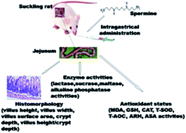
This study is part of a larger study that involved determining the metabolomic effects of spermine and extended spermine administration in suckling rats.13 In this work, the effects of spermine supplementation and its extended administration on the jejunum development and antioxidant status of lactating rats is evaluated. In particular, histomorphological structures, digestive enzyme activities, and antioxidant parameters in the jejunum were examined. The results can provide scientific evidence of the ability of spermine to promote intestinal development and alter the intestinal antioxidant status and may pave the way for the development of spermine as a functional food.
Materials and methods
Materials
Pregnant Sprague-Dawley (SD) rats and their food were supplied by Dossy Experimental Animals Co., Ltd (Chengdu, China). Spermine (S3256-1G) was purchased from Sigma Chemical Co. (St. Louis, MO, USA). All antioxidative reagents and enzyme assay kits used in this work were obtained from Nanjing Jiancheng Bioengineering Institute (China).
Animal experiment and sample collection
The animal experimental procedures were conducted in accordance with the Animal Care and Use Committee of the Animal Nutrition Institute of Sichuan Agricultural University and performed according to the guidelines approved by the Care and Use of Laboratory Animals of the National Research Council. A total of forty eight-day-old suckling SD rats were randomly divided into four groups (n = 10 per group): control-3 days (group 1), control-7 days (group 2), spermine-3 days (group 3), and spermine-7 days (group 4). Ten litters from each of four male pups were employed and habituated in individual cages. The day of birth was designated as day 0. Mothers were provided ad libitum access to food and drinking water. Half of the treated pups in each litter received intragastric supplementation of spermine (0.2 μmol g−1 body weight; spermine was dissolved in physiological saline) for 3 or 7 d. The remaining pups from the same litter were considered controls and received normal saline in the same manner once a day for 3 or 7 d. At the end of the 3 and 7 day feeding experiments (by this time, the pups were 11 and 15 days old, respectively), jejunum samples (5 cm from end of the duodenojejunal junction) were immediately harvested from the small intestine under ether anesthesia, washed in cold saline (0.9% NaCl; 4 °C), frozen in liquid nitrogen, and then transferred to storage at −80 °C for enzymatic and antioxidant status analysis. The spermine dosage and time of spermine administration were set according to a prior experiment.14,15 Ambient temperatures within the range of 23–25 °C, 50%–70% relative humidity, and a 12 h light/12 h dark cycle were maintained throughout the whole period of the study.
Jejunum histomorphological studies
A 1 cm-long jejunum segment (5 cm from end of the duodenojejunal junction) was fixed in 10% buffered neutral formaldehyde. The fixed tissue samples were dehydrated with normal saline and then embedded in paraffin. Cross sections of each sample were prepared, stained with hematoxylin and eosin, and then sealed by neutral resin size. Ultrathin sections of the jejunum samples were used to examine the villus height, crypt depth, and width with image processing and analysis system (Image Pro Plus, Media Cybernetics, Bethesda, MD, USA). Villus height was examined from the tip of the villi to the villus-crypt junction, and width was measured at half of the villus height.16 Crypt depth was expressed as the invaginated depth between adjacent villi. A total of 10 intact, well-oriented crypt-villus units were analyzed in triplicate per segment. The villus surface area was calculated as described by the following formula.
Villus surface area (mm2) = 2π(villus width/2) × (villus height) |
Biochemical analysis of jejunum
Tissue preparation. Jejunum sample preparation was completed using the method of Zhang et al.17 About 0.1 g of jejunum was quickly weighed, thawed, and homogenized in 10 volumes (w/v) of ice-cold normal saline (0.7 g mL−1). The mixture of jejunum and physiological saline was centrifuged at 6000 × g for 20 min at 4 °C. The supernatant was acquired and stored at −20 °C used for enzyme activity examination.
Assay of disaccharidase activities. The specific activities of sucrase, lactase, and maltase in the jejunum supernatant were estimated by using the corresponding diagnostic kits (Nanjing Institute of Jiancheng Biological Engineering, China) according to the instructions of the manufacturer and measurement methods described in a previous study.18 Sucrase, lactase, and maltase activities were determined by a microplate reader (SpectraMax M2, Molecular Devices, USA) at 450, 450, and 505 nm, respectively. One unit of maltase is defined as the amount of enzyme that hydrolyzes 1 nmol of maltase per milligram of protein per minute at pH 6.0 and 37 °C. Sucrase and lactase units were similarly defined. The protein content of the supernatant was assayed by using a protein quantification kit (Coomassie brilliant blue) with bovine serum-albumin as protein standard; results are presented as milligrams per gram of wet weight of intestine.
Alkaline phosphatase activity examination. Alkaline phosphatase (APK) activity in the jejunum was detected using disodium phenyl phosphate as the substrate with APK test kits purchased from Jiancheng Bioengineering Institute (Nanjing, China) at 520 nm. One alkaline phosphatase unit was defined as the quantity of enzyme required to produce 1 mg of phenol per 15 min at 37 °C.
Measurements of antioxidant-related enzymes. The activities of jejunum antioxidant-related enzymes, such as catalase (CAT), anti-superoxide anion (ASA), glutathione (GSH), malondialdehyde (MDA), total superoxide dismutase (T-SOD), total antioxidant capacity (T-AOC), and anti-hydroxyl radical (AHR), were determined, and all corresponding detection kits were supplied by the Nanjing Institute of Jiancheng Biological Engineering (China). ASA and AHR abilities were measured in accordance with the method described by Jiang et al.19 Superoxide radicals (O2−) was generated by the reaction of xanthine and xanthine oxidase; here, a coloration reaction was developed using the Griess reagent, with the addition of an electron acceptor. The coloration degree can directly express the quantity of superoxide anion in the reaction. One ASA unit is defined as the quantity of superoxide anion free radicals required to scavenge 1 milligram of tissue protein for 40 min at 37 °C. Hydroxyl free radicals (OH−) are generated on the basis of the Fenton reaction (Fe2+ + H2O2 → Fe3+ + OH− + ·OH), and, upon addition of the electron acceptor, a coloration reaction is developed using the Griess reagent. The degree of coloration in this system is directly proportional to the quantity of hydroxyl radicals in the reaction. AHR activity was expressed as units per milligram of protein. MDA content was analyzed followed the protocol described by Livingstone et al. via the thiobarbituric acid reaction.20 MDA content was detected at 532 nm, and results are presented as nanomoles per milligram of protein. SOD activity was measured according to the method of Zhang et al.17 and one unit of this enzyme was defined as the quantity of enzyme required to produce 50% inhibition of nitric ion production. CAT activity was determined through decomposition of hydrogen peroxide;21 one unit of CAT activity was defined as the amount of enzyme required to decompose 1 mmol L−1 H2O2 within 1 s per milligram of tissue protein at 37 °C. T-AOC was measured using the colorimetric technique according to the method described by Nicholas et al.22 All antioxidants can reduce Fe3+ to Fe2+, and the latter develops colored and stable chelates when combined with phenanthroline. The T-AOC of jejunum was expressed as units per milligram of protein. GSH content, which was expressed as milligram per gram of protein, was assayed in terms of the formation of 5-thio-2-nitrobenzoate and detected spectrophotometrically at 412 nm.23 Commercial GSH was used as a standard.
Statistical analysis. All experimental data were statistically analyzed by two-way ANOVA using the general linear model procedure of SPSS 17.0 (SPSS Inc., Chicago, IL, USA) and are expressed as mean ± SD. The main effects of the model included spermine level (0 or 0.2 μmol g−1 body weight) and treatment time (3 or 7 d). Statistical differences were determined among treatments by the Duncan method. Differences were considered statistically significant at P < 0.05.
Results
Morphological structure in suckling rat jejunum
Table 1 presents the morphological indices of jejunum. The villus height, villus width, surface area, and crypt depth of the rat jejunum were increased by 11.2%, 18.2%, 5.9%, and 50%, respectively [duration extension of spermine supplementation (7 days) relative to 3 days] (P < 0.05). However, differences between the spermine and control groups were not significant (P > 0.05).
Table 1 Effects of spermine and its duration extension on morphology of suckling rat jejunuma
Groups |
Spermine (μmol g−1 BW) |
Time extension of spermine (days) |
Villus height (μm) |
Villus width (μm) |
Crypt depth (μm) |
Villus height/crypt depth (μm) |
Villus surface area (mm2) |
Data are expressed as mean ± SD. Different superscript letters (b and c) show significant difference (P < 0.05) between groups (group1, group2, group3, group4), between spermine (0, 0.2 μmol g−1 BW, main effects), and between time extension of spermine (3, 7 days, main effects) for one of all parameters (villus height, villus width, crypt depth, villus width/crypt depth, villus surface area), respectively. |
1 |
0 |
3 |
264.93 ± 8.77 |
71.70 ± 3.11b |
68.45 ± 0.99 |
3.87 ± 0.13 |
0.06 ± 0.00b |
2 |
0 |
7 |
303.86 ± 20.58 |
71.60 ± 3.66b |
70.34 ± 2.56 |
4.32 ± 0.26 |
0.07 ± 0.01bc |
3 |
0.2 |
3 |
316.98 ± 10.37 |
82.27 ± 4.02c |
72.13 ± 1.50 |
4.24 ± 0.10 |
0.08 ± 0.01c |
4 |
0.2 |
7 |
315.24 ± 14.02 |
87.09 ± 2.54c |
74.90 ± 1.96 |
4.22 ± 0.21 |
0.09 ± 0.01c |
![[thin space (1/6-em)]](https://www.rsc.org/images/entities/char_2009.gif) |
Main effects |
Spermine |
0 |
|
290.95 ± 10.03 |
76.99 ± 2.39 |
70.29 ± 1.31 |
4.06 ± 0.14 |
0.07 ± 0.00 |
0.2 |
|
309.55 ± 10.03 |
79.34 ± 2.39 |
72.62 ± 1.31 |
4.27 ± 0.13 |
0.08 ± 0.00 |
Time extension of spermine |
|
3 |
284.39 ± 10.03b |
71.65 ± 2.39b |
69.40 ± 1.31b |
4.10 ± 0.13 |
0.06 ± 0.00b |
|
7 |
316.11 ± 10.03c |
84.68 ± 2.39c |
73.52 ± 1.31c |
4.23 ± 0.14 |
0.09 ± 0.00c |
![[thin space (1/6-em)]](https://www.rsc.org/images/entities/char_2009.gif) |
P value |
Spermine |
|
|
0.200 |
0.492 |
0.217 |
0.267 |
0.251 |
Time extension of spermine |
|
|
0.033 |
0.001 |
0.034 |
0.483 |
0.002 |
Spermine × time extension of spermine |
|
|
0.163 |
0.472 |
0.814 |
0.232 |
0.664 |
Digestive enzyme activities of jejunum in suckling rat
The specific activity of maltase in rat jejunum was significantly increased by 105.5%, whereas lactase activity was significantly decreased by 34.8% (P < 0.05) for spermine supplementation relative to the control group (Table 2); but protein contents and APK and sucrase activities were not affected (P > 0.05) by spermine supplementation relative to the control group (Table 2). Sucrase activity and protein contents of the rat jejunum were higher (by 424.2% and 23.1%, respectively), and the specific activities of lactase, maltase, and APK were lower (by 26.8%, 36.4%, and 41.3%, respectively) in 7 d of spermine supplementation than in 3 d of spermine supplementation (P < 0.05).
Table 2 Effects of spermine and its duration extension on the specific enzymes activities of suckling rat jejunuma
Groups |
Spermine (μmol g−1 BW) |
Time extension of spermine (days) |
Protein content (mg g−1 tissue) |
Lactase activity (U mg−1 protein) |
Sucrase activity (U mg−1 protein) |
Maltase activity (U mg−1 protein) |
Alkaline phosphatase activity (U mg−1 protein) |
Data are expressed as mean ± SD. Different superscript letters (b–d) show significant difference (P < 0.05) between groups (group1, group2, group3, group4), between spermine (0, 0.2 μmol g−1 BW, main effects), and between time extension of spermine (3, 7 days, main effects) for one of all parameters (protein content, lactase activity, sucrase activity, maltase activity, alkaline phosphatase activity), respectively. |
1 |
0 |
3 |
79.8 ± 2.4b |
108 ± 12b |
0.47 ± 0.29b |
44.4 ± 4.4b |
2572 ± 203b |
2 |
0 |
7 |
87.7 ± 3.3c |
58.6 ± 8.7c |
2.17 ± 0.56b |
91.9 ± 16.8c |
2354 ± 219b |
3 |
0.2 |
3 |
104 ± 1d |
66.5 ± 7.6c |
3.60 ± 2.64b |
28.7 ± 6.1b |
1542 ± 203c |
4 |
0.2 |
7 |
101 ± 2d |
55.1 ± 4.8c |
10.3 ± 3.1c |
58.0 ± 10.0b |
1350 ± 153c |
![[thin space (1/6-em)]](https://www.rsc.org/images/entities/char_2009.gif) |
Main effects |
Spermine |
0 |
|
91.9 ± 1.8 |
87.1 ± 6.2b |
2.03 ± 1.45 |
36.5 ± 8.1b |
2057 ± 136 |
0.2 |
|
94.6 ± 1.7 |
56.8 ± 6.2c |
6.21 ± 1.45 |
75.0 ± 7.5c |
1852 ± 141 |
Time extension of spermine |
|
3 |
83.7 ± 1.7b |
83.1 ± 6.2b |
1.32 ± 1.45b |
68.2 ± 7.8b |
2463 ± 141b |
|
7 |
103 ± 2c |
60.8 ± 6.2c |
6.92 ± 1.45c |
43.4 ± 7.8c |
1446 ± 136c |
![[thin space (1/6-em)]](https://www.rsc.org/images/entities/char_2009.gif) |
P value |
Spermine |
|
|
0.288 |
0.002 |
0.053 |
0.002 |
0.303 |
Time extension of spermine |
|
|
0.000 |
0.016 |
0.012 |
0.033 |
0.000 |
Spermine × time extension of spermine |
|
|
0.044 |
0.039 |
0.239 |
0.419 |
0.948 |
Activities of antioxidant parameters in rat jejunum
Based on the results listed in Table 3, spermine supplementation promoted a increase in T-AOC activity (by 11.1%, P < 0.05), but had no effect on MDA and GSH contents or CAT, ASA, T-AOC and AHR activities of the rat jejunum relative to the controls (P > 0.05). Compared with results obtained from 3 d of spermine administration, the activities of CAT, T-SOD, and ASA in the jejunum were significantly increased by 45.7%, 11.5%, and 26.4%, respectively, but MDA content was decreased by 26.0% (P < 0.05) after 7 d of spermine administration.
Table 3 Effects of spermine and its duration extension on the antioxidant status of suckling rat jejunuma
Groups |
Spermine (μmol g−1 BW) |
Time extension of spermine (days) |
MDA (nmol mg−1 protein) |
CAT (U mg−1 protein) |
GSH (mg g−1 protein) |
T-AOC (U mg−1 protein) |
T-SOD (U mg−1 protein) |
ASA (U g−1 protein) |
AHR (U mg−1 protein) |
Data are expressed as mean ± SD. CAT, catalase; ASA, anti-superoxide anion; GSH, glutathione; MDA, malondialdehyde; T-SOD, total superoxide dismutase; T-AOC total antioxidant capacity; AHR, anti-hydroxyl radical. Different superscript letters (b and c) show significant difference (P < 0.05) between groups (group1, group2, group3, group4), between spermine (0, 0.2 μmol g−1 BW, main effects), and between time extension of spermine (3, 7 days, main effects) for one of all parameters (CAT, GSH, MDA, T-SOD, T-AOC, ASA, AHR), respectively. |
1 |
0 |
3 |
0.51 ± 0.14 |
2.85 ± 1.40 |
6.77 ± 2.21 |
1.15 ± 0.18b |
30.0 ± 3.7 |
142 ± 21 |
101 ± 23 |
2 |
0 |
7 |
0.48 ± 0.21 |
2.97 ± 0.63 |
8.54 ± 4.46 |
1.37 ± 0.13c |
31.7 ± 4.8 |
153 ± 25 |
111 ± 38 |
3 |
0.2 |
3 |
0.41 ± 0.08 |
3.88 ± 1.11 |
9.30 ± 4.00 |
1.36 ± 0.22c |
32.5 ± 3.1 |
185 ± 29 |
106 ± 31 |
4 |
0.2 |
7 |
0.34 ± 0.09 |
4.61 ± 1.58 |
10.5 ± 4.1 |
1.42 ± 0.15c |
36.4 ± 4.6 |
190 ± 20 |
117 ± 49 |
![[thin space (1/6-em)]](https://www.rsc.org/images/entities/char_2009.gif) |
Main effects |
Spermine |
0 |
|
0.45 ± 0.04 |
3.42 ± 0.33 |
8.03 ± 1.07 |
1.26 ± 0.05b |
31.3 ± 1.1 |
163 ± 6 |
104 ± 9 |
0.2 |
|
0.42 ± 0.04 |
3.73 ± 0.33 |
9.52 ± 1.03 |
1.40 ± 0.05c |
34.1 ± 1.1 |
171 ± 6 |
114 ± 9 |
Time extension of spermine |
|
3 |
0.50 ± 0.04b |
2.91 ± 0.33b |
7.66 ± 1.07 |
1.26 ± 0.05 |
30.9 ± 1.1b |
147 ± 6b |
106 ± 9 |
|
7 |
0.37 ± 0.04c |
4.24 ± 0.33c |
9.89 ± 1.03 |
1.39 ± 0.05 |
34.5 ± 1.1c |
187 ± 6c |
113 ± 9 |
![[thin space (1/6-em)]](https://www.rsc.org/images/entities/char_2009.gif) |
P value |
Spermine |
|
|
0.653 |
0.519 |
0.326 |
0.039 |
0.085 |
0.393 |
0.441 |
Time extension of spermine |
|
|
0.035 |
0.009 |
0.144 |
0.053 |
0.031 |
0.000 |
0.705 |
Spermine × time extension of spermine |
|
|
0.337 |
0.368 |
0.847 |
0.203 |
0.482 |
0.754 |
0.997 |
Discussion
Spermine intake and time extension of spermine supplementation induce changes to the morphological structure of the jejunum
The morphological structure of the small intestine is closely related to its functional status, and the integrity of the intestinal morphology allows the small intestine to perform normal physiological functions. Proliferation of small intestine involves increases in the gut surface area and intestinal epithelial cell quantity; villi and crypt hyperplasia are also embodied in intestinal morphology.24,25 While villi and crypts exhibit proliferative activity during the embryonic period, the proliferative ability of intestinal epithelial cells is confined to the crypt when the animal is born.26 Crypt proliferation is significantly and positively correlated with its depth and responsible for reflecting the status of enterotype proliferation.27 Hyperplasia of the villus and crypt can lead to secretion of more digestive enzymes into the intestinal tract and enlargement of the intestinal mucosa area, both of which enhance intestinal digestion and absorption and allow the gut to accommodate additional nutrients. Intestinal villi play an important role in digesting and absorbing nutrients, and the villus height and width are highly associated with the amount of mature intestinal epithelia available; intestinal epithelia are responsible for digesting and absorbing various nutritional substances.28 The villus surface area is considered an important morphological parameter for nutrient digestion and absorption because it is directly related to the absorption area of the intestinal mucosa.29 In the present study, spermine supplementation did not significantly affect the villus height, width, surface area, crypt depth, or villus/crypt ratio in the jejunum, which is not agree with our observation in the ileum (data not shown). This may be related to the time that spermine administration affects different intestinal segments, since jejunum is the proximal part of the small intestine which leads spermine to stay at the jejunum for a short time. However, we observed the 7 d of spermine administration results in higher values of villus height, width, surface area, and crypt depth compared with those obtained after 3 d of administration. These results indicate that time extension of spermine administration can change the morphological structure of the rat jejunum and promote the development of the small intestine in suckling rats. Together, the effects of spermine on the development of the small intestine partly depend on its action time. The morphological changes observed in the small intestine are intimately associated with alterations in enzyme activities observed.
Spermine administration and duration extension of spermine ingestion alter jejunum digestive enzyme activities
Intestinal disaccharidases are crucial for disaccharide decomposition; thus, disaccharides existing in the body cannot be directly absorbed without the digestive functions of the intestinal digestive enzymes. The hydrolytic activities of disaccharidases in the neonatal rat intestine are specific for, and mainly limited to, the components of maternal milk. Maternal milk is the primary source of nutritive materials for neonatal animals in the stages of breast feeding; this milk is relatively low in total carbohydrates and mainly composed of lactose. Lactase is an essential disaccharidase that reaches maximal activity during the first week after birth and decreases dramatically thereafter. Thus, decreases in lactase activity can be established as an intestinal development maturation marker for juvenile animals.30 Sucrase and maltase are intestinal brush border glycoside hydrolases responsible for the final steps of carbohydrate digestion, and changes of their activities allow the young animal to accommodate dietary changes after weaning. Increases in the specific activities of sucrase and maltase are believed to be the signs of intestinal development and maturation.31,32 The results of this study show that maltase activity was significantly increased in the jejunum by oral administration of spermine; by contrast, spermine administration significantly decreased the specific activity of lactase. These results demonstrate that spermine supplementation can induce small intestinal epithelial cell maturation and promote the development of intestines in rat, thereby agreeing with the results of previous studies.33,34 Time extension of spermine intake significantly enhanced sucrase activity and decreased the specific activities of lactase and maltose in the rat jejunum. This finding is also consistent with that of a previous work,34 which suggested that time extension of spermine supplementation can facilitate better intestinal development in suckling rat.
Brush border enzymes, such as APK, help animals, especially younger ones, utilize ingested nutrients for growth and development. APK is enterocyte differentiation-dependent; this enzyme, which can be observed along the crypt-villus unit and in the microvilli of enterocytes, accelerates nutrient intake and transport, is associated with the complete development of the digestive system, and plays major roles in fat absorption from milk or food as well as intestinal homeostasis and protection.35,36 Consequently, APK is regarded as a key marker enzyme of changes in the primary digestive and absorptive functions of the small intestine.36 The results of the present study reveal that APK activity in the rat jejunum is not significantly affected by spermine ingestion, although its levels decrease during extended spermine administration. This finding contrasts theoretical beliefs but is similar to previous observations in suckling rats.33 We speculate that the decrease in APK activity observed may be attributed to reductions in maltose activity; the relationship between APK and maltose activities requires further investigation.
The intestinal cellular membrane is mainly composed of intestinal proteins, and protein contents can reflect the degree of intestinal cell differentiation and maturity of an enterocyte. In the current study, extended spermine supplementation significantly enhanced protein contents in jejunum tissue; however, no effect was observed during regular (3 d) spermine intake, which suggests that duration extension of spermine administration can promote body protein synthesis and protect the integrity of the intestinal cytomembrane. Taken together, results demonstrate that oral spermine intake and time extension of spermine supplementation can promote the development of the small intestine in rats and improve intestinal digestive and absorptive functions for nutrient substances.
Spermine supplementation and time extension of spermine administration cause alterations in the antioxidant status of the jejunum
Suckling animals frequently suffer from oxidative stress induced by a number of factors, such as xenobiotics, pathogens, and other environmental factors.37 Several studies have further confirmed that oxidative stress changes the cellularity of rat digestive organs.23 To investigate the effects of spermine and time extension of spermine administration on the antioxidative reactions of jejunum in suckling rats, we assayed MDA contents, free radical scavenging abilities (ASA and AHR), and the activities of both enzymatic (CAT and T-SOD) and non-enzymatic (GSH and T-AOC) antioxidants in the rat jejunum. MDA is the end-product of lipid peroxidation and a highly reactive aldehyde; thus, MDA contents may be used as a marker of lipid peroxidation in the body.38 In the current study, time extension of spermine supplementation significantly decreased MDA contents in the jejunum, although no such effects were observed after regular (3 d) spermine intake. These findings demonstrate that time extension of spermine administration can improve the anti-lipid peroxidation capacity of the rat jejunum, which may be due to the role of spermine binding to membranes that can suppress lipid peroxidation through forming a compound with phospholipid polar head.
Lipid peroxidation damage is primarily caused by superoxide anions and hydroperoxyl radicals.39 ASA and AHR abilities are two indices that may be used to assess the total capacity of an organ to scavenge superoxide anions and hydroperoxyl radical, respectively.40 ASA activity was significantly elevated by duration extension of spermine administration in the jejunum of suckling rats, although no such effects in AHR capacity were observed. Regular (3 d) spermine supplementation did not affect the activities of ASA and AHR in the jejunum, which may be correlated with the tissue differences as described by the results in weaned rats we had investigated (data not listed) and this hypothesis warrants further study. These results suggest that enhancement of free radical scavenging ability partly depends on the extension of spermine supplementation.
Free radical scavenging abilities are beneficial to enzymatic and non-enzymatic antioxidant defense systems.39 The major enzymatic antioxidants are represented by T-SOD and CAT. SOD plays an important role in maintaining balance between body oxidation and antioxidation because it acts as a major endogenous antioxidant to protect cells against toxic compounds and oxygen radicals; this protection mechanism is achieved by converting two superoxide anions into H2O2 and O2, which present lower toxicity to the body, in the presence of CAT.41,42 CAT is considered the main enzyme responsible for the clearance of hydroxyl radicals because it decomposes H2O2 into oxygen and water.43 In the present study, time extension of spermine ingestion dramatically improved T-SOD and CAT activities in the jejunum compared with the corresponding values detected after 3 d of treatment, although no such result was determine after regular (3 d) spermine administration, which may be associated with the effects of spermine administration on ASA and ARH activities. These results reveal that time extension of spermine supplementation can improve the functions of antioxidant recovery systems to different extents via improving enzymatic antioxidant activities in suckling rats.
GSH and T-AOC are regarded as non-enzymatic antioxidants. GSH can protect the body from damage induced by free radicals both intracellularly and extracellularly in conjunction with many enzymatic processes that deplete H2O2 by converting GSH into oxidized glutathione and other mixed disulfides.44,45 T-AOC is associated with free radical scavenging capacity and can be used as an integrative marker of the total antioxidant capacity of the body and the protective ability of the non-enzymatic antioxidant defense system.46 Spermine administration and extended spermine supplementation demonstrated no significant influence on GSH content, which may be due to the AHR activity observed in the present study; thus, the relationship between GSH content and AHR activity requires further investigation. Moreover, T-AOC in the spermine groups showed significant enhancements compared with the controls. This result suggests that spermine intake can improve the antioxidant capacity of suckling rats. Taken together, the results demonstrate that spermine supplementation and time extension of spermine administration can maintain the redox balance to a certain extent in suckling rats, thereby providing evidence of improvements in small intestinal antioxidant ability in response to spermine administration and extended spermine supplementation.
Conclusions
This study suggests that time extension of spermine administration can promote the development and maturation of the jejunum structure in suckling rats, as evidenced by the improved villus height, villus width, villus surface area, and crypt depth observed in the present study. Spermine administration and extended spermine supplementation can enhance the ability of suckling rats to utilize digestive and absorptive nutrient substances for growth by enhancing or decreasing the specific activities of digestive enzymes and brush border enzymes in the jejenum. Non-antioxidant enzyme activities increased in the rat jejunum after spermine administration. Time extension of spermine supplementation exerted improved effects on the gradual development, maturation, and antioxidant status of suckling rat jejunum. Therefore, spermine may be considered a beneficial feed supplement for animals. To the best of our knowledge, this study is the first to report the response of animal intestinal antioxidant defense systems to spermine administration and its extended duration in vivo. Further study is necessary to determine the mechanism of the effects of spermine supplementation and extended spermine administration on the antioxidant capacity of animals.
Acknowledgements
We wish to thank the personnel of our teams for their ongoing assistance. We also would like to thank the National Natural Science Foundation of China (No. 31301986), and Specific Research Supporting Program for Discipline Construction in Sichuan Agricultural University (to G. Liu) for financial support.
References
- R. Zabielski, M. M. Godlewski and P. Guilloteau, J. Physiol. Pharmacol., 2008, 59, 35–54 Search PubMed.
- Y. Wang, E. Holmes, E. M. Comelli, G. Fotopoulos, G. Dorta, H. Tang, M. J. Rantalainen, J. C. Lindon, I. E. Corthésy-Theulaz, L. B. Fay, S. Kochhar and J. K. Nicholson, J. Proteome Res., 2007, 6, 3944–3951 CrossRef CAS PubMed.
- S. C. Bischoff, BMC Med., 2011, 9, 24 CrossRef PubMed.
- T. Y. Aw, Toxicol. Appl. Pharmacol., 2005, 204, 320–328 CrossRef CAS PubMed.
- P. Guilloteau, R. Zabielski, H. M. Hammon and C. C. Metges, Nutr. Res. Rev., 2010, 23, 4–22 CrossRef PubMed.
- C. Domeneghini, A. D. Giancamillo, G. Bosi and S. Arrighi, Vet. Res. Commun., 2006, 30, 331–342 CrossRef CAS PubMed.
- A. E. Pegg, IUBMB Life, 2014, 66, 8–18 CrossRef CAS PubMed.
- P. Busselen, J. Mol. Cell. Cardiol., 1991, 23, 237–247 CrossRef CAS.
- T. Kusano, T. Berberich, C. Tateda and Y. Takahashi, Planta, 2008, 228, 367–381 CrossRef CAS PubMed.
- O. Peulen, P. Deloyer, C. Grandfils, S. Loret and G. Dandrifosse, Livest. Prod. Sci., 2000, 66, 109–120 CrossRef.
- O. Peulen, C. Pirlet, M. Klimek, G. Goffinet and G. Dandrifosse, Arch. Physiol. Biochem., 1998, 106, 46–55 CrossRef CAS PubMed.
- E. Lovaas and G. Carlin, Free Radical Biol. Med., 1991, 11, 455–461 CrossRef CAS.
- G. M. Liu, T. Yan, T. T. Fang, G. Jia, X. L. Chen, H. Zhao, J. Wang and C. M. Wu, RSC Adv., 2015, 5, 48769–48778 RSC.
- G. M. Liu, T. T. Fang, T. Yan, G. Jia, H. Zhao, Z. Q. Huang, X. L. Chen, J. Wang and B. Xue, J. Agric. Food Chem., 2014, 62, 9035–9042 CrossRef CAS PubMed.
- P. Deloyer, O. Peulen and G. Dandrifosse, Exp. Physiol., 2005, 90, 901–908 CrossRef CAS PubMed.
- X. Y. Dong, Y. M. Wang, L. Dai, M. M. M. Azzam, C. Wang and X. T. Zou, Poult. Sci., 2012, 91, 1886–1892 CrossRef CAS PubMed.
- X. D. Zhang, Y. F. Zhu, L. S. Cai and T. X. Wu, Aquaculture, 2008, 280, 136–139 CrossRef CAS PubMed.
- A. Dalqhvist, Anal. Biochem., 1964, 7, 18–25 CrossRef.
- J. Jiang, T. Zheng, X. Q. Zhou, Y. Liu and L. Feng, Aquacult. Nutr., 2009, 15, 409–414 CrossRef CAS PubMed.
- D. R. Livingstone, P. G. Martinez, X. Michel, J. F. Narbonne, S. O'hara, D. Ribera and G. W. Winston, Funct. Ecol., 1990, 4, 415–424 CrossRef.
- H. Abei, Methods Enzymol., 1984, 105, 121–126 Search PubMed.
- N. J. Miller, C. Rice-Evans, M. J. Davies, V. Gopinathan and A. Milner, Clin. Sci., 1993, 84, 407 CrossRef CAS.
- N. Vardi, H. Parlakpinar, F. Ozturk, B. Ates, M. Gul, A. Cetin, A. Erdogan and A. Otlu, Food Chem. Toxicol., 2008, 46, 3015–3022 CrossRef CAS PubMed.
- F. M. Thompson, A. G. Catto-Smith, D. Moore, G. Davidson and A. G. Cummins, J. Pediatr. Gastroenterol. Nutr., 1998, 26, 506–512 CrossRef CAS.
- J. J. Herbst and P. Sunshin, Pediatr. Res., 1969, 3, 27–33 CrossRef CAS.
- M. S. Hedemann, S. Hojsgaard and B. B. Jensen, J. Anim. Physiol. Anim. Nutr., 2003, 87, 32–41 CrossRef CAS.
- D. J. Hampson, Res. Vet. Sci., 1986, 40, 32–40 CAS.
- A. B. R. Thomson and M. Keelan, Can. J. Physiol. Pharmacol., 1986, 64, 13–29 CrossRef CAS.
- W. F. Caspary, Am. J. Clin. Nutr., 1992, 55, 299–308 Search PubMed.
- E. Nsi-Emvo, J. F. Launay and F. Raul, Cell. Mol. Biol., 1987, 33, 335–344 CAS.
- S. J. Henning, Annu. Rev. Physiol., 1985, 47, 231–245 CrossRef CAS PubMed.
- G. Galand, Comp. Biochem. Physiol., Part B: Biochem. Mol. Biol., 1989, 94, 1–11 CrossRef CAS.
- O. Peulen, P. Deloyer and G. Dandrifosse, Reprod., Nutr., Dev., 2004, 44, 353–364 CrossRef CAS.
- E. Harada, Y. Hashimoto and B. Syuto, Comp. Biochem. Physiol., Part A: Mol. Integr. Physiol., 1994, 109, 667–673 CrossRef CAS.
- J. P. Lallès, Nutr. Rev., 2010, 68, 323–332 CrossRef PubMed.
- D. Lackeyram, C. B. Yang, T. Archbold, K. C. Swanson and M. Z. Fan, J. Nutr., 2010, 140, 461–468 CrossRef CAS PubMed.
- J. Yin, W. Ren, G. Liu, J. L. Duan, G. Yang and L. Wu, Free Radical Res., 2013, 47, 1027–1035 CrossRef CAS PubMed.
- M. A. Mansour, Life Sci., 2000, 66, 2583–2591 CrossRef CAS.
- M. Valko, D. Leibfritz, J. Moncol, M. T. D. Cronin, M. Mazur and J. Telser, Int. J. Biochem. Cell Biol., 2007, 39, 44–84 CrossRef CAS PubMed.
- W. D. Jiang, L. Feng, Y. Liu, J. Jiang and X. Q. Zhou, Aquacult. Res., 2009, 40, 955–962 CrossRef CAS PubMed.
- D. A. Nita, V. Nita, S. Spulber, M. Moldovan, D. P. Popa, A. M. Zagrean and L. Zagrean, J. Cell. Mol. Med., 2001, 5, 163–170 CrossRef CAS PubMed.
- I. Fridovich, Annu. Rev. Biochem., 1995, 64, 97–112 CrossRef CAS PubMed.
- M. Z. Jiang, H. Yan, Y. Wen and X. M. Li, Afr. J. Pharm. Pharmacol., 2011, 5, 2079–2085 CAS.
- M. Y. Hung, T. Y. C. Fu, P. H. Shih, C. P. Lee and G. C. Yen, Food Chem. Toxicol., 2006, 44, 1424–1431 CrossRef CAS PubMed.
- S. C. Das, U. P. Isichei, A. Z. Mohammed, A. A. Otokwula and A. Emokpae, Indian J. Clin. Biochem., 2005, 20, 35–42 Search PubMed.
- W. K. Ren, Y. L. Yin, G. Liu, X. L. Yu, Y. H. Li, G. Yang, T. J. Li and G. Y. Wu, Amino Acids, 2012, 42, 2089–2094 CrossRef CAS PubMed.
|
This journal is © The Royal Society of Chemistry 2015 |
Click here to see how this site uses Cookies. View our privacy policy here.