Cork extracts reduce UV-mediated DNA fragmentation and cell death

Ana R. Araújo*ab, David M. Pereira ab, Ivo M. Arosoab, Tânia Santosc, Maria T. Batistac, Mariana T. Cerqueiraab, Alexandra P. Marquesab, Rui L. Reisab and Ricardo A. Pires*ab
a3B's Research Group – Biomaterials, Biodegradables and Biomimetics, University of Minho, Headquarters of the European Institute of Excellence on Tissue Engineering and Regenerative Medicine, AvePark, 4806-909 Taipas, Guimarães, Portugal. E-mail: rpires@dep.uminho.pt; anarita.araujo@dep.uminho.pt
bICVS/3B's – PT Government Associate Laboratory, Braga, Guimarães, Portugal
cCentro de Estudos Farmacêuticos – Faculdade de Farmácia, Universidade de Coimbra, Pólo das Ciências da Saúde, Azinhaga de Santa Comba, 3000-548 Coimbra, Portugal

Received 5th August 2015 , Accepted 2nd November 2015

First published on 3rd November 2015


Abstract

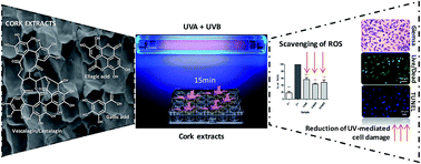
UV radiation is known to induce the premature aging of human skin and to contribute to the occurrence of different skin cancers. High doses of UVA (which is able to penetrate through the epidermis into the dermis) and/or UVB radiation (which only affects the epidermis) lead to cellular oxidative damage compromising the recovery of the normal functions of the cells. This cellular damage is mainly driven by the generation of reactive oxygen species (ROS) that alter the redox status of the intracellular milieu, affecting the cellular metabolic activity, leading to DNA damage, apoptosis and, consequently, to a drastic decrease in the number of live cells, compromising the function of the skin. A series of polyphenolic fractions were extracted from the outer bark (cork) of Quercus suber L., and tested for their capacity to reduce the cellular damage promoted by the ROS produced during UV exposure. This was evaluated after exposing L929 fibroblasts to UV radiation in the presence and absence of the cork extracts. In all the cases the extracts at the concentration of 75 μg ml−1 demonstrated the capacity to preserve cell metabolic activity and their typical morphology, as well as to avoid DNA fragmentation after exposure to UV radiation. We were also able to correlate these findings with the intracellular reduction of ROS species and the presence of higher proportions of castalagin and vescalagin in the extracts. Our data prove that cork is a relevant source of antioxidant compounds able to act in the cellular environment, protecting cells against oxidation, reducing the number of ROS species and limiting the negative impact of UV radiation. These extracts can be further exploited in the preparation of anti-UV formulations for skin protection.


Introduction

At the sea level, solar radiation is composed of ∼3% of Ultra-Violet (UV) light (100–400 nm), ∼44% of visible light (400–700 nm), and ∼53% of infra-red light (700–1440 nm).1 The UV section of the solar spectrum is composed of three types of UV rays, such as: UVA (315–400 nm); UVB (280–315 nm); and UVC (<280 nm).2 The ozone layer is able to adsorb the UVC wavelengths and a significant part of the UVB radiation that reaches the Earth. However, the small quantity of UVB that cross the ozone barrier is enough to interfere with the physiological balance (homeostasis) of tissues and cells.3 In the case of the UVA radiation, it has been associated to skin premature aging or long-term skin damage4 (e.g. wrinkles), as well as with skin cancer in individuals that presented a history of long periods of sunlight exposure. In fact, several epidemiological studies link the combined UVA and UVB exposure to an increased risk of occurrence of skin cancer.5,6

At the cellular level, UV irradiation induces the direct or indirect DNA degradation. This has been reported for the UVB range and for the high-energy artificial UVC radiation, through the dimerization of the pyrimidines nucleobases.7 Apoptosis is one of the hallmarks of UV cell damage.5,8 This UV-mediated cell death is the result of several biochemical processes, such as: formation of pyrimidine dimers and photoproducts,8,9 activation of death receptors (including CD95, i.e. Fas/APO-1),10 and formation of reactive oxygen species (ROS).9,11 ROS, such as, superoxide radical (˙O2), hydrogen peroxide (H2O2) and hydroxyl radical (˙OH), are known as key mediators of DNA and protein oxidative damage in skin cells. However, they also act in the oxidation of membrane lipids via the generation of lipids' peroxides and in the fast reduction in the activity of several endogenous skin enzymes, such as, reductase and catalase, as well as lowering the concentration of cellular antioxidants (e.g. glutathione).12,13

The reported relation between UV-mediated skin damage and the increment of ROS in the cellular milieu, led several authors to propose the use of phenolic compounds to neutralize the ROS generated by the UV radiation14–17 and to protect the skin cells from mutations or death.18–20 Cork (the outer bark of Quercus suber L.) is a suberized cellular tissue that present ∼15% of extractives,21 which can be easily obtained using suitable solvents.21–23 Several studies on the chemical composition of such extracts have reported the presence of phenolic acids, such as, gallic, protocatechuic, caffeic, and ferulic acids, which as natural antioxidants (AO) have been proven relevant in the biomedical area.22,24 Moreover, antibacterial, antiviral, anticarcinogenic, anti-inflammatory and antiallergic activities have been also attributed to these phenolic compounds.25

Considering the presence of these AOs in cork and the above-mentioned correlation between UV-mediated cell damage and the increased concentration of ROS in the cellular environment, we hypothesized that the cork polyphenols had the capacity to reduce the ROS oxidative damage generated in skin cells by UV radiation. In this context we evaluated the capacity of cork's AOs to reduce the negative impact of UVA and UVB radiation on cell function. This study was centred in the use of L929 fibroblast cell line due to the fact that fibroblasts are one of the main cell type present in skin, namely in the dermis, which is responsible for the generation of connective tissue, and for the skin's recovery from injury (since they synthesize most of the dermis's extracellular matrix).26–30 The cork extracts were characterized by HPLC to quantify the most abundant compounds. Its total content in phenolic acids was determined and then correlated with the AO activity of the extracts. The AO activity of the cork extracts and consequently their capacity to act as anti-UV agents was confirmed by their capacity to preserve cells' viability; depicting their typical morphology and, more importantly, to prevent DNA fragmentation.

Materials and methods

Unless otherwise stated, chemicals were acquired from Sigma-Aldrich and used without further purification.

Preparation of cork extracts

Two different raw extracts were obtained by maintaining cork powder (Amorim Cork Composites, Portugal) in contact with water (producing the CWE) or a mixture of water and ethanol (50[thin space (1/6-em)]:[thin space (1/6-em)]50 v/v, producing the CWEE), under reflux conditions, for a period of 6 h. After cooling, the liquid fraction was recovered by filtration, the solvent was partially removed by vacuum evaporation, and the final solid extracts were recovered upon freeze-drying. The CWEE was loaded to a chromatographic column, packed with Sephadex LH-20 as stationary phase and stabilized in water. The elution was performed using water, followed by mixtures of water and increasing percentages of ethanol, yielding fCWEE.

Chemical characterization of cork extracts

Reagents. Folin–Ciocalteu phenol reagent, gallic acid, 2,2-diphenyl-1-picrylhydrazyl (DPPH), 2,2-azobis (2-methylpropionamide) dihydrochloride (AAPH), fluorescein sodium salt and 6-hydroxy-2,5,7,8-tetramethylchromane-2-carboxylic acid (trolox) and ellagic acid were purchased from Sigma-Aldrich while methanol was acquired from Fluka. All reagents were used as received. Vescalagin and castalagin were obtained internally by preparative HPLC and identified by mass spectrometry.
Total phenolic content (TPC) and antioxidant (AO) activity. The TPC was determined according to the Folin–Ciocalteu method31 and adapted from Santos et al.23 Briefly, 50 μl of sample solution (1 mg ml−1) was added to 500 μl of Folin reagent (diluted 1[thin space (1/6-em)]:[thin space (1/6-em)]10) and 450 μl of sodium carbonate solution (75 g l−1). After heating to 50 °C for 5 min, the absorbance at 756 nm was read in a microplate reader (Synergy HT, BioTek, USA). The TPC was calculated from a calibration curve obtained with gallic acid standards and expressed as mg of gallic acid equivalents per g of extract.

The AO activity was determined by the DPPH radical scavenging activity and the oxygen radical absorbance capacity (ORAC) assays. The DPPH scavenging was determined following the methodology reported by Santos et al.23 Briefly, 50 μl of suitable sample dilutions was added to 900 μl of DPPH (6.2 mg in 100 ml methanol, initial absorbance of 1.02 ± 0.03) and the absorbance was measured at 517 nm in the microplate reader. The EC50 was calculated as the concentration of extract necessary to reduce the initial absorbance by 50%. The ORAC assay was adapted from the methodology described by Huang et al.32 The reaction mixture consisted of 25 μl of sample or trolox (as standard), 25 μl of 250 mM AAPH and 150 μl of fluorescein at a concentration of 0.025 μM. Fluorescence measurements (excitation wavelength at 485 nm and emission wavelength at 520 nm) were performed in the microplate reader at 37 °C, with readings taken at 2 min intervals and with agitation in between each measurement. The results are expressed as mg of trolox equivalents (TE) per g of extract.

High performance liquid chromatography (HPLC) analysis. The HPLC analysis was used to quantify the proportion of castalagin, vescalagin, gallic acid and ellagic acid present in CWE, CWEE and fCWEE. The analysis were performed on a Knauer apparatus equipped with a photodiode array detector (PDA) model Smartline UV detector 2600 and using an Atlantis T3 5 μm (4.6 × 250 mm) column protected by an Atlantis T3 5 μm (4.6 × 20 mm) guard column. The mobile phase consisted of water[thin space (1/6-em)]:[thin space (1/6-em)]methanol[thin space (1/6-em)]:[thin space (1/6-em)]acetic acid 88[thin space (1/6-em)]:[thin space (1/6-em)]10[thin space (1/6-em)]:[thin space (1/6-em)]2 (v/v/v) (A) and methanol[thin space (1/6-em)]:[thin space (1/6-em)]acetic acid 98[thin space (1/6-em)]:[thin space (1/6-em)]2 (v/v) (B) and the elution program was as follows: 0% B (0–2 min), 0–100% B (2–50 min), 100% B (50–60 min), 100–0% B (60–70 min) and 0% B (70–80 min). The flow rate was 1 ml min−1 and the injection volume was 100 μl. The identification was performed by mass spectrometry or PDA and the quantification was performed using calibration curves of the standards, i.e. vescalagin and castalagin purified in our lab by semi-preparative HPLC (Atlantis T3 column, 25 × 250 mm and injection volume of 2 ml) using the same eluent gradients as the analytical program; and gallic acid and ellagic acid obtained from Sigma-Aldrich.
Cell culture. L929 cells (passages 10 to 12) were maintained in DMEM, supplemented with 10% FBS (ALFAGENE) and 1% penicillin/streptomycin (ALFAGENE). Cells were plated at a concentration of 45[thin space (1/6-em)]000 cells per ml, in 24 well plates; incubated at 37 °C under 5% CO2 with the sample extracts at the 75 μg ml−1 (for each extract) for 24 h. Afterwards, the cell-culture plates were positioned over an ice dish and irradiated with a 400 W HPA lamp (Hapro Summer Glow HB 404, Germany) during 15 min. The UV dose during exposure was measured using a radiometer (UVP UVX, USA) at different wavelengths: 365 nm (UVA), 302 nm (UVB) and 254 nm (UVC). Only UVA and UVB were detected at an exposure dose of 17.1 J cm−2 and 4.1 J cm−2, respectively. After an additional 24 h of cell culture under standard conditions, cells were evaluated for several parameters as described in the next subsections.
Viability and metabolic assays. Cellular metabolic activity was assessed using a commercial MTS assay kit (Promega, USA). Briefly, a tetrazolium derivative, subsequently reduced to a water-soluble brown formazan product by viable cells, was added to the culture wells. After an incubation period of 3 h, the absorbance at 490 nm was read in the microplate reader and the results expressed in relation to control, i.e. cells without UV irradiation.

Cell viability was evaluated by Live/Dead assay respectively after calcein AM–propidium iodide (PI) staining. Cells were incubated for 20 min with both dyes and then observed under a fluorescence microscope (Axio Imager Z1m, Zeiss). Viable cells were stained green and dead cells were stained red.

Giemsa staining was used to evaluate the morphology of the cells. Cells were fixed with cold methanol, washed, stained with Giemsa solution, mounted and analysed by microscopy (Axio Imager Z1m, Zeiss).

Reactive oxygen species. Intracellular ROS were evaluated as described before by Pereira et al.33 Cells, 10[thin space (1/6-em)]000 per well, were seeded in 96-well black plates according to the above-mentioned cell culture conditions. After irradiation, cells were incubated with 25 μM of DCDHF-DA (2′,7′-dichlorodihydrofluorescein diacetate) and its fluorescence recorded in a microplate reader after 90 min (at an emission wavelength of 520 nm and at an excitation wavelength of 490 nm). Cells in DPBS were used as negative control. Results are expressed in relation to the positive control (100% of ROS), i.e. irradiated cells in the absence of cork extracts.
Tunel assay. The in situ cell death detection kit (Roche) was used to detect apoptotic cell death at the single-cell level by fluorescence microscopy. Cells were fixed with formalin 10% and permeabilized using 0.1% Triton X-100 in 0.1% sodium citrate for 5 min. The Tunel reaction mixture was then added to each sample and incubated for 1 h at 37 °C. The negative control was only incubated with labelling solution without terminal transferase and the positive control was incubated with recombinant DNase I to induce DNA strand breaks. Finally, cells were stained with DAPI at a concentration of 1[thin space (1/6-em)]:[thin space (1/6-em)]1000 for 20 min and analysed by fluorescence microscopy (Axio Imager Z1m, Zeiss).
Statistical analysis. All the quantitative results were obtained after analysis of at least six measurements per sample. Initially, a Shapiro–Wilk test was used to ascertain the normality of the data. All the collected data followed a normal distribution, and the results were presented as mean ± standard deviation (SD). Student's t-tests for independent samples were performed to test differences among the samples. Throughout the following discussion, the differences were considered significant if p < 0.05.

Results and discussion

Chemical composition of cork extracts

Cork powder was extracted with water (CWE) and a mixture of water[thin space (1/6-em)]:[thin space (1/6-em)]ethanol (50[thin space (1/6-em)]:[thin space (1/6-em)]50) (CWEE). Both CWE and CWEE were characterized for their extraction yield, TPC and AO activity (Table 1). The latter one seems to be related with the anti-UV activity due to the fact that the protection of tissues from damage by UV radiation is usually associated with the capacity to quench oxygen radical species and scavenge those radicals.
Table 1 Yield of extraction, TPC and AO activity of the cork extracts (CWE and CWEE) and chromatographically fractionated sample (fCWEE)b
Sample Yield (%m m−1) TPC (mgGAeq/gextract) DPPH, EC50 (μg ml−1) ORAC (mgTEeq/gextract)
a m/m of the CWEE sample.b All values are presented as average ± standard deviation.
CWE 6.7 ± 1.0 486 ± 6 6.33 ± 0.38 1.21 ± 0.25
CWEE 9.3 ± 0.2 570 ± 39 5.32 ± 0.45 2.11 ± 0.24
fCWEE 28a 866 ± 30 4.04 ± 0.25 2.23 ± 0.32


Our results revealed that the use of a mixture of water and ethanol (50[thin space (1/6-em)]:[thin space (1/6-em)]50 v/v; CWEE) generates higher yields of extraction and an extract with higher TPC and AO activity than the extract obtained only with water, i.e. CWE (as indicated by both DPPH and ORAC assays). This data led us to fractionate the CWEE using chromatographic techniques, yielding fCWEE.

This fractionated sample (fCWEE), presented a significantly higher TPC and lower EC50. However, no significant differences were observed between AO activity of CWEE and fCWEE in the ORAC assay. This latter assay measures the efficiency of a given AO molecule to capture ROS, with origin on the thermal decomposition of AAPH and in the presence of fluorescein that functions as a probe. Therefore, the AO efficiency depends not only on the chemical ability to react with ROS (through a hydrogen bond transfer reaction) but also to do so on a timely manner. On the other hand, the DPPH assay is based on the reaction of the AO molecule with the stable radical DPPH, through an electron transfer reaction. Therefore, the fractionation of CWEE into fCWEE results in a sample with a higher capacity to act as AO through radical scavenging. However, this fractionation did not impart a noticeable improvement in the capacity of the extract to neutralize the ROS by hydrogen atom transfer.

In order to establish a link between the chemical composition and the detected AO activity, we acquired the HPLC chromatographic profile of each sample (Fig. S1), leading us to conclude that the 3 samples are mostly composed of ellagitannins and phenolic acids, namely: vescalagin, castalagin, gallic acid and ellagic acid (identification by PDA and MS presented in Table S1). HPLC was also used to quantify their weight percentage in the composition of the extracts (results presented in Table 2).

Table 2 Quantitative analysis of the cork extracts (CWE and CWEE) and the fractioned sample (fCWEE), expressed as mg of compound/g of extract
Sample Compounds
Vescalagin Castalagin Gallic acid Ellagic acid
CWE 32.1 40.3 4.2 6.5
CWEE 22.4 46.9 2.9 26.7
fCWEE 140.9 200.3 3.8 3.4


The highest content on ellagic acid was observed for CWEE, while gallic acid was more abundant in the CWE. The content on castalagin was slightly higher in CWEE, while vescalagin was found in higher amounts in CWE. This result reflects the lower solubility of vescalagin in ethanol than that of castalagin.34 In sample fCWEE the concentration of vescalagin and castalagin increased by 4 and 6 times, respectively, in relation to the unfractioned sample (CWEE). In addition, the content on ellagic acid decreased from 26.7 to 3.4 mg/g of extract. Combining this characterization with the results obtained for the AO activity (Table 1), our data suggests that vescalagin and castalagin (the main components of fCWEE) are responsible for the improvement of the DPPH AO activity, i.e. they are efficient in stabilizing free radicals through an electron transfer mechanism. However, they do not seem to be as effective AOs through mechanisms related with hydrogen atom transfer (ORAC assay).

Cork extracts protect L929 cells from UV-induced cell death

In order to evaluate the capacity of the cork extracts to protect the L929 fibroblasts from UV-induced cell death, we conducted initial experiments to determine the cytotoxicity limit of the extracts (Fig. S2) and the timeframe of UV exposure capable of inducing a noticeable reduction of cellular metabolic activity (Fig. 1). This initial screening revealed that cork extracts, at a concentration of 75 μg ml−1, did not elicit any noticeable cytotoxicity. Thus, this concentration was established for all the subsequent biological tests. Regarding the time of UV exposure, a 15 min timeframe (in the absence of cork extracts, that corresponds to a dose of 17.1 J cm−2 of UVA and 4.1 J cm−2 of UVB) led to a reduction of the cell metabolic activity to, approximately, 60%, leading us to select this timeframe for the whole set of experiments. Longer time periods (e.g. 30 min) already generated irreversible loss of cell viability.35
image file: c5ra15712a-f1.tif
Fig. 1 Metabolic activity of L929 cells measured 27 h after exposure to UV radiation for different timeframes/UV doses (upon UV exposure, cells were pre-incubated for 24 h in the absence of cork extracts, and an additional 3 h for MTS incubation). Results are expressed in relation to the negative control C−, i.e., 100% corresponds to the metabolic activity of non-exposed cells that were pre-incubated in the absence of cork extracts. Statistical significance corresponds to **p < 0.01 or ***p < 0.001.

The structural diversity of the compounds (and their proportions) detected in the 3 samples (as well as their different TPC and AO activity) could lead us to expect differences in the biological AO potential and anti-UV activity. However, while the presence of the cork samples significantly reduce the negative impact of the UV-mediated cell damage, our data also indicates that, in the cellular environment, the variations observed between the 3 cork samples in the metabolic activity are not statistically significant.

As it can be seen in Fig. 2, under the selected conditions (UVA dose of 17.1 J cm−2, UVB dose of 4.1 J cm−2 and 75 μg ml−1 of each cork extract), the ∼40% loss of viability caused by UV-irradiation was partially avoided (between 40% and 60%) when the cells were pre-incubated with CWE, CWEE and fCWEE. No significant differences were detected between the different samples.


image file: c5ra15712a-f2.tif
Fig. 2 Metabolic activity of L929 cells pre-incubated with 75 μg ml−1 of each cork extract for 24 h, followed by a UVA dose of 17.1 J cm−2 in combination with a UVB dose of 4.1 J cm−2. C+ corresponds to the control sample prepared in the absence of cork extracts. Results are expressed and normalized in relation to C−, i.e. 100% corresponds to the metabolic activity of non-exposed cells that were pre-incubated in the absence of cork extracts. Statistical significance corresponds to **p < 0.01 and ***p < 0.001.

Cork extracts prevent the UV-triggered increase in cellular ROS

The increment of oxidative stress promoted by the UV radiation is known to promote the generation of ROS. These species play a pivotal role in cellular damage, arising from their interaction with the cellular macromolecules.

As depicted in Fig. 3, the UV irradiation significantly increased the amount of intracellular ROS in the L929 cell culture. However, the pre-incubation of the cells with the cork extracts lowered the ROS levels in the intracellular environment by ∼50% (higher efficiency observed with CWEE and fCWEE). These results are in accordance with the performed AO tests that also indicated a higher AO activity in the case of the CWEE and fCWEE. It is important to notice that in a non-irradiated state (C−), the amount of ROS detected in the medium is within the range of the physiological levels originated from the normal metabolism of cells.


image file: c5ra15712a-f3.tif
Fig. 3 Intracellular ROS levels assessed with the DCDHF-DA probe. Results are expressed in relation to the positive control C+ (100% of ROS), i.e. UV-exposed cells in the absence of cork extracts. C− corresponds to cells not exposed to the UV radiation and pre-incubated in the absence of cork extracts. Statistical significance corresponds to ***p < 0.001 in relation to C+.

Cell morphology and viability after UV irradiation

UV irradiation also elicited marked changes in the cellular morphology compatible with cell death (Fig. 4). Shrinked cells, condensed chromatin and pycnotic nuclei were clearly observed in the positive control. The majority of these features were not observed in the cells pre-incubated with cork extracts and exposed to UV (the same results were obtained with the cells pre-incubated with cork extracts and without UV exposure, Fig. S3). The only exception was observed for the cells pre-incubated with CWEE (and UV exposed) where several irregularities in the membrane of the cells were observed. The purified fraction fCWEE improved this scenario. In this case, the cells show a normal morphology. This observation seems to be related with the increase in the proportion of vescalagin and castalagin (Table 2) after the fractionation of CWEE. This can be also related with the improvement on the AO activity of the fCWEE (Table 1 – DPPH and ORAC assay) protecting the cells from UV-mediated oxidative damage. The Live/Dead assay also confirmed that in the positive control the majority of the cells were dead, while the percentage of these in the presence of the extracts was similar to what was observed for the negative control. Thus, a significant cell death induced by UV irradiation was avoided by pre-incubation with the cork extracts.
image file: c5ra15712a-f4.tif
Fig. 4 Assessment of the morphology (Giemsa staining), viability (Live (green)/Dead (red)) and apoptosis level (Tunel assay, nuclei (blue) and DNA fragmentation (green)) upon UV irradiation, in the absence (C+) and presence of cork extracts (at a concentration of 75 μg ml−1). C− corresponds to cells cultured in the absence of cork extracts and not exposed to UV radiation.

The Tunel assay allowed us to evaluate the presence of significant DNA fragmentation and of apoptotic cells. In this case, limited UV-mediated DNA damage was observed in the presence cork extracts, in opposition to what was observed for the positive control (i.e. cells irradiated in the absence of cork samples).

Overall, CWEE exhibited a lower capacity to: maintain the cell typical morphology; rescue cell viability; and prevent DNA fragmentation. Its fractionated sample, fCWEE, was able to act as a more efficient anti-UV extract potentially due to the higher AO activity resulting from the higher content of vescalagin and castalagin. Although lower AO activity was detected for the CWE, its capacity to avoid cell death, and to protect DNA from fragmentation, suggests that the activity of these extracts is not only driven by their AO capacity.36 Other biochemical mechanisms, whose origin is still unclear, may have a pivotal role in the case of the CWE activity. In fact, while UV radiation has been linked primarily to generation of ROS species, it is also reported its capacity to break single-strand DNA; disrupt DNA synthesis; deplete the intracellular glutathione and induce the peroxidation of biomembrane lipids. CWE might interfere with some of these events in a way that is still to be evaluated.13,37,38

Conclusions

A series of cork fractions (obtained by water or a water[thin space (1/6-em)]:[thin space (1/6-em)]ethanol 50[thin space (1/6-em)]:[thin space (1/6-em)]50 mixture) were tested for their capacity to prevent UV-mediated cellular damage. Their capacity to reduce the concentration of ROS in the cellular milieu was correlated to their AO capacity. Our data showed that the sample with highest AO activity (fCWEE), and highest proportion of vescalagin and castalagin, is the one that is able to better prevent UV-mediated cellular damage. However, the CWE fraction, the one with lower AO activity, also presented protection against UV-mediated cell death, morphological modification and DNA fragmentation at levels similar to the ones presented by the fCWEE sample. It seems that CWE is also able to reduce the UV-mediated cell damage, although through ROS-independent pathways. Therefore, CWE and fCWEE are vescalagin/castalagin-rich extracts that can be exploited in the preparation of pharmaceutical or cosmetic skin-care products with the capacity to prevent UV-mediated cellular damage.

Acknowledgements

The authors are grateful to Amorim Cork Composites for providing the cork powder raw material and for the financing by the COMPETE/QREN/EU funding program through the project with acronym BioActiveCork (QREN FCOMP-01-0202-FEDER-005455). Ivo M. Aroso and Mariana T. Cerqueira acknowledge the financial support of FCT through the grants SFRH/BD/42273/2007 and SFRH/BPD/96611/2013, respectively. Funding was also granted from the European Union's Seventh Framework Programme (FP7/2007–2013) under grant agreement no. REGPOT-CT2012-316331-POLARIS and from project “Novel smart and biomimetic materials for innovative regenerative medicine approaches (Ref.: RL1-ABMR-NORTE-01-0124-FEDER-000016)” co-financed by North Portugal Regional Operational Programme (ON.2 and “O Novo Norte”), under the National Strategic Reference Framework (NSRF), through the European Regional Development Fund (ERDF) as appropriate.

Notes and references

  1. J. E. Frederick, H. E. Snell and E. K. Haywood, Photochem. Photobiol., 1989, 50, 443–450 CrossRef CAS.
  2. F. R. de Gruijl, Eur. J. Canc. Care, 1999, 35, 2003–2009 CrossRef CAS.
  3. F. R. de Gruijl, H. J. van Kranen and L. H. F. Mullenders, J. Photochem. Photobiol., B, 2001, 63, 19–27 CrossRef CAS.
  4. Z. H. Wu, M. R. Wang, Q. C. Yan, W. Pu and J. S. Zhang, Zhonghua Yanke Zazhi, 2006, 42, 1002–1007 CAS.
  5. M. A. Tucker, Cancer Epidemiol., Biomarkers Prev., 2008, 17, 467–468 CrossRef PubMed.
  6. R. Greinert, Pathobiology, 2009, 76, 64–81 CrossRef PubMed.
  7. G. P. Pfeifer and A. Besaratinia, Photochem. Photobiol. Sci., 2012, 11, 90–97 CAS.
  8. S. K. Katiyar, S. K. Mantena and S. M. Meeran, PLoS One, 2011, 6, e21410 CAS.
  9. D. Kulms, B. Poppelmann, D. Yarosh, T. A. Luger, J. Krutmann and T. Schwarz, Proc. Natl. Acad. Sci. U. S. A., 1999, 96, 7974–7979 CrossRef CAS.
  10. Y. Aragane, D. Kulms, D. Metze, G. Wilkes, B. Poppelmann, T. Luger and T. Schwarz, J. Invest. Dermatol., 1998, 110, 490 Search PubMed.
  11. K. Scharffetter-Kochanek, M. Wlaschek, P. Brenneisen, M. Schauen, R. Blaudschun and J. Wenk, Biol. Chem., 1997, 378, 1247–1257 CAS.
  12. M. Ichihashi, M. Ueda, A. Budiyanto, T. Bito, M. Oka, M. Fukunaga, K. Tsuru and T. Horikawa, Toxicology, 2003, 189, 21–39 CrossRef CAS PubMed.
  13. F. Liebel, S. Kaur, E. Ruvolo, N. Kollias and M. D. Southall, J. Invest. Dermatol., 2012, 132, 1901–1907 CrossRef CAS PubMed.
  14. C.-C. Huang, J.-Y. Fang, W.-B. Wu, H.-S. Chiang, Y.-J. Wei and C.-F. Hung, Arch. Dermatol. Res., 2005, 296, 473–481 CrossRef CAS PubMed.
  15. J. Psotova, A. Svobodova, H. Kolarova and D. Walterova, J. Photochem. Photobiol., B, 2006, 84, 167–174 CrossRef CAS PubMed.
  16. A. Svobodová, A. Zdařilová, J. Mališková, H. Mikulková, D. Walterová and J. Vostalová, J. Dermatol. Sci., 2007, 46, 21–30 CrossRef PubMed.
  17. J. P. Silva, A. C. Gomes and O. P. Coutinho, Eur. J. Pharmacol., 2008, 601, 50–60 CrossRef CAS PubMed.
  18. H. Y. Aboul-Enein, I. Kruk, A. Kladna, K. Lichszteld and T. Michalska, Biopolymers, 2007, 86, 222–230 CrossRef CAS PubMed.
  19. A. Svobodova, J. Psotova and D. Walterova, Biomed. Pap., 2003, 147, 137–145 CrossRef CAS.
  20. W. Stahl and H. Sies, Mol. Nutr. Food Res., 2012, 56, 287–295 CAS.
  21. N. Cordeiro, M. N. Belgacem, A. J. D. Silvestre, C. P. Neto and A. Gandini, Int. J. Biol. Macromol., 1998, 22, 71–80 CrossRef CAS PubMed.
  22. A. Fernandes, I. Fernandes, L. Cruz, N. Mateus, M. Cabral and V. de Freitas, J. Agric. Food Chem., 2009, 57, 11154–11160 CrossRef CAS PubMed.
  23. S. A. O. Santos, P. C. R. O. Pinto, A. J. D. Silvestre and C. P. Neto, Ind. Crops Prod., 2010, 31, 521–526 CrossRef CAS.
  24. A. Fernandes, A. Sousa, N. Mateus, M. Cabral and V. de Freitas, Food Chem., 2011, 125, 1398–1405 CrossRef CAS.
  25. L. Hooper and A. Cassidy, J. Sci. Food Agric., 2006, 86, 1805–1813 CrossRef CAS.
  26. C. Agyare, M. Lechtenberg, A. Deters, F. Petereit and A. Hensel, Phytomedicine, 2011, 18, 617–624 CrossRef CAS PubMed.
  27. J. L. Alonso-Lebrero, C. Dominguez-Jimenez, R. Tejedor, A. Brieva and J. P. Pivel, J. Photochem. Photobiol., B, 2003, 70, 31–37 CrossRef CAS.
  28. S. Basu-Modak, M. J. Gordon, L. H. Dobson, J. P. Spencer, C. Rice-Evans and R. M. Tyrrell, Free Radical Biol. Med., 2003, 35, 910–921 CrossRef CAS PubMed.
  29. L. Rittié and G. J. Fisher, Ageing Res. Rev., 2002, 1, 705–720 CrossRef.
  30. M. Y. Seo, S. Y. Chung, W. K. Choi, Y. K. Seo, S. H. Jung, J. M. Park, M. J. Seo, J. K. Park, J. W. Kim and C. S. Park, J. Biosci. Bioeng., 2009, 107, 266–271 CrossRef CAS PubMed.
  31. V. L. Singleton, J. Agr. Biol. Environ. Stat., 1985, 18 Search PubMed.
  32. D. J. Huang, B. X. Ou, M. Hampsch-Woodill, J. A. Flanagan and R. L. Prior, J. Agric. Food Chem., 2002, 50, 4437–4444 CrossRef CAS PubMed.
  33. D. M. Pereira, G. Correia-da-Silva, P. Valentao, N. Teixeira and P. B. Andrade, Mar. Drugs, 2014, 12, 54–68 CrossRef PubMed.
  34. N. Vivas, M. Laguerre, I. Pianet de Boissel, N. Vivas de Gaulejac and M. F. Nonier, J. Agric. Food Chem., 2004, 52, 2073–2078 CrossRef CAS PubMed.
  35. F. Giampieri, J. M. Alvarez-Suarez, S. Tulipani, A. M. Gonzales-Paramas, C. Santos-Buelga, S. Bompadre, J. L. Quiles, B. Mezzetti and M. Battino, J. Agric. Food Chem., 2012, 60, 2322–2327 CrossRef CAS PubMed.
  36. J. A. Nichols and S. K. Katiyar, Arch. Dermatol. Res., 2010, 302, 71–83 CrossRef CAS PubMed.
  37. L. Marrot and J.-R. Meunier, JAAD, 2008, 58, S139–S148 CrossRef PubMed.
  38. D. Trachootham, W. Lu, M. A. Ogasawara, N. R.-D. Valle and P. Huang, Antioxid. Redox Signaling, 2008, 10, 1343–1374 CrossRef CAS PubMed.

Footnotes

Electronic supplementary information (ESI) available: Identification of main components of the extracts and cytotoxicity assays. See DOI: 10.1039/c5ra15712a
Present address: REQUIMTE/LAQV, Laboratório de Farmacognosia, Departamento de Ciências Químicas, Faculdade de Farmácia, Universidade do Porto, Rua de Jorge Viterbo Ferreira, no. 228, 4050-313 Porto, Portugal.

This journal is © The Royal Society of Chemistry 2015
Click here to see how this site uses Cookies. View our privacy policy here.